External carotid artery

Last updated
External carotid artery
Blausen 0170 CarotidArteries.png
The external carotid artery arises from the common carotid artery and supplies structures in the face and neck.
Details
Precursor 1 and 2. aortic arches
Source Common carotid artery
Branches Superior thyroid, lingual, facial, ascending pharyngeal, occipital, posterior auricular, maxillary, superficial temporal
Identifiers
Latin arteria carotis externa
MeSH D002342
TA98 A12.2.05.001
TA2 4369
FMA 10635
Anatomical terminology

The external carotid artery is the major artery of the head and upper neck. It arises from the common carotid artery. It terminates by splitting into the superficial temporal and maxillary artery within the parotid gland. [1]

Contents

Structure

Origin

The external carotid artery arises from the common carotid artery just inferior to the inferior border of the thyroid cartilage. [1]

At its origin, this artery is closer to the skin and more medial than the internal carotid, and is situated within the carotid triangle.

Course and fate

It curves to pass anterosuperiorly before inclining posterior-ward to reach the space posterior the neck of the mandible, where it divides into the superficial temporal and maxillary artery within the parotid gland.

It rapidly diminishes in size as it travels up the neck, owing to the number and large size of its branches.

Relations

At the origin, external carotid artery is more medial than internal carotid artery. When external carotid artery ascends the neck, it lies more lateral than internal carotid artery. [2]

The external carotid artery is covered by the skin, superficial fascia, platysma muscle, deep fascia, and anterior margin of the sternocleidomastoid; it is crossed by the hypoglossal nerve, by the lingual, ranine, common facial, and superior thyroid veins; and by the digastricus and stylohyoideus muscles; higher up it passes deeply into the substance of the parotid gland, where it lies deep to the facial nerve and the junction of the temporal and internal maxillary veins.

Medial to it are the hyoid bone, the wall of the pharynx, the superior laryngeal nerve, and a portion of the parotid gland.

Posterior to it, near its origin, is the superior laryngeal nerve; and higher up, it is separated from the internal carotid by the styloglossus and stylopharyngeus muscles, the glossopharyngeal nerve, the pharyngeal branch of the vagus, and part of the parotid gland.

Branches

Branches of external carotid artery. External carotid artery.png
Branches of external carotid artery.

As the artery travels upwards, it gives the following branches:

The external carotid artery terminates as two branches:

Anastomoses

The superior thyroid artery anastomoses with inferior thyroid artery, where the latter arises from thyrocervical trunk of the subclavian artery. [2]

Terminal branch of facial artery anastomose with ophthalmic artery of internal carotid artery. [2]

Posterior auricular artery anastomose with occipital artery, another branch of external carotid artery. [2]

One of the branches of superficial temporal artery anastomose with lacrimal and palpebral branches of ophthalmic artery. [2]

Development

In children, the external carotid artery is smaller than the internal carotid; but in the adult, the two vessels are of nearly equal size.

Diagnostics

Left external carotid artery with normal spectral doppler ultrasound Left external carotid artery with normal spectral doppler.jpg
Left external carotid artery with normal spectral doppler ultrasound

The condition and health of the external carotid arteries is usually evaluated using Doppler ultrasound, CT angiogram or phase contrast magnetic resonance imaging (PC-MRI). Typically, blood flow velocities in the external carotid artery are measured as peak systolic velocity (PSV) and end diastolic velocity (EDV). [4]

PSV values greater than 200 cm/s are considered to be predictive of more than 50% of external carotid artery stenosis. [5]

Additional images

Related Research Articles

Articles related to anatomy include:

<span class="mw-page-title-main">Parotid gland</span> Major salivary gland in many animals

The parotid gland is a major salivary gland in many animals. In humans, the two parotid glands are present on either side of the mouth and in front of both ears. They are the largest of the salivary glands. Each parotid is wrapped around the mandibular ramus, and secretes serous saliva through the parotid duct into the mouth, to facilitate mastication and swallowing and to begin the digestion of starches. There are also two other types of salivary glands; they are submandibular and sublingual glands. Sometimes accessory parotid glands are found close to the main parotid glands.

<span class="mw-page-title-main">Internal carotid artery</span> Artery of the human brain

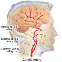
The internal carotid artery is an artery in the neck which supplies the anterior circulation of the brain.

<span class="mw-page-title-main">Scalp</span> Anatomical area bordered by the face at the front, and by the neck at the sides and back

The scalp is the area of the head where head hair grows. It is made up of skin, layers of connective and fibrous tissues, and the membrane of the skull. Anatomically, the scalp is part of the epicranium, a collection of structures covering the cranium. The scalp is bordered by the face at the front, and by the neck at the sides and back. The scientific study of hair and scalp is called trichology.

<span class="mw-page-title-main">Auriculotemporal nerve</span> Branch of the mandibular nerve

The auriculotemporal nerve is a sensory branch of the mandibular nerve (CN V3) that runs with the superficial temporal artery and vein, and provides sensory innervation to parts of the external ear, scalp, and temporomandibular joint. The nerve also conveys post-ganglionic parasympathetic fibres from the otic ganglion to the parotid gland.
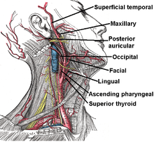
<span class="mw-page-title-main">Facial artery</span> Blood vessel supplying blood to the superficial face

The facial artery is a branch of the external carotid artery that supplies structures of the superficial face.

<span class="mw-page-title-main">Common carotid artery</span> One of the two arteries that supply the head and neck with blood

In anatomy, the left and right common carotid arteries (carotids) are arteries that supply the head and neck with oxygenated blood; they divide in the neck to form the external and internal carotid arteries.

<span class="mw-page-title-main">Superficial temporal artery</span> Major artery of the head

In human anatomy, the superficial temporal artery is a major artery of the head. It arises from the external carotid artery when it splits into the superficial temporal artery and maxillary artery.

<span class="mw-page-title-main">Occipital artery</span>

The occipital artery is a branch of the external carotid artery that provides arterial supply to the back of the scalp, sternocleidomastoid muscles, and deep muscles of the back and neck.

<span class="mw-page-title-main">Superior thyroid artery</span>

The superior thyroid artery arises from the external carotid artery just below the level of the greater cornu of the hyoid bone and ends in the thyroid gland.

<span class="mw-page-title-main">Inferior thyroid artery</span> Artery of the neck

The inferior thyroid artery is an artery in the neck. It arises from the thyrocervical trunk and passes upward, in front of the vertebral artery and longus colli muscle. It then turns medially behind the carotid sheath and its contents, and also behind the sympathetic trunk, the middle cervical ganglion resting upon the vessel.

<span class="mw-page-title-main">Posterior auricular artery</span> Small artery

The posterior auricular artery is a small artery that arises from the external carotid artery. It ascends along the side of the head. It supplies several muscles of the neck and several structures of the head.

<span class="mw-page-title-main">Maxillary artery</span> Artery supplying face structures in humans

The maxillary artery supplies deep structures of the face. It branches from the external carotid artery just deep to the neck of the mandible.

<span class="mw-page-title-main">Retromandibular vein</span> Major face vein

The retromandibular vein is a major vein of the face. It is formed within the parotid gland by the confluence of the maxillary vein, and superficial temporal vein. It descends in the gland and splits into two branches upon emerging from the gland. Its anterior branch then joins the (anterior) facial vein forming the common facial vein, while its posterior branch joins the posterior auricular vein forming the external jugular vein.

In anatomy, arterial tree is used to refer to all arteries and/or the branching pattern of the arteries. This article regards the human arterial tree. Starting from the aorta:

<span class="mw-page-title-main">Submandibular triangle</span>

The submandibular triangle corresponds to the region of the neck immediately beneath the body of the mandible.

<span class="mw-page-title-main">Carotid triangle</span>

The carotid triangle is a portion of the anterior triangle of the neck.

<span class="mw-page-title-main">Outline of human anatomy</span> Overview of and topical guide to human anatomy

The following outline is provided as an overview of and topical guide to human anatomy:

<span class="mw-page-title-main">Parapharyngeal space</span>

The parapharyngeal space, is a potential space in the head and the neck. It has clinical importance in otolaryngology due to parapharyngeal space tumours and parapharyngeal abscess developing in this area. It is also a key anatomic landmark for localizing disease processes in the surrounding spaces of the neck; the direction of its displacement indirectly reflects the site of origin for masses or infection in adjacent areas, and consequently their appropriate differential diagnosis.

The parotid fascia is a tough fascia enclosing the parotid gland. It has a superficial layer and a deep layer.

References

  1. 1 2 "Dictionnaire médical de l'Académie de Médecine". www.academie-medecine.fr. Retrieved 2024-05-09.
  2. 1 2 3 4 5 Ryan, Stephanie (2011). "Chapter 1". Anatomy for diagnostic imaging (Third ed.). Elsevier Ltd. pp. 45–50. ISBN   9780702029714.
  3. Human Anatomy – Lab 25 Step 12
  4. Lee, Whal (January 2014). "General principles of carotid Doppler ultrasonography". Ultrasonography. 33 (1): 11–17. doi:10.14366/usg.13018. PMC   4058969 . PMID   24936490.
  5. Kronick, Matthew D.; Chopra, Atish; Swamy, Shivam; Brar, Varneet; Jung, Enjae; Abraham, Cherrie Z.; Liem, Timothy K.; Landry, Gregory J.; Moneta, Gregory L. (September 2020). "Peak systolic velocity and color aliasing are important in the development of duplex ultrasound criteria for external carotid artery stenosis". Journal of Vascular Surgery. 72 (3): 951–957. doi: 10.1016/j.jvs.2019.10.099 . ISSN   1097-6809. PMID   31964570.