Carotid triangle

Last updated
Carotid triangle
Musculi coli base, my edits for tringles, Carotid T.svg
Carotid triangle
Details
Identifiers
Latin trigonum caroticum
TA98 A01.2.02.004
TA2 235
FMA 61598
Anatomical terminology

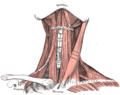
The carotid triangle (or superior carotid triangle) is a portion of the anterior triangle of the neck.

Contents

Anatomy

Boundaries

It is bounded:

Roof

The roof is formed by:

Floor

The floor is formed by (parts of) the:

Contents

Arteries

Veins

Nerves

Superficial to the carotid sheath lies the hypoglossal nerve, and ansa cervicalis of the cervical plexus.

The hypoglossal nerve crosses both the internal and external carotids, curving around the origin of the occipital artery.

Within the sheath, between the artery and vein, and behind both, is the vagus nerve; behind the sheath, the sympathetic trunk.

On the lateral side of the vessels, the accessory nerve runs for a short distance before it pierces the Sternocleidomastoideus; and on the medial side of the external carotid, just below the hyoid bone, the internal branch of the superior laryngeal nerve may be seen; and, still more inferiorly, the external branch of the same nerve.

Other

The superior portion of the larynx and inferior portion of the pharynx are also found in the anterior portion part of this space.

See also

Additional images

Related Research Articles

Articles related to anatomy include:

<span class="mw-page-title-main">External carotid artery</span> Major artery of the head and neck

The external carotid artery is a major artery of the head and neck. It arises from the common carotid artery when it splits into the external and internal carotid artery. The external carotid artery supplies blood to the face and neck.

<span class="mw-page-title-main">Internal carotid artery</span> Artery of the human brain

The internal carotid artery is an artery in the neck which supplies the anterior circulation of the brain.

<span class="mw-page-title-main">Jugular vein</span> Veins that bring deoxygenated blood from the head back to the heart

The jugular veins are veins that take deoxygenated blood from the head back to the heart via the superior vena cava. The internal jugular vein descends next to the internal carotid artery and continues posteriorly to the sternocleidomastoid muscle.

<span class="mw-page-title-main">Internal jugular vein</span> Blood vessel that drains the head

The internal jugular vein is a paired jugular vein that collects blood from the brain and the superficial parts of the face and neck. This vein runs in the carotid sheath with the common carotid artery and vagus nerve.

<span class="mw-page-title-main">Facial artery</span>

The facial artery is a branch of the external carotid artery that supplies structures of the superficial face.

<span class="mw-page-title-main">Common carotid artery</span> One of the two arteries that supply the head and neck with blood

In anatomy, the left and right common carotid arteries (carotids) are arteries that supply the head and neck with oxygenated blood; they divide in the neck to form the external and internal carotid arteries.

<span class="mw-page-title-main">Ansa cervicalis</span> From cervical plexus

The ansa cervicalis is a loop formed by muscular branches of the cervical plexus formed by branches of cervical spinal nerves C1-C3. The ansa cervicalis has two roots - a superior root and an inferior root - that unite distally, forming a loop. It is situated within the carotid sheath.

<span class="mw-page-title-main">Carotid sheath</span>

The carotid sheath is a condensation of the deep cervical fascia enveloping multiple vital neurovascular structures of the neck, including the common and internal carotid arteries, the internal jugular vein, the vagus nerve, and ansa cervicalis. The carotid sheath helps protects the structures contained therein.

<span class="mw-page-title-main">Occipital artery</span>

The occipital artery arises from the external carotid artery opposite the facial artery. Its path is below the posterior belly of digastric to the occipital region. This artery supplies blood to the back of the scalp and sternocleidomastoid muscles, and deep muscles in the back and neck.

<span class="mw-page-title-main">Posterior triangle of the neck</span>

The posterior triangle is a region of the neck.

<span class="mw-page-title-main">Superior thyroid artery</span>

The superior thyroid artery arises from the external carotid artery just below the level of the greater cornu of the hyoid bone and ends in the thyroid gland.

<span class="mw-page-title-main">Inferior thyroid artery</span> Artery of the neck

The inferior thyroid artery is an artery in the neck. It arises from the thyrocervical trunk and passes upward, in front of the vertebral artery and longus colli muscle. It then turns medially behind the carotid sheath and its contents, and also behind the sympathetic trunk, the middle cervical ganglion resting upon the vessel.

<span class="mw-page-title-main">Ascending pharyngeal artery</span>

The ascending pharyngeal artery is an artery in the neck that supplies the pharynx, developing from the proximal part of the embryonic second aortic arch.

<span class="mw-page-title-main">Anterior triangle of the neck</span>

The anterior triangle is a region of the neck.

<span class="mw-page-title-main">Deep cervical fascia</span>

The deep cervical fascia lies under cover of the platysma, and invests the muscles of the neck; it also forms sheaths for the carotid vessels, and for the structures situated in front of the vertebral column. Its attachment to the hyoid bone prevents the formation of a dewlap.

<span class="mw-page-title-main">Muscular triangle</span>

The inferior carotid triangle, is bounded, in front, by the median line of the neck from the hyoid bone to the sternum; behind, by the anterior margin of the sternocleidomastoid; above, by the superior belly of the omohyoid.

<span class="mw-page-title-main">Prevertebral fascia</span> Layer of deep cervical fascia that surrounds the vertebral column

The prevertebral fascia is the layer of deep cervical fascia that surrounds the vertebral column. It contains the sympathetic trunk, brachial plexus, phrenic nerve, prevertebral muscles, and the cervical vertebral column.

<span class="mw-page-title-main">Outline of human anatomy</span> Overview of and topical guide to human anatomy

The following outline is provided as an overview of and topical guide to human anatomy:

<span class="mw-page-title-main">Parapharyngeal space</span>

The parapharyngeal space, is a potential space in the head and the neck. It has clinical importance in otolaryngology due to parapharyngeal space tumours and parapharyngeal abscess developing in this area. It is also a key anatomic landmark for localizing disease processes in the surrounding spaces of the neck; the direction of its displacement indirectly reflects the site of origin for masses or infection in adjacent areas, and consequently their appropriate differential diagnosis.

References

PD-icon.svgThis article incorporates text in the public domain from page 564 of the 20th edition of Gray's Anatomy (1918)