In vertebrate anatomy, ribs are the long curved bones which form the rib cage, part of the axial skeleton. In most tetrapods, ribs surround the chest, enabling the lungs to expand and thus facilitate breathing by expanding the chest cavity. They serve to protect the lungs, heart, and other internal organs of the thorax. In some animals, especially snakes, ribs may provide support and protection for the entire body.
The rib cage is an endoskeletal enclosure in the thorax of most vertebrate animals that comprises the ribs, vertebral column and sternum, which protects vital organs such as the heart, lungs and great vessels. The circumferential enclosure formed by left and right rib cages, together known as the thoracic cage, is a semi-rigid bony and cartilaginous structure which surrounds the thoracic cavity and supports the shoulder girdles to form the core part of the axial skeleton.

The thoracic cavity is the chamber of the body of vertebrates that is protected by the thoracic wall. The central compartment of the thoracic cavity is the mediastinum. There are two openings of the thoracic cavity, a superior thoracic aperture known as the thoracic inlet and a lower inferior thoracic aperture known as the thoracic outlet.

The thorax or chest is a part of the anatomy of humans, mammals, and other tetrapod animals located between the neck and the abdomen. In insects, crustaceans, and the extinct trilobites, the thorax is one of the three main divisions of the creature's body, each of which is in turn composed of multiple segments.

The axial skeleton is the part of the skeleton that consists of the bones of the head and trunk of a vertebrate. In the human skeleton, it consists of 80 bones and is composed of six parts; the skull, also the ossicles of the middle ear, the hyoid bone, the rib cage, sternum and the vertebral column. The axial skeleton together with the appendicular skeleton form the complete skeleton. Another definition of axial skeleton is the bones including the vertebrae, sacrum, coccyx, skull, ribs, and sternum.

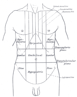
The rectus abdominis muscle, also known as the "abdominal muscle" or simply the "abs", is a pair of segmented skeletal muscle on the ventral aspect of a person's abdomen. The paired muscles are separated at the midline by a band of dense connective tissue called the linea alba, and the connective tissue defining each lateral margin of the rectus abdominus is the linea semilunaris. The muscle extends from the pubic symphysis, pubic crest and pubic tubercle inferiorly, to the xiphoid process and costal cartilages of the 5th–7th ribs superiorly.

The rib eye or ribeye is a boneless rib steak from the rib section.

The transversus thoracis muscle, also known as triangularis sterni, lies internal to the thoracic cage, anteriorly. It is usually a thin plane of muscular and tendinous fibers, however on athletic individuals it can be a thick 'slab of meat', situated upon the inner surface of the front wall of the chest. It is in the same layer as the subcostal muscles and the innermost intercostal muscles.

Pork ribs are a cut of pork popular in Western and Asian cuisines. The ribcage of a domestic pig, meat and bones together, is cut into usable pieces, prepared by smoking, grilling, or baking – usually with a sauce, often barbecue – and then served.
The costochondral joints are the joints between the ribs and costal cartilage in the front of the rib cage. They are hyaline cartilaginous joints. Each rib has a depression shaped like a cup that the costal cartilage articulates with. There is normally no movement at these joints. Joints between costal cartilages of the sixth and ninth rib are plane synovial joints. Articulation between costal cartilage of the ninth rib and tenth rib is fibrous.

Flail chest is a life-threatening medical condition that occurs when a segment of the rib cage breaks due to trauma and becomes detached from the rest of the chest wall. Two of the symptoms of flail chest are chest pain and shortness of breath.

An hourglass corset is a garment that produces a silhouette resembling an hourglass shape characterized by wide hips, narrow waist, and wide bust.

Spare ribs are a variety of ribs cut from the lower portion of a pig, specifically the belly and breastbone, behind the shoulder, and include 11 to 13 long bones. Meat and fat cover the bones. Spare ribs (pork) are distinguished from short ribs, which are beef. Spareribs are typically cooked low and slow, either smoked, grilled, or braised.

In French, entrecôte is a premium cut of beef used for steaks and roasts.

The muscles of respiration are the muscles that contribute to inhalation and exhalation, by aiding in the expansion and contraction of the thoracic cavity. The diaphragm and, to a lesser extent, the intercostal muscles drive respiration during quiet breathing. The elasticity of these muscles is crucial to the health of the respiratory system and to maximize its functional capabilities.

Asphyxiating thoracic dysplasia (ATD), also known as Jeune syndrome, is a rare inherited bone growth disorder that primarily affects the thoracic region. It was first described in 1955 by the French pediatrician Mathis Jeune. Common signs and symptoms can include a narrow chest, short ribs, shortened bones in the arms and legs, short stature, and extra fingers and toes (polydactyly). The restricted growth and expansion of the lungs caused by this disorder results in life-threatening breathing difficulties; occurring in 1 in every 100,000-130,000 live births in the United States.

The sternum or breastbone is a long flat bone located in the central part of the chest. It connects to the ribs via cartilage and forms the front of the rib cage, thus helping to protect the heart, lungs, and major blood vessels from injury. Shaped roughly like a necktie, it is one of the largest and longest flat bones of the body. Its three regions are the manubrium, the body, and the xiphoid process. The word sternum originates from Ancient Greek στέρνον (stérnon) 'chest'.
Eocasea is an extinct genus of caseid synapsids from the Late Pennsylvanian of Kansas. It is known from a single type species, Eocasea martini.
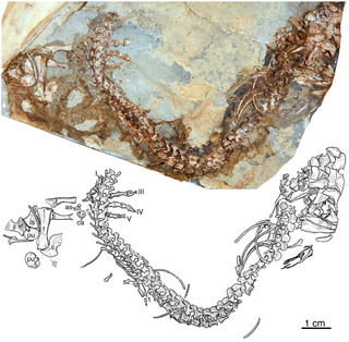
Parahupehsuchus is an extinct genus of hupehsuchian marine reptiles from the Early Triassic of China. The genus is monotypic, known from the single species Parahupehsuchus longus and based on a single specimen. Like other hupehsuchians, it had an elongated torso, a tail nearly as long as the rest of the body, short and paddle-like limbs, extra bones in the fore- and hind limbs, thick ribs and gastralia, neural spines of the vertebrae split into two parts, and bony plates over the neural spines. It differs from other hupehsuchians in having an even more elongated body and wider ribs that touch along their edges and have no spaces between them. The ribs connect with gastralia on the underside of the torso to form a bony "tube" around the body wall.

Baluse or Baloese is a traditional shield of the Nias people originating from Nias, an island off the west coast of North Sumatra, Indonesia. Baluse in the Northern Nias is somewhat smaller than those of the rest of the island.