The cubital fossa, antecubital fossa, chelidon, inside of elbow, or, humorously, wagina,[1] is the area on the anterior side of the upper part between the arm and forearm of a human or other hominid animals. It lies anteriorly to the elbow (antecubital) (Latin cubitus) when in standard anatomical position. The cubital fossa is a triangular area having three borders.[2]
The cubital fossa contains four main vertical structures (from lateral to medial):
The radial nerve passes underneath the brachioradialis muscle where it divides into deep and superficial branches. It is not always considered part of the cubital fossa but is in the vicinity.[2]
The brachial artery. The artery usually bifurcates near the apex (inferior part) of the cubital fossa into the radial artery (superficial) and ulnar artery (deeper)
Several veins are also in the area (for example, the median cubital vein, cephalic vein, and basilic vein) but these are usually considered superficial to the cubital fossa, and not part of its contents.
From lateral to medial, the order of the contents within the cubital fossa can be described by the acronym TAN: tendon, artery, nerve
Like other flexion surfaces of large joints (groin, popliteal fossa, armpit and essentially the anterior part of the neck), it is an area where blood vessels and nerves pass relatively superficially, and with an increased amount of lymph nodes.
During blood pressuremeasurements, the stethoscope is placed over the brachial artery in the cubital fossa. The artery runs medial to the biceps tendon. The brachial pulse may be palpated in the cubital fossa just medial to the tendon.
Historically, during bloodletting, the bicipital aponeurosis (the ceiling of the cubital fossa) was known as the "grace of God tendon" because it separated and protected the more important contents of the fossa such as the brachial artery and the median nerve.[2]
Statistically, the antecubital fossa is the least tender region for peripheral intravenous access, although it provides a greater risk for venous thrombosis.
Additional images
Superficial veins of the upper limb.
Front of right upper extremity.
Front of right upper extremity, showing surface markings for bones, arteries, and nerves.
This page is based on this Wikipedia article Text is available under the CC BY-SA 4.0 license; additional terms may apply. Images, videos and audio are available under their respective licenses.


 Superficial veins of the upper limb.
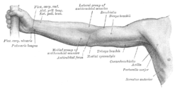 Front of right upper extremity.
 Front of right upper extremity, showing surface markings for bones, arteries, and nerves.


