Ulnar nerve

Last updated
Ulnar nerve
Brachial plexus 2.svg
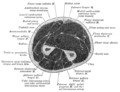
Click image to enlarge - ulnar nerve is visible in lower left
Nerves of the left upper extremity.gif
Nerves of the left upper extremity. (Ulnar labeled at center left.)
Details
FromC8, T1 (branch from medial cord)
Innervates Flexor carpi ulnaris
flexor digitorum profundus
lumbrical muscles
opponens digiti minimi
flexor digiti minimi
abductor digiti minimi
interossei
adductor pollicis
Identifiers
Latin nervus ulnaris
MeSH D014459
TA98 A14.2.03.040
TA2 6449
FMA 37319
Anatomical terms of neuroanatomy

In human anatomy, the ulnar nerve is a nerve that runs near the ulna bone. The ulnar collateral ligament of elbow joint is in relation with the ulnar nerve. The nerve is the largest in the human body unprotected by muscle or bone, so injury is common. [1] This nerve is directly connected to the little finger, and the adjacent half of the ring finger, innervating the palmar aspect of these fingers, including both front and back of the tips, perhaps as far back as the fingernail beds.

Contents

This nerve can cause an electric shock-like sensation by striking the medial epicondyle of the humerus posteriorly, or inferiorly with the elbow flexed. The ulnar nerve is trapped between the bone and the overlying skin at this point. This is commonly referred to as bumping one's "funny bone". This name is thought to be a pun, based on the sound resemblance between the name of the bone of the upper arm, the humerus, and the word "humorous". [2] Alternatively, according to the Oxford English Dictionary, it may refer to "the peculiar sensation experienced when it is struck". [3]

Structure

Arm

The ulnar nerve originates from the C8-T1 nerve roots (and occasionally carries C7 fibers which arise from the lateral cord), [4] [5] which then form part of the medial cord of the brachial plexus, and descends medial to the brachial artery, up until the insertion point of coracobrachialis muscle (middle 5 cm over the medial border of the humerus). Then, it pierces the medial intermuscular septum and enters the posterior compartment of the arm, accompanied by superior ulnar collateral vessels. It runs at the posteromedial aspects of the humerus, passing behind the medial epicondyle (in the cubital tunnel) at the elbow, where it can be palpated by hand. [6]

Forearm

The ulnar nerve is not a content of the cubital fossa. It enters the anterior (flexor) compartment of the forearm between the two heads of flexor carpi ulnaris, [5] and lies along the lateral border of the flexor carpi ulnaris. [5] The ulnar nerve runs between the flexor digitorum superficialis (laterally) and flexor digitorum profundus medially. Near the wrist, it courses superficial to the flexor retinaculum of hand, but covered by volar carpal ligament to enter the hand. [6]

In the forearm it gives off the following branches: [7] :700

Hand

Branches of the ulnar nerve in hand Guyon canal syndrome zones.svg
Branches of the ulnar nerve in hand

Ulnar nerve enters the palm of the hand via the Guyon's canal, superficial to the flexor retinaculum and lateral to the pisiform bone. [6]

Here it gives off the following branches: [7]

Function

Ulnar nerve is also known as "musician's nerve" as it controls the fine movements of the fingers. [6]

Sensory

Cutaneous innervation of the right upper extremity. Areas innervated by the ulnar nerve are the areas on the hand colored in light blue. Gray812and814.PNG
Cutaneous innervation of the right upper extremity. Areas innervated by the ulnar nerve are the areas on the hand colored in light blue.

The ulnar nerve also provides sensory innervation to the fifth digit and the medial half of the fourth digit, and the corresponding part of the palm:

Motor

The ulnar nerve and its branches innervate the following muscles in the forearm and hand:

Clinical significance

The ulnar nerve can suffer injury anywhere between its proximal origin of the brachial plexus all the way to its distal branches in the hand. It is the most commonly injured nerve around the elbow. [8] [9] Although it can be damaged under various circumstances, it is commonly injured by local trauma or physical impingement ("pinched nerve"). Injury of the ulnar nerve at different levels causes specific motor and sensory deficits.

At the elbow

At the wrist

In severe cases, surgery may be performed to relocate or "release" the nerve to prevent further injury.

Additional images

See also

Related Research Articles

<span class="mw-page-title-main">Arm</span> Proximal part of the free upper limb between the shoulder and the elbow

In human anatomy, the arm refers to the upper limb in common usage, although academically the term specifically means the upper arm between the glenohumeral joint and the elbow joint. The distal part of the upper limb between the elbow and the radiocarpal joint is known as the forearm or "lower" arm, and the extremity beyond the wrist is the hand.

<span class="mw-page-title-main">Carpal bones</span> Eight small bones that make up the wrist (or carpus) that connects the hand to the forearm

The carpal bones are the eight small bones that make up the wrist (carpus) that connects the hand to the forearm. The term "carpus" and "carpal" is derived from the Latin carpus and the Greek καρπός (karpós), meaning "wrist". In human anatomy, the main role of the carpal bones is to articulate with the radial and ulnar heads to form a highly mobile condyloid joint, to provide attachments for thenar and hypothenar muscles, and to form part of the rigid carpal tunnel which allows the median nerve and tendons of the anterior forearm muscles to be transmitted to the hand and fingers.

<span class="mw-page-title-main">Radial nerve</span> Nerve in the human body that supplies the posterior portion of the upper limb

The radial nerve is a nerve in the human body that supplies the posterior portion of the upper limb. It innervates the medial and lateral heads of the triceps brachii muscle of the arm, as well as all 12 muscles in the posterior osteofascial compartment of the forearm and the associated joints and overlying skin.

<span class="mw-page-title-main">Median nerve</span> Nerve of the upper limb

The median nerve is a nerve in humans and other animals in the upper limb. It is one of the five main nerves originating from the brachial plexus.

The forearm is the region of the upper limb between the elbow and the wrist. The term forearm is used in anatomy to distinguish it from the arm, a word which is used to describe the entire appendage of the upper limb, but which in anatomy, technically, means only the region of the upper arm, whereas the lower "arm" is called the forearm. It is homologous to the region of the leg that lies between the knee and the ankle joints, the crus.

In human anatomy, the extensor carpi ulnaris is a skeletal muscle located on the ulnar side of the forearm. The extensor carpi ulnaris acts to extend and adduct at the carpus/wrist from anatomical position.

<span class="mw-page-title-main">Flexor carpi radialis muscle</span> Superficial muscle in the anterior side of the forearm

In anatomy, flexor carpi radialis is a muscle of the human forearm that acts to flex and (radially) abduct the hand. The Latin carpus means wrist; hence flexor carpi is a flexor of the wrist.

<span class="mw-page-title-main">Wrist drop</span> Medical condition

Wrist drop is a medical condition in which the wrist and the fingers cannot extend at the metacarpophalangeal joints. The wrist remains partially flexed due to an opposing action of flexor muscles of the forearm. As a result, the extensor muscles in the posterior compartment remain paralyzed.

<span class="mw-page-title-main">Upper limb</span> Consists of the arm, forearm, and hand

The upper limbs or upper extremities are the forelimbs of an upright-postured tetrapod vertebrate, extending from the scapulae and clavicles down to and including the digits, including all the musculatures and ligaments involved with the shoulder, elbow, wrist and knuckle joints. In humans, each upper limb is divided into the arm, forearm and hand, and is primarily used for climbing, lifting and manipulating objects.

<span class="mw-page-title-main">Ulnar artery</span> Artery of the forearm

The ulnar artery is the main blood vessel, with oxygenated blood, of the medial aspects of the forearm. It arises from the brachial artery and terminates in the superficial palmar arch, which joins with the superficial branch of the radial artery. It is palpable on the anterior and medial aspect of the wrist.

<span class="mw-page-title-main">Flexor carpi ulnaris muscle</span> Muscle of the forearm

The flexor carpi ulnaris (FCU) is a muscle of the forearm that flexes and adducts at the wrist joint.

<span class="mw-page-title-main">Extensor digitorum muscle</span> Muscle of the posterior forearm

The extensor digitorum muscle is a muscle of the posterior forearm present in humans and other animals. It extends the medial four digits of the hand. Extensor digitorum is innervated by the posterior interosseous nerve, which is a branch of the radial nerve.

The pronator teres is a muscle that, along with the pronator quadratus, serves to pronate the forearm. It has two origins, at the medial humeral supracondylar ridge and the ulnar tuberosity, and inserts near the middle of the radius.

<span class="mw-page-title-main">Dorsal interossei of the hand</span> Muscles between the metacarpals

In human anatomy, the dorsal interossei (DI) are four muscles in the back of the hand that act to abduct (spread) the index, middle, and ring fingers away from hand's midline and assist in flexion at the metacarpophalangeal joints and extension at the interphalangeal joints of the index, middle and ring fingers.

<span class="mw-page-title-main">Abductor digiti minimi muscle of hand</span> Muscle in the hypothenar compartment

In human anatomy, the abductor digiti minimi is a skeletal muscle situated on the ulnar border of the palm of the hand. It forms the ulnar border of the palm and its spindle-like shape defines the hypothenar eminence of the palm together with the skin, connective tissue, and fat surrounding it. Its main function is to pull the little finger away from the other fingers.

<span class="mw-page-title-main">Flexor digiti minimi brevis muscle of hand</span> Muscle in the hypothenar compartment

The flexor digiti minimi brevis is a hypothenar muscle in the hand that flexes the little finger at the metacarpophalangeal joint. It lies lateral to the abductor digiti minimi when the hand is in anatomical position.

The posterior compartment of the forearm contains twelve muscles which primarily extend the wrist and digits. It is separated from the anterior compartment by the interosseous membrane between the radius and ulna.

<span class="mw-page-title-main">Cervical spinal nerve 8</span> Spinal nerve of the cervical segment

The cervical spinal nerve 8 (C8) is a spinal nerve of the cervical segment.

<span class="mw-page-title-main">Ulnar claw</span> Deformity of the hand that develops due to ulnar nerve damage

An ulnar claw, also known as claw hand or ‘Spinster’s Claw’, is a deformity or an abnormal attitude of the hand that develops due to ulnar nerve damage causing paralysis of the lumbricals. A claw hand presents with a hyperextension at the metacarpophalangeal joints and flexion at the proximal and distal interphalangeal joints of the 4th and 5th fingers. The patients with this condition can make a full fist but when they extend their fingers, the hand posture is referred to as claw hand. The ring- and little finger can usually not fully extend at the proximal interphalangeal joint (PIP).

<span class="mw-page-title-main">Extrinsic extensor muscles of the hand</span>

The extrinsic extensor muscles of the hand are located in the back of the forearm and have long tendons connecting them to bones in the hand, where they exert their action. Extrinsic denotes their location outside the hand. Extensor denotes their action which is to extend, or open flat, joints in the hand. They include the extensor carpi radialis longus (ECRL), extensor carpi radialis brevis (ECRB), extensor digitorum (ED), extensor digiti minimi (EDM), extensor carpi ulnaris (ECU), abductor pollicis longus (APL), extensor pollicis brevis (EPB), extensor pollicis longus (EPL), and extensor indicis (EI).

References

  1. N, Catena; Mg, Calevo; D, Fracassetti; D, Moharamzadeh; C, Origo; M, De Pellegrin (2019). "Risk of Ulnar Nerve Injury During Cross-Pinning in Supine and Prone Position for Supracondylar Humeral Fractures in Children: A Recent Literature Review". European Journal of Orthopaedic Surgery & Traumatology: Orthopedie Traumatologie. 29 (6): 1169–1175. doi:10.1007/s00590-019-02444-0. PMID   31037406. S2CID   139108013 . Retrieved 2020-05-22.
  2. Hendrickson, Robert A. (2004). The Facts on File Encyclopedia of Word and Phrase Origins (Facts on File Writer's Library). New York: Checkmark Books. p. 281. ISBN   0-8160-5992-6.
  3. "Welcome to the new OED Online: Oxford English Dictionary". Dictionary.oed.com. Retrieved 2012-03-20.
  4. 1 2 3 4 Bonfiglioli, Roberta; Mattioli, Stefano; Violante, Francesco S. (2015-01-01), Lotti, Marcello; Bleecker, Margit L. (eds.), "Chapter 22 - Occupational mononeuropathies in industry", Handbook of Clinical Neurology, Occupational Neurology, 131, Elsevier: 411–426, doi:10.1016/b978-0-444-62627-1.00021-4, ISBN   9780444626271, PMID   26563800 , retrieved 2020-10-25
  5. 1 2 3 4 Rea, Paul (2016-01-01), Rea, Paul (ed.), "Chapter 3 - Neck", Essential Clinically Applied Anatomy of the Peripheral Nervous System in the Head and Neck, Academic Press, pp. 131–183, doi:10.1016/b978-0-12-803633-4.00003-x, ISBN   978-0-12-803633-4 , retrieved 2020-10-25
  6. 1 2 3 4 5 6 7 8 9 10 11 Krishna, Garg (2010). "8 - Arm". BD Chaurasia's Human Anatomy (Regional and Applied Dissection and Clinical) Volume 1 - Upper limb and thorax (Fifth ed.). India: CBS Publishers and Distributors Pvt Ltd. p. 91,110,111. ISBN   978-81-239-1863-1.
  7. 1 2 Ellis, Harold; Susan Standring; Gray, Henry David (2005). Gray's anatomy: the anatomical basis of clinical practice. St. Louis, Mo: Elsevier Churchill Livingstone. p.  726. ISBN   0-443-07168-3.
  8. Selby, Ronald; Safran, Marc; O'brien, Stephen (2007). "Practical Orthopaedic Sports Medicine & Arthroscopy, 1st edition: Elbow Injuries". msdlatinamerica.com. Lippincott Williams & Wilkins. Archived from the original on 2014-10-06. Retrieved 2014-09-30.
  9. Minieka, Michael; Nishida, Takashi (2005-01-01), Benzon, Honorio T.; Raja, Srinivasa N.; Molloy, Robert E.; Liu, Spencer S. (eds.), "Chapter 54 - Entrapment Neuropathies", Essentials of Pain Medicine and Regional Anesthesia (Second Edition), Philadelphia: Churchill Livingstone, pp. 426–432, doi:10.1016/b978-0-443-06651-1.50058-7, ISBN   978-0-443-06651-1 , retrieved 2020-10-25
  10. Waldman, Steven D. (2014-01-01), Waldman, Steven D. (ed.), "Chapter 44 - Driver's Elbow", Atlas of Uncommon Pain Syndromes (Third Edition), Philadelphia: W.B. Saunders, pp. 126–129, doi:10.1016/b978-1-4557-0999-1.00044-7, ISBN   978-1-4557-0999-1 , retrieved 2020-10-25
  11. Fuller, Geraint; Manford, Mark (2010-01-01), Fuller, Geraint; Manford, Mark (eds.), "Common peripheral nerve lesions", Neurology (Third Edition), Churchill Livingstone, pp. 106–107, doi:10.1016/b978-0-7020-3224-0.00054-9, ISBN   978-0-7020-3224-0, S2CID   88836902 , retrieved 2020-10-25