Dorsal scapular nerve

Last updated

Dorsal scapular nerve
Gray808.png
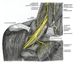
The right brachial plexus with its short branches, viewed from in front. (Dorsalis scapulae labeled at left, second from top.)
Details
FromC5 of brachial plexus
Innervates Rhomboid minor, rhomboid major, levator scapulae
Identifiers
Latin nervus dorsalis scapulae
TA98 A14.2.03.011
TA2 6409
FMA 65279
Anatomical terms of neuroanatomy

The dorsal scapular nerve is a branch of the brachial plexus, usually derived from the ventral ramus of cervical nerve C5. It provides motor innervation to the rhomboid major muscle, rhomboid minor muscle, and levator scapulae muscle.

Contents

Dorsal scapular nerve syndrome can cause a winged scapula, with pain and limited motion.

Structure

Origin

The dorsal scapular nerve arises from the brachial plexus, usually from the plexus root (anterior (ventral) ramus) of cervical nerve C5. [1] [2] [3]

Course and relations

Once the nerve leaves C5 it commonly pierces the middle scalene muscle. [1] It continues deep to levator scapulae muscle and the rhomboids (minor superior to major). [1]

The nerve is accompanied by dorsal scapular artery. [4]

Function

The dorsal scapular nerve provides motor innervation to the two rhomboid muscles, [2] [3] and the levator scapulae muscle. [2] [3]

Clinical significance

Injury to the dorsal scapular nerve is usually apparent on inspection when the scapula on the injured side is located farther from the midline than the uninjured scapula. The patient would be unable to pull their shoulder back, as when standing at attention. Isolated dorsal scapular nerve injury is uncommon, but case reports usually involve injury to the scalene muscles. [5]

The dorsal scapular nerve is typically not anaesthetised during a supraclavicular nerve block. [6] This can cause pain after some surgeries. [6]

Dorsal scapular nerve syndrome

Dorsal scapular nerve syndrome can be caused by nerve compression syndrome. A winged scapula is the most common symptom. [7] Shoulder pain may occur. [7] It causes weakness in rhomboid major muscle, rhomboid minor muscle, and levator scapulae muscle. [7] The range of motion of the shoulder may be limited. [7] Treatment is usually conservative. [7]

See also

Additional images

Related Research Articles

<span class="mw-page-title-main">Trapezius</span> Muscle between the lower spine and the shoulder blade

The trapezius is a large paired trapezoid-shaped surface muscle that extends longitudinally from the occipital bone to the lower thoracic vertebrae of the spine and laterally to the spine of the scapula. It moves the scapula and supports the arm.

<span class="mw-page-title-main">Brachial plexus</span> Network of nerves

The brachial plexus is a network of nerves formed by the anterior rami of the lower four cervical nerves and first thoracic nerve. This plexus extends from the spinal cord, through the cervicoaxillary canal in the neck, over the first rib, and into the armpit, it supplies afferent and efferent nerve fibers to the chest, shoulder, arm, forearm, and hand.

<span class="mw-page-title-main">Long thoracic nerve</span> Large nerve

The long thoracic nerve is a branch of the brachial plexus derived from cervical nerves C5-C7 that innervates the serratus anterior muscle.

<span class="mw-page-title-main">Subclavian artery</span> Major arteries of the upper thorax, below the clavicle

In human anatomy, the subclavian arteries are paired major arteries of the upper thorax, below the clavicle. They receive blood from the aortic arch. The left subclavian artery supplies blood to the left arm and the right subclavian artery supplies blood to the right arm, with some branches supplying the head and thorax. On the left side of the body, the subclavian comes directly off the aortic arch, while on the right side it arises from the relatively short brachiocephalic artery when it bifurcates into the subclavian and the right common carotid artery.

<span class="mw-page-title-main">Levator scapulae muscle</span> Slender skeletal muscle at the back and side of the neck

The levator scapulae is a slender skeletal muscle situated at the back and side of the neck. It originates from the transverse processes of the four uppermost cervical vertebrae; it inserts onto the upper portion of the medial border of the scapula. It is innervated by the cervical nerves C3-C4, and frequently also by the dorsal scapular nerve. As the Latin name suggests, its main function is to lift the scapula.

<span class="mw-page-title-main">Rhomboid major muscle</span> Skeletal muscle in the human back

The rhomboid major is a skeletal muscle of the back that connects the scapula with the vertebrae of the spinal column. It originates from the spinous processes of the thoracic vertebrae T2–T5 and supraspinous ligament; it inserts onto the lower portion of the medial border of the scapula. It acts together with the rhomboid minor to keep the scapula pressed against thoracic wall and to retract the scapula toward the vertebral column.

<span class="mw-page-title-main">Rhomboid minor muscle</span> Skeletal muscle of the upper back

In human anatomy, the rhomboid minor is a small skeletal muscle of the back that connects the scapula to the vertebrae of the spinal column. It arises from the nuchal ligament, and the 7th cervical and 1st thoracic vertebrae and intervening supraspinous ligaments; it inserts onto the medial border of the scapula. It is innervated by the dorsal scapular nerve.

<span class="mw-page-title-main">Cervical plexus</span> Network of nerves in the neck

The cervical plexus is a nerve plexus of the anterior rami of the first four cervical spinal nerves C1-C4. The cervical plexus provides motor innervation to some muscles of the neck, and the diaphragm; it provides sensory innervation to parts of the head, neck, and chest.

<span class="mw-page-title-main">Suprascapular nerve</span> Mixed nerve of the upper limb

The suprascapular nerve is a mixed nerve that branches from the upper trunk of the brachial plexus. It is derived from the ventral rami of cervical nerves C5-C6. It provides motor innervation to the supraspinatus muscle, and the infraspinatus muscle.

<span class="mw-page-title-main">Serratus anterior muscle</span> Muscle on the surface of the ribs

The serratus anterior is a muscle of the chest. It originates at the side of the chest from the upper 8 or 9 ribs; it inserts along the entire length of the anterior aspect of the medial border of the scapula. It is innervated by the long thoracic nerve from the brachial plexus. The serratus anterior acts to pull the scapula forward around the thorax.

<span class="mw-page-title-main">Scalene muscles</span> Muscles on the sides of the neck

The scalene muscles are a group of three muscles on each side of the neck, identified as the anterior, the middle, and the posterior. They are innervated by the third to the eighth cervical spinal nerves (C3-C8).

<span class="mw-page-title-main">Intercostal nerves</span> Nerves in the thorax and abdomen

The intercostal nerves are part of the somatic nervous system, and arise from the anterior rami of the thoracic spinal nerves from T1 to T11. The intercostal nerves are distributed chiefly to the thoracic pleura and abdominal peritoneum, and differ from the anterior rami of the other spinal nerves in that each pursues an independent course without plexus formation.

<span class="mw-page-title-main">Medial pectoral nerve</span>

The medial pectoral nerve is (typically) a branch of the medial cord of the brachial plexus and is derived from spinal nerve roots C8-T1. It provides motor innervation to the pectoralis minor muscle, and the lower half of the pectoralis major muscle. It runs along the inferior border of the pectoralis minor muscle.

<span class="mw-page-title-main">Thoracodorsal nerve</span> Nerve that supplies the latissimus dorsi

The thoracodorsal nerve is a nerve present in humans and other animals, also known as the middle subscapular nerve or the long subscapular nerve. It supplies the latissimus dorsi muscle.

<span class="mw-page-title-main">Posterior triangle of the neck</span> Region of the neck

The posterior triangle is a region of the neck.

<span class="mw-page-title-main">Transverse cervical artery</span> Artery of the neck

The transverse cervical artery is an artery in the neck and a branch of the thyrocervical trunk, running at a higher level than the suprascapular artery.

<span class="mw-page-title-main">Lower subscapular nerve</span>

The lower subscapular nerve, also known as the inferior subscapular nerve, is the third branch of the posterior cord of the brachial plexus. It innervates the inferior portion of the subscapularis muscle and the teres major muscle.

<span class="mw-page-title-main">Lateral pectoral nerve</span>

The lateral pectoral nerve arises from the lateral cord of the brachial plexus, and through it from the C5-7.

<span class="mw-page-title-main">Piriformis nerve</span>

The piriformis nerve, also known as the nerve to piriformis, is the peripheral nerve that provides motor innervation to the piriformis muscle.

The Eden–Lange procedure is an orthopedic procedure to alleviate the symptoms of trapezius palsy when more conservative measures, such as spontaneous resolution and surgical nerve repair are not promising. The rhomboid major, rhomboid minor, and levator scapulae muscles are transferred laterally along the scapula to replace the functions of the lower, middle, and upper fibers of the trapezius, respectively. The transferred muscles hold the scapula in a more medial and upwardly rotated position, without winging.

References

  1. 1 2 3 Tubbs, R. Shane; Goodrich, Dylan; Watanabe, Koichi; Loukas, Marios (January 1, 2015), Tubbs, R. Shane; Rizk, Elias; Shoja, Mohammadali M.; Loukas, Marios (eds.), "Chapter 43 - Anatomic Landmarks for Selected Nerves of the Head, Neck, and Upper and Lower Limbs", Nerves and Nerve Injuries, San Diego: Academic Press, pp. 575–588, doi:10.1016/b978-0-12-410390-0.00045-7, ISBN   978-0-12-410390-0 , retrieved November 2, 2020
  2. 1 2 3 Felten, David L.; O'Banion, M. Kerry; Maida, Mary Summo (2016), "Peripheral Nervous System", Netter's Atlas of Neuroscience, Elsevier, pp. 153–231, doi:10.1016/b978-0-323-26511-9.00009-6, ISBN   978-0-323-26511-9 , retrieved November 2, 2020
  3. 1 2 3 Ma, Yun-tao (January 1, 2011), Ma, Yun-tao (ed.), "CHAPTER 8 - Neuroanatomy of Acu-Reflex Points", Acupuncture for Sports and Trauma Rehabilitation, Saint Louis: Churchill Livingstone, pp. 88–131, doi:10.1016/b978-1-4377-0927-8.00008-7, ISBN   978-1-4377-0927-8 , retrieved November 2, 2020
  4. Chang, Ke-Vin; Lin, Chih-Peng; Lin, Chia-Shiang; Wu, Wei-Ting; Karmakar, Manoj K; Özçakar, Levent (January 4, 2017). "Sonographic tracking of trunk nerves: essential for ultrasound-guided pain management and research". Journal of Pain Research. 10: 79–88. doi: 10.2147/JPR.S123828 . ISSN   1178-7090. PMC   5222601 . PMID   28115867.
  5. Akuthota, Venu; Herring, Stanley A. (May 28, 2009). Nerve and Vascular Injuries in Sports Medicine. Springer Science & Business Media. ISBN   9780387766003 via Google Books.
  6. 1 2 Williams, Brain A.; Neumann, Krystof J.; Goel, Shiv K.; Wu, Christopher L. (2008). "14 - Postoperative Pain and Other Acute Pain Syndromes". Raj's Practical Management of Pain (4th ed.). Mosby. pp. 299–334. doi:10.1016/B978-032304184-3.50017-0. ISBN   978-0-323-04184-3.
  7. 1 2 3 4 5 Rea, Paul (January 1, 2015). "Chapter 2 - Upper Limb Nerve Supply". Essential Clinically Applied Anatomy of the Peripheral Nervous System in the Limbs. Academic Press. pp. 41–100. doi:10.1016/B978-0-12-803062-2.00002-4. ISBN   978-0-12-803062-2.{{cite book}}: CS1 maint: date and year (link)