| Medial brachial cutaneous nerve | |
|---|---|
 Diagram of segmental distribution of the cutaneous nerves of the right upper extremity. Anterior view. "Medial Brachial Cutan. T1-T2" labeled near center, in yellow. | |
 Cutaneous nerves of right upper extremity. | |
| Details | |
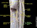
| From | T1 (medial cord) |
| Identifiers | |
| Latin | n. cutaneus brachii medialis |
| TA98 | A14.2.03.027 |
| TA2 | 6445 |
| FMA | 65246 |
| Anatomical terms of neuroanatomy | |
The medial brachial cutaneous nerve (lesser internal cutaneous nerve; medial cutaneous nerve of arm) is a sensory branch of the medial cord of the brachial plexus derived from spinal nerves C8-T1. It provides sensory innervation to the medial arm. It descends accompanied by the basilic vein. [1]