The thumb is the first digit of the hand, next to the index finger. When a person is standing in the medical anatomical position, the thumb is the outermost digit. The Medical Latin English noun for thumb is pollex, and the corresponding adjective for thumb is pollical.

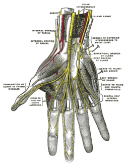
The radial nerve is a nerve in the human body that supplies the posterior portion of the upper limb. It innervates the medial and lateral heads of the triceps brachii muscle of the arm, as well as all 12 muscles in the posterior osteofascial compartment of the forearm and the associated joints and overlying skin.

The median nerve is a nerve in humans and other animals in the upper limb. It is one of the five main nerves originating from the brachial plexus.

The thenar eminence is the mound formed at the base of the thumb on the palm of the hand by the intrinsic group of muscles of the thumb. The skin overlying this region is the area stimulated when trying to elicit a palmomental reflex. The word thenar comes from Ancient Greek θέναρ (thenar) 'palm of the hand'.

In human anatomy, the ulnar nerve is a nerve that runs near the ulna bone. The ulnar collateral ligament of elbow joint is in relation with the ulnar nerve. The nerve is the largest in the human body unprotected by muscle or bone, so injury is common. This nerve is directly connected to the little finger, and the adjacent half of the ring finger, innervating the palmar aspect of these fingers, including both front and back of the tips, perhaps as far back as the fingernail beds.

The upper limbs or upper extremities are the forelimbs of an upright-postured tetrapod vertebrate, extending from the scapulae and clavicles down to and including the digits, including all the musculatures and ligaments involved with the shoulder, elbow, wrist and knuckle joints. In humans, each upper limb is divided into the arm, forearm and hand, and is primarily used for climbing, lifting and manipulating objects.
In human anatomy, the extensor pollicis longus muscle (EPL) is a skeletal muscle located dorsally on the forearm. It is much larger than the extensor pollicis brevis, the origin of which it partly covers and acts to stretch the thumb together with this muscle.

The flexor pollicis brevis is a muscle in the hand that flexes the thumb. It is one of three thenar muscles. It has both a superficial part and a deep part.
The flexor pollicis longus is a muscle in the forearm and hand that flexes the thumb. It lies in the same plane as the flexor digitorum profundus. This muscle is unique to humans, being either rudimentary or absent in other primates. A meta-analysis indicated accessory flexor pollicis longus is present in around 48% of the population.
In human anatomy, the adductor pollicis muscle is a muscle in the hand that functions to adduct the thumb. It has two heads: transverse and oblique.

In human anatomy, the palmar or volar interossei are four muscles, one on the thumb that is occasionally missing, and three small, unipennate, central muscles in the hand that lie between the metacarpal bones and are attached to the index, ring, and little fingers. They are smaller than the dorsal interossei of the hand.

In human anatomy, the abductor digiti minimi is a skeletal muscle situated on the ulnar border of the palm of the hand. It forms the ulnar border of the palm and its spindle-like shape defines the hypothenar eminence of the palm together with the skin, connective tissue, and fat surrounding it. Its main function is to pull the little finger away from the other fingers.

The palmar aponeurosis invests the muscles of the palm, and consists of central, lateral, and medial portions.

The lateral cutaneous nerve of forearm is a sensory nerve representing the continuation of the musculocutaneous nerve beyond the lateral edge of the tendon of the biceps brachii muscle. The lateral cutaneous nerve provides sensory innervation to the skin of the lateral forearm. It pierces the deep fascia of forearm to enter the subcutaneous compartment before splitting into a volar branch and a dorsal branch.

The superficial branch of the radial nerve passes along the front of the radial side of the forearm to the commencement of its lower third. It is a sensory nerve.

The medial plantar nerve is the larger of the two terminal divisions of the tibial nerve, which accompanies the medial plantar artery.

In the palm of the hand, the median nerve is covered by the skin and the palmar aponeurosis, and rests on the tendons of the Flexor muscles. Immediately after emerging from under the transverse carpal ligament the median nerve becomes enlarged and flattened and splits into a smaller, lateral, and a larger, medial portion.

The cervical spinal nerve 8 (C8) is a spinal nerve of the cervical segment.

The extrinsic extensor muscles of the hand are located in the back of the forearm and have long tendons connecting them to bones in the hand, where they exert their action. Extrinsic denotes their location outside the hand. Extensor denotes their action which is to extend, or open flat, joints in the hand. They include the extensor carpi radialis longus (ECRL), extensor carpi radialis brevis (ECRB), extensor digitorum (ED), extensor digiti minimi (EDM), extensor carpi ulnaris (ECU), abductor pollicis longus (APL), extensor pollicis brevis (EPB), extensor pollicis longus (EPL), and extensor indicis (EI).

The muscles of the thumb are nine skeletal muscles located in the hand and forearm. The muscles allow for flexion, extension, adduction, abduction and opposition of the thumb. The muscles acting on the thumb can be divided into two groups: The extrinsic hand muscles, with their muscle bellies located in the forearm, and the intrinsic hand muscles, with their muscles bellies located in the hand proper.