Thoracodorsal nerve

Last updated
Thoracodorsal nerve
Gray807.png
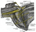
Plan of brachial plexus. (Label for thoracodorsal nerve at bottom center.)
Latissimus dorsi.png
Latissimus dorsi
Details
From Posterior cord (C6-C8)
Innervates Latissimus dorsi muscle
Identifiers
Latin nervus thoracodorsalis
TA98 A14.2.03.016
TA2 6430
FMA 65290
Anatomical terms of neuroanatomy

The thoracodorsal nerve is a nerve present in humans and other animals, also known as the middle subscapular nerve or the long subscapular nerve. It supplies the latissimus dorsi muscle. [1] [2]

Contents

Structure

The thoracodorsal nerve arises from the brachial plexus. It derives its fibers from the sixth, seventh, and eighth cervical nerves. It is derived from their ventral rami, in spite of the fact that the latissimus dorsi is found in the back. The thoracodorsal nerve is a branch of the posterior cord of the brachial plexus, [1] [3] and is made up of fibres from the posterior divisions of all three trunks of the brachial plexus. [4]

It follows the course of the subscapular artery, along the posterior wall of the axilla to the latissimus dorsi muscle, [1] in which it may be traced as far as the lower border of the muscle.

Function

The thoracodorsal nerve innervates the latissimus dorsi muscle on its deep surface. [1]

Clinical Significance

The latissimus dorsi is occasionally used for transplantation, and for augmentation of systole in cardiac failure. In these cases, the nerve supply is preserved, and transplanted with the muscle (for example, with facial reanimation). [5]

Posterior cord lesions can result in the loss of adduction of the shoulder joint, as innervation to latissimus dorsi is lost. [3]

Additional images

Related Research Articles

<span class="mw-page-title-main">Radial nerve</span> Nerve in the human body that supplies the posterior portion of the upper limb

The radial nerve is a nerve in the human body that supplies the posterior portion of the upper limb. It innervates the medial and lateral heads of the triceps brachii muscle of the arm, as well as all 12 muscles in the posterior osteofascial compartment of the forearm and the associated joints and overlying skin.

<span class="mw-page-title-main">Latissimus dorsi muscle</span> Large, flat back muscle

The latissimus dorsi is a large, flat muscle on the back that stretches to the sides, behind the arm, and is partly covered by the trapezius on the back near the midline. The word latissimus dorsi comes from Latin and means "broadest [muscle] of the back", from "latissimus" and "dorsum". The pair of muscles are commonly known as "lats", especially among bodybuilders. The latissimus dorsi is the largest muscle in the upper body.

<span class="mw-page-title-main">Median nerve</span> Nerve of the upper limb

The median nerve is a nerve in humans and other animals in the upper limb. It is one of the five main nerves originating from the brachial plexus.

<span class="mw-page-title-main">Brachial plexus</span> Network of nerves

The brachial plexus is a network of nerves formed by the anterior rami of the lower four cervical nerves and first thoracic nerve. This plexus extends from the spinal cord, through the cervicoaxillary canal in the neck, over the first rib, and into the armpit, it supplies afferent and efferent nerve fibers to the chest, shoulder, arm, forearm, and hand.

<span class="mw-page-title-main">Spinal nerve</span> Nerve that carries signals between the spinal cord and the body

A spinal nerve is a mixed nerve, which carries motor, sensory, and autonomic signals between the spinal cord and the body. In the human body there are 31 pairs of spinal nerves, one on each side of the vertebral column. These are grouped into the corresponding cervical, thoracic, lumbar, sacral and coccygeal regions of the spine. There are eight pairs of cervical nerves, twelve pairs of thoracic nerves, five pairs of lumbar nerves, five pairs of sacral nerves, and one pair of coccygeal nerves. The spinal nerves are part of the peripheral nervous system.

<span class="mw-page-title-main">Axillary nerve</span> Nerve of the human body near the armpit

The axillary nerve or the circumflex nerve is a nerve of the human body, that originates from the brachial plexus at the level of the axilla (armpit) and carries nerve fibers from C5 and C6. The axillary nerve travels through the quadrangular space with the posterior circumflex humeral artery and vein to innervate the deltoid and teres minor.

<span class="mw-page-title-main">Ulnar nerve</span> Nerve which runs near the ulna bone

In human anatomy, the ulnar nerve is a nerve that runs near the ulna bone. The ulnar collateral ligament of elbow joint is in relation with the ulnar nerve. The nerve is the largest in the human body unprotected by muscle or bone, so injury is common. This nerve is directly connected to the little finger, and the adjacent half of the ring finger, innervating the palmar aspect of these fingers, including both front and back of the tips, perhaps as far back as the fingernail beds.

<span class="mw-page-title-main">Long thoracic nerve</span> Large nerve

The long thoracic nerve is a branch of the brachial plexus derived from cervical nerves C5-C7 that innervates the serratus anterior muscle.

<span class="mw-page-title-main">Deltoid muscle</span> Shoulder muscle

The deltoid muscle is the muscle forming the rounded contour of the human shoulder. It is also known as the 'common shoulder muscle', particularly in other animals such as the domestic cat. Anatomically, the deltoid muscle appears to be made up of three distinct sets of muscle fibers, namely the

  1. anterior or clavicular part
  2. posterior or scapular part
  3. intermediate or acromial part
<span class="mw-page-title-main">Cervical plexus</span> Network of nerves in the neck

The cervical plexus is a nerve plexus of the anterior rami of the first four cervical spinal nerves C1-C4. The cervical plexus provides motor innervation to some muscles of the neck, and the diaphragm; it provides sensory innervation to parts of the head, neck, and chest.

<span class="mw-page-title-main">Teres major muscle</span> Muscle of the upper limb

The teres major muscle is a muscle of the upper limb. It attaches to the scapula and the humerus and is one of the seven scapulohumeral muscles. It is a thick but somewhat flattened muscle.

<span class="mw-page-title-main">Intercostal nerves</span> Nerves in the thorax and abdomen

The intercostal nerves are part of the somatic nervous system, and arise from the anterior rami of the thoracic spinal nerves from T1 to T11. The intercostal nerves are distributed chiefly to the thoracic pleura and abdominal peritoneum, and differ from the anterior rami of the other spinal nerves in that each pursues an independent course without plexus formation.

<span class="mw-page-title-main">Medial cord</span>

The medial cord is the part of the brachial plexus formed by of the anterior division of the lower trunk (C8-T1). Its name comes from it being medial to the axillary artery as it passes through the axilla. The other cords of the brachial plexus are the posterior cord and lateral cord.

<span class="mw-page-title-main">Medial pectoral nerve</span>

The medial pectoral nerve is (typically) a branch of the medial cord of the brachial plexus and is derived from spinal nerve roots C8-T1. It provides motor innervation to the pectoralis minor muscle, and the lower half of the pectoralis major muscle. It runs along the inferior border of the pectoralis minor muscle.

<span class="mw-page-title-main">Anterior superior iliac spine</span> Bony projection of the iliac bone

The anterior superior iliac spine (ASIS) is a bony projection of the iliac bone, and an important landmark of surface anatomy. It refers to the anterior extremity of the iliac crest of the pelvis. It provides attachment for the inguinal ligament, and the sartorius muscle. The tensor fasciae latae muscle attaches to the lateral aspect of the superior anterior iliac spine, and also about 5 cm away at the iliac tubercle.

<span class="mw-page-title-main">Lower subscapular nerve</span>

The lower subscapular nerve, also known as the inferior subscapular nerve, is the third branch of the posterior cord of the brachial plexus. It innervates the inferior portion of the subscapularis muscle and the teres major muscle.

<span class="mw-page-title-main">Upper subscapular nerve</span> Nerve of upper limb

The upper (superior) subscapular nerve is the first branch of the posterior cord of the brachial plexus. The upper subscapular nerve contains axons from the ventral rami of the C5 and C6 cervical spinal nerves. It innervates the superior portion of the subscapularis muscle. The inferior portion of the subscapularis is innervated by the lower subscapular nerve.

<span class="mw-page-title-main">Subscapular nerves</span> Nerves that control the shoulder blade

Subscapular nerves can refer to:

<span class="mw-page-title-main">Lateral pectoral nerve</span>

The lateral pectoral nerve arises from the lateral cord of the brachial plexus, and through it from the C5-7.

<span class="mw-page-title-main">Lumbosacral trunk</span> Nervous tissue that connects the lumbar plexus with the sacral plexus

The lumbosacral trunk is nervous tissue that connects the lumbar plexus with the sacral plexus. It is formed by the union of parts of the fourth and fifth lumbar nerves and descends to join the sacral plexus.

References

PD-icon.svgThis article incorporates text in the public domain from page 934 of the 20th edition of Gray's Anatomy (1918)

  1. 1 2 3 4 Nicole Bentley, J.; Yang, Lynda J. -S. (2015-01-01), Tubbs, R. Shane; Rizk, Elias; Shoja, Mohammadali M.; Loukas, Marios (eds.), "Chapter 42 - Anatomy of the Posterior Cord and Its Branches", Nerves and Nerve Injuries, San Diego: Academic Press, pp. 563–574, doi:10.1016/b978-0-12-410390-0.00012-3, ISBN   978-0-12-410390-0 , retrieved 2020-11-01
  2. Katirji, Bashar (2007-01-01), Katirji, Bashar (ed.), "Case 11", Electromyography in Clinical Practice (Second Edition), Philadelphia: Mosby, pp. 175–187, doi:10.1016/b978-0-323-02899-8.50020-3, ISBN   978-0-323-02899-8 , retrieved 2020-11-01
  3. 1 2 Preston, David C.; Shapiro, Barbara E. (2013-01-01), Preston, David C.; Shapiro, Barbara E. (eds.), "30 - Brachial Plexopathy", Electromyography and Neuromuscular Disorders (Third Edition), London: W.B. Saunders, pp. 468–486, doi:10.1016/b978-1-4557-2672-1.00030-1, ISBN   978-1-4557-2672-1 , retrieved 2020-11-01
  4. Bertorini, Tulio E. (2008-01-01), Bertorini, Tulio E. (ed.), "1 - Neuromuscular Anatomy and Function", Neuromuscular Case Studies, Philadelphia: Butterworth-Heinemann, pp. 1–25, doi:10.1016/b978-0-7506-7332-7.50005-2, ISBN   978-0-7506-7332-7 , retrieved 2020-11-01
  5. Rea, Paul (2015-01-01), Rea, Paul (ed.), "Chapter 2 - Upper Limb Nerve Supply", Essential Clinically Applied Anatomy of the Peripheral Nervous System in the Limbs, Academic Press, pp. 41–100, doi:10.1016/b978-0-12-803062-2.00002-4, ISBN   978-0-12-803062-2 , retrieved 2020-11-01