A dorsal fin is a fin located on the back of most marine and freshwater vertebrates within various taxa of the animal kingdom. Many species of animals possessing dorsal fins are not particularly closely related to each other, though through convergent evolution they have independently evolved external superficial fish-like body plans adapted to their marine environments, including most numerously fish, but also mammals such as cetaceans, and even extinct ancient marine reptiles such as various known species of ichthyosaurs. Most species have only one dorsal fin, but some have two or three.

In human anatomy, the metacarpal bones or metacarpus, also known as the "palm bones", are the appendicular bones that form the intermediate part of the hand between the phalanges (fingers) and the carpal bones, which articulate with the forearm. The metacarpal bones are homologous to the metatarsal bones in the foot.
In human anatomy, the extensor pollicis longus muscle (EPL) is a skeletal muscle located dorsally on the forearm. It is much larger than the extensor pollicis brevis, the origin of which it partly covers and acts to stretch the thumb together with this muscle.

The superficial fibular nerve is a mixed nerve that provides motor innervation to the fibularis longus and fibularis brevis muscles, and sensory innervation to skin over the antero-lateral aspect of the leg along with the greater part of the dorsum of the foot.

The superficial branch of the radial nerve passes along the front of the radial side of the forearm to the commencement of its lower third. It is a sensory nerve.

Most of the dorsal metacarpal arteries arise from the dorsal carpal arch and run downward on the second, third, and fourth dorsal interossei of the hand and bifurcate into the dorsal digital arteries. Near their origin, they anastomose with the deep palmar arch by perforating arteries. They also anastomose with common palmar digital arteries, also via perforating arteries.

In the palm of the hand the median nerve is covered by the skin and the palmar aponeurosis, and rests on the tendons of the flexor muscles. Immediately after emerging from under the transverse carpal ligament the median nerve becomes enlarged and flattened and splits into a smaller, lateral, and a larger, medial portion.

An extensor expansion is the special connective attachments by which the extensor tendons insert into the phalanges.

The proper palmar digital arteries travel along the sides of the phalanges, each artery lying just below its corresponding digital nerve.

The dorsal venous network of the hand is a venous network on the dorsum (backside) of hand. It is formed by the dorsal metacarpal veins, a dorsal digital vein from the radial side of the index finger and one from the ulnar side of the little finger, and both dorsal digital veins of the thumb. The venous network gives rise to the cephalic vein and the basilic vein; an accessory cephalic vein may arise from it as well.

The intermediate dorsal cutaneous nerve, the smaller, passes along the lateral part of the dorsum of the foot, and divides into dorsal digital branches, which supply the contiguous sides of the third and fourth, and of the fourth and fifth toes.
The medial dorsal cutaneous nerve passes in front of the ankle-joint, and divides into two dorsal digital branches, one of which supplies the medial side of the great toe, the other, the adjacent side of the second and third toes.
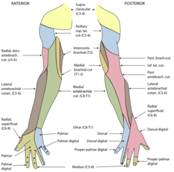
Cutaneous innervation of the upper limbs is the nerve supply to areas of the skin of the upper limbs which are supplied by specific cutaneous nerves.
The arcuate artery of the foot gives off the second, third, and fourth dorsal metatarsal arteries, which run forward upon the corresponding Interossei dorsales; in the clefts between the toes, each divides into two dorsal digital branches for the adjoining toes.
The lateral dorsal cutaneous nerve is a cutaneous branch of the foot.
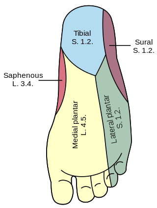
Dorsal digital nerves of foot are branches of the intermediate dorsal cutaneous nerve, medial dorsal cutaneous nerve, sural nerve and deep fibular nerve.

Dorsal digital nerves of ulnar nerve are branches on the dorsum of the hand. The dorsal branch of the ulnar nerve divides into two dorsal digital branches; one supplies the ulnar side of the little finger; the other, the adjacent sides of the little and ring fingers. It also sends a twig to join that given by the superficial branch of the radial nerve for the adjoining sides of the middle and ring fingers, and assists in supplying them.
The dorsal digital arteries of foot are small arteries which supply the toes.

The spinal cord is a long, thin, tubular structure made up of nervous tissue that extends from the medulla oblongata in the brainstem to the lumbar region of the vertebral column (backbone) of vertebrate animals. The center of the spinal cord is hollow and contains a structure called the central canal, which contains cerebrospinal fluid. The spinal cord is also covered by meninges and enclosed by the neural arches. Together, the brain and spinal cord make up the central nervous system.
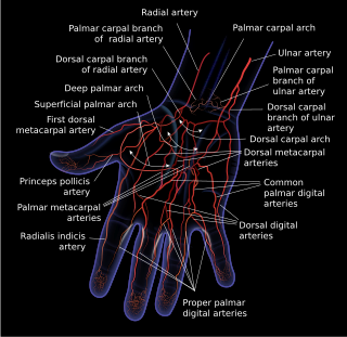
Dorsal digital arteries arise from the bifurcation of dorsal metacarpal arteries. They travel along the sides and dorsal aspects of the phalanges of the middle finger, ring finger, and little finger. They communicate with the proper palmar digital arteries.