Median nerve

Last updated
Median nerve
Nerves of the left upper extremity.gif
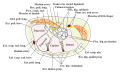
Diagram from Gray's anatomy, depicting the peripheral nerves of the upper extremity, amongst others the median nerve
Details
From Lateral cord and medial cord
Innervates Anterior compartment of the forearm (with two exceptions), thenar eminence, lumbricals, skin of the hand
Identifiers
Latin nervus medianus
MeSH D008475
TA98 A14.2.03.031
TA2 6459
FMA 14385
Anatomical terms of neuroanatomy

The median nerve is a nerve in humans and other animals in the upper limb. It is one of the five main nerves originating from the brachial plexus.

Contents

The median nerve originates from the lateral and medial cords of the brachial plexus, [1] and has contributions from ventral roots of C6-C7 (lateral cord) and C8 and T1 (medial cord). [1] [2]

The median nerve is the only nerve that passes through the carpal tunnel. Carpal tunnel syndrome is the disability that results from the median nerve being pressed in the carpal tunnel.

Structure

The median nerve arises from the branches from lateral and medial cords of the brachial plexus, courses through the anterior part of arm, forearm, and hand, and terminates by supplying the muscles of the hand.

Arm

After receiving inputs from both the lateral and medial cords of the brachial plexus, the median nerve enters the arm from the axilla at the inferior margin of the teres major muscle. It then passes vertically down and courses lateral to the brachial artery between biceps brachii (above) and brachialis (below). At first, it is lateral to the artery and lies anterior to the shoulder joint; it then crosses anteriorly to run medial to the artery in the distal arm and into the cubital fossa. Inside the cubital fossa, the median nerve passes medial to the brachial artery. [1] The median nerve gives off an articular branch to the elbow joint. A branch to pronator teres muscle arise from the median nerve immediately above the elbow joint. [3]

Forearm

The median nerve continues in the cubital fossa medial to the brachial artery and passes between the two heads of the pronator teres, deep to the bicipital aponeurosis (aponeurosis of biceps) and superficial the brachialis muscle. It crosses the ulnar artery (branch of brachial artery) while being separated by the deep head of the pronator teres. It then travels between the flexor digitorum superficialis (above) and flexor digitorum profundus (below). The median nerve is accompanied by the median artery (a branch of anterior interosseous artery) during this course. Then, about 5 cm above the flexor retinaculum (wrist), it emerges between the flexor digitorum superficialis (medially) and the flexor carpi radialis (laterally) into the hand. [3]

The main trunk of the median nerve innervates the superficial and deep groups of the muscles in the anterior compartment of the forearm with the exception of flexor carpi ulnaris. The median nerve does this by giving off two branches as it courses through the forearm:

The median nerve also gives off sensory and other branches in the forearm. The palmar cutaneous branch of the median nerve arises at the distal part of the forearm. It supplies sensory innervation to the thenar eminence of the palm and the central palm. Articular branches are given to the elbow joint and proximal radioulnar joint. Vascular branches supply the radial and ulnar arteries. Meanwhile, a communicating branch is given to the ulnar nerve. [1] [3]

Hand

The median nerve enters the hand through the carpal tunnel, deep to the flexor retinaculum along with the tendons of flexor digitorum superficialis, flexor digitorum profundus, and flexor pollicis longus. From there, it is divided into recurrent muscular branch and digital cutaneous branch: [3]

Variation

The naturally occurring anomalies of the median nerve are:

Function

The median nerve is the main nerve of the front of the forearm. It supplies the muscles of the front of the forearm and muscles of the thenar eminence, thus controlling the coarse movements of the hand. Therefore, it is also called "labourer's nerve". [3]

Arm

The median nerve has no voluntary motor or cutaneous function in the brachium. It gives vascular branches to the wall of the brachial artery. These vascular branches carry sympathetic fibers.

Forearm

It innervates all of the flexors in the forearm, except flexor carpi ulnaris and that part of flexor digitorum profundus that supplies the fourth and fifth digits. [8] The latter two muscles are supplied by the ulnar nerve (specifically the muscular branches of ulnar nerve).

The main portion of the median nerve supplies these muscles:

Superficial group:

Intermediate group:

The anterior interosseus branch of the median nerve supplies these muscles:

Deep group:

Hand

In the hand, the median nerve supplies motor innervation to the first and second lumbrical muscles. It also supplies the muscles of the thenar eminence by a recurrent thenar branch. [8] The rest of the intrinsic muscles of the hand are supplied by the ulnar nerve.

The median nerve innervates the skin of the palmar (volar) side of the index finger, thumb, middle finger, and half the ring finger, and the nail bed. The radial aspect of the palm is supplied by the palmar cutaneous branch of the median nerve, which leaves the nerve proximal to the wrist creases. This palmar cutaneous branch travels in a separate fascial groove adjacent to the flexor carpi radialis and then superficial to the flexor retinaculum. It is, therefore, spared in carpal tunnel syndrome.


Clinical significance

Injury

Injury of median nerve at different levels causes different syndromes with varying motor and sensory deficits.

At the shoulder

Above the elbow

At the elbow

Within the proximal forearm: anterior interosseous syndrome

At the wrist

Within the wrist: carpal tunnel syndrome

Assessment

When symptoms of tingling, numbness, pain, or muscle weakness occur, various assessments are conducted, including patient-reported subjective surveys, provocative tests meant to elicit or exacerbate symptoms, sensory and motor function tests, and diagnostic equipment.

Combinations of these assessments are useful for ruling out other conditions, identifying the location of nerve injury or compression, uncovering underlying causes of symptoms (such as tendonitis), determining the severity of the nerve injury, and developing individualized treatment plans based on patient characteristics. [16] Such combinations of median nerve evaluations have been most widely validated in the assessment of carpal tunnel syndrome. [17] [18]

Additional images

See also

Related Research Articles

<span class="mw-page-title-main">Radial nerve</span> Nerve in the human body that supplies the posterior portion of the upper limb

The radial nerve is a nerve in the human body that supplies the posterior portion of the upper limb. It innervates the medial and lateral heads of the triceps brachii muscle of the arm, as well as all 12 muscles in the posterior osteofascial compartment of the forearm and the associated joints and overlying skin.

The flexor digitorum profundus is a muscle in the forearm of humans that flexes the fingers. It is considered an extrinsic hand muscle because it acts on the hand while its muscle belly is located in the forearm.

<span class="mw-page-title-main">Flexor digitorum superficialis muscle</span> Forearm muscle that flexes the fingers

Flexor digitorum superficialis is an extrinsic flexor muscle of the fingers at the proximal interphalangeal joints.

The forearm is the region of the upper limb between the elbow and the wrist. The term forearm is used in anatomy to distinguish it from the arm, a word which is used to describe the entire appendage of the upper limb, but which in anatomy, technically, means only the region of the upper arm, whereas the lower "arm" is called the forearm. It is homologous to the region of the leg that lies between the knee and the ankle joints, the crus.

<span class="mw-page-title-main">Ulnar nerve</span> Nerve which runs near the ulna bone

In human anatomy, the ulnar nerve is a nerve that runs near the ulna bone. The ulnar collateral ligament of elbow joint is in relation with the ulnar nerve. The nerve is the largest in the human body unprotected by muscle or bone, so injury is common. This nerve is directly connected to the little finger, and the adjacent half of the ring finger, innervating the palmar aspect of these fingers, including both front and back of the tips, perhaps as far back as the fingernail beds.

<span class="mw-page-title-main">Wrist drop</span> Medical condition

Wrist drop is a medical condition in which the wrist and the fingers cannot extend at the metacarpophalangeal joints. The wrist remains partially flexed due to an opposing action of flexor muscles of the forearm. As a result, the extensor muscles in the posterior compartment remain paralyzed.

<span class="mw-page-title-main">Upper limb</span> Consists of the arm, forearm, and hand

The upper limbs or upper extremities are the forelimbs of an upright-postured tetrapod vertebrate, extending from the scapulae and clavicles down to and including the digits, including all the musculatures and ligaments involved with the shoulder, elbow, wrist and knuckle joints. In humans, each upper limb is divided into the arm, forearm and hand, and is primarily used for climbing, lifting and manipulating objects.

<span class="mw-page-title-main">Ulnar artery</span> Artery of the forearm

The ulnar artery is the main blood vessel, with oxygenated blood, of the medial aspects of the forearm. It arises from the brachial artery and terminates in the superficial palmar arch, which joins with the superficial branch of the radial artery. It is palpable on the anterior and medial aspect of the wrist.

The flexor pollicis longus is a muscle in the forearm and hand that flexes the thumb. It lies in the same plane as the flexor digitorum profundus. This muscle is unique to humans, being either rudimentary or absent in other primates. A meta-analysis indicated accessory flexor pollicis longus is present in around 48% of the population.

The pronator teres is a muscle that, along with the pronator quadratus, serves to pronate the forearm. It has two origins, at the medial humeral supracondylar ridge and the ulnar tuberosity, and inserts near the middle of the radius.

<span class="mw-page-title-main">Anterior interosseous artery</span>

The anterior interosseous artery is an artery in the forearm. It is a branch of the common interosseous artery.

<span class="mw-page-title-main">Anterior interosseous nerve</span>

The anterior interosseous nerve is a branch of the median nerve that supplies the deep muscles on the anterior of the forearm, except the ulnar (medial) half of the flexor digitorum profundus. Its nerve roots come from C8 and T1.

<span class="mw-page-title-main">Carpal tunnel</span> Structure of human wrist

In the human body, the carpal tunnel or carpal canal is a flattened body cavity on the flexor (palmar/volar) side of the wrist, bounded by the carpal bones and flexor retinaculum. It forms the passageway that transmits the median nerve and the tendons of the extrinsic flexor muscles of the hand from the forearm to the hand. There are described cases of the anatomical variant median artery occurrence.

<span class="mw-page-title-main">Cervical spinal nerve 8</span> Spinal nerve of the cervical segment

The cervical spinal nerve 8 (C8) is a spinal nerve of the cervical segment.

<span class="mw-page-title-main">Hand</span> Extremity at the end of an arm or forelimb

A hand is a prehensile, multi-fingered appendage located at the end of the forearm or forelimb of primates such as humans, chimpanzees, monkeys, and lemurs. A few other vertebrates such as the koala are often described as having "hands" instead of paws on their front limbs. The raccoon is usually described as having "hands" though opposable thumbs are lacking.

Anterior interosseous syndrome is a medical condition in which damage to the anterior interosseous nerve (AIN), a distal motor and sensory branch of the median nerve, classically with severe weakness of the pincer movement of the thumb and index finger, and can cause transient pain in the wrist.

<span class="mw-page-title-main">Pronator teres syndrome</span> Medical condition

Pronator teres syndrome is a compression neuropathy of the median nerve at the elbow. It is rare compared to compression at the wrist or isolated injury of the anterior interosseous branch of the median nerve.

<span class="mw-page-title-main">Median nerve palsy</span> Medical condition

Injuries to the arm, forearm or wrist area can lead to various nerve disorders. One such disorder is median nerve palsy. The median nerve controls the majority of the muscles in the forearm. It controls abduction of the thumb, flexion of hand at wrist, flexion of digital phalanx of the fingers, is the sensory nerve for the first three fingers, etc. Because of this major role of the median nerve, it is also called the eye of the hand. If the median nerve is damaged, the ability to abduct and oppose the thumb may be lost due to paralysis of the thenar muscles. Various other symptoms can occur which may be repaired through surgery and tendon transfers. Tendon transfers have been very successful in restoring motor function and improving functional outcomes in patients with median nerve palsy.

<span class="mw-page-title-main">Muscles of the thumb</span>

The muscles of the thumb are nine skeletal muscles located in the hand and forearm. The muscles allow for flexion, extension, adduction, abduction and opposition of the thumb. The muscles acting on the thumb can be divided into two groups: The extrinsic hand muscles, with their muscle bellies located in the forearm, and the intrinsic hand muscles, with their muscles bellies located in the hand proper.

<span class="mw-page-title-main">Palmaris profundus muscle</span>

Palmaris profundus is a rare anatomical variant in the anterior compartment of forearm. It was first described in 1908. It is usually found incidentally in cadaveric dissection or surgery.

References

PD-icon.svgThis article incorporates text in the public domain from page 938 of the 20th edition of Gray's Anatomy (1918)

  1. 1 2 3 4 Rea, Paul (2016-01-01), Rea, Paul (ed.), "Chapter 3 - Neck", Essential Clinically Applied Anatomy of the Peripheral Nervous System in the Head and Neck, Academic Press, pp. 131–183, doi:10.1016/b978-0-12-803633-4.00003-x, ISBN   978-0-12-803633-4 , retrieved 2020-10-25
  2. 1 2 3 4 5 6 7 8 Ryan, Monique M.; Jones, H. Royden (2015-01-01), Darras, Basil T.; Jones, H. Royden; Ryan, Monique M.; De Vivo, Darryl C. (eds.), "Chapter 14 – Mononeuropathies", Neuromuscular Disorders of Infancy, Childhood, and Adolescence (Second Edition), San Diego: Academic Press, pp. 243–273, doi:10.1016/b978-0-12-417044-5.00014-7, ISBN   978-0-12-417044-5 , retrieved 2020-10-25
  3. 1 2 3 4 5 6 7 8 9 Krishna, Garg (2010). "8 - Arm". BD Chaurasia's Human Anatomy (Regional and Applied Dissection and Clinical) Volume 1 - Upper limb and thorax (Fifth ed.). India: CBS Publishers and Distributors Pvt Ltd. pp. 91, 109–110, 112. ISBN   978-81-239-1863-1.
  4. 1 2 Waldman, Steven D. (2014-01-01), Waldman, Steven D. (ed.), "Chapter 45 - Anterior Interosseous Syndrome", Atlas of Uncommon Pain Syndromes (Third Edition), Philadelphia: W.B. Saunders, pp. 130–132, doi:10.1016/b978-1-4557-0999-1.00045-9, ISBN   978-1-4557-0999-1 , retrieved 2020-10-25
  5. 1 2 Roll, Shawn C.; Evans, Kevin D. (2011). "Sonographic Representation of Bifid Median Nerve and Persistent Median Artery". Journal of Diagnostic Medical Sonography. 27 (2): 89–94. doi:10.1177/8756479311399763. S2CID   56670411.
  6. Cavalheiro, Cristina Schmitt; Filho, Mauro Razuk; Pedro, Gabriel; Caetano, Maurício Ferreira; Vieira, Luiz Angelo; Caetano, Edie Benedito (2016-02-23). "Clinical repercussions of Martin-Gruber anastomosis: anatomical study". Revista Brasileira de Ortopedia. 51 (2): 214–223. doi:10.1016/j.rboe.2016.02.003. ISSN   2255-4971. PMC   4812040 . PMID   27069892.
  7. Caetano, Edie Benedito; Vieira, Luiz Angelo; Sabongi, João José; Caetano, Maurício Ferreira; Sabongi, Rodrigo Guerra (2019-11-14). "Riché-Cannieu Anastomosis: Structure, Function, and Clinical Significance". Revista Brasileira de Ortopedia. 54 (5): 564–571. doi:10.1016/j.rbo.2017.12.019. ISSN   0102-3616. PMC   6855928 . PMID   31736524.
  8. 1 2 Bonfiglioli, Roberta; Mattioli, Stefano; Violante, Francesco S. (2015-01-01), Lotti, Marcello; Bleecker, Margit L. (eds.), "Chapter 22 - Occupational mononeuropathies in industry", Handbook of Clinical Neurology, Occupational Neurology, 131, Elsevier: 411–426, doi:10.1016/b978-0-444-62627-1.00021-4, ISBN   9780444626271, PMID   26563800 , retrieved 2020-10-25
  9. Neal, Sara (2015-01-01), Tubbs, R. Shane; Rizk, Elias; Shoja, Mohammadali M.; Loukas, Marios (eds.), "Chapter 33 - Peripheral Nerve Injury of the Upper Extremity", Nerves and Nerve Injuries, San Diego: Academic Press, pp. 505–524, doi:10.1016/b978-0-12-802653-3.00082-8, ISBN   978-0-12-802653-3 , retrieved 2020-10-25
  10. Figure 6.86, Moore Clinically Orientated Anatomy 7th Edition
  11. Multanen, Juhani; Ylinen, Jari; Karjalainen, Teemu; Ikonen, Joona; Häkkinen, Arja; Repo, Jussi P. (2020). "Structural validity of the Boston Carpal Tunnel Questionnaire and its short version, the 6-Item CTS symptoms scale: A Rasch analysis one year after surgery". BMC Musculoskeletal Disorders. 21 (1): 609. doi: 10.1186/s12891-020-03626-2 . PMC   7488577 . PMID   32919457.
  12. Hudak, Pamela L.; Amadio, Peter C.; Bombardier, Claire; Beaton, Dorcas; Cole, Donald; Davis, Aileen; Hawker, Gillian; Katz, Jeffrey N.; Makela, Matti; Marx, Robert G.; Punnett, Laura; Wright, James (1996). "Development of an upper extremity outcome measure: The DASH (Disabilities of the arm, shoulder, and head)". American Journal of Industrial Medicine. 29 (6): 602–608. doi:10.1002/(sici)1097-0274(199606)29:6<602::aid-ajim4>3.0.co;2-l. PMID   8773720.
  13. Gabel, C. Philip; Michener, Lori A.; Burkett, Brendan; Neller, Anne (2006). "The Upper Limb Functional Index: Development and Determination of Reliability, Validity, and Responsiveness". Journal of Hand Therapy. 19 (3): 328–349. doi:10.1197/j.jht.2006.04.001. PMID   16861132.
  14. Roll, Shawn C.; Takata, Sandy C.; Yao, Buwen; Kysh, Lynn; Mack, Wendy J. (2023). "Sonographic Reference Values for Median Nerve Cross-sectional Area: A Meta-analysis of Data from Healthy Individuals". Journal of Diagnostic Medical Sonography. doi:10.1177/87564793231176009. S2CID   259789268.
  15. Cartwright, Michael S.; Hobson-Webb, Lisa D.; Boon, Andrea J.; Alter, Katharine E.; Hunt, Christopher H.; Flores, Victor H.; Werner, Robert A.; Shook, Steven J.; Thomas, T. Darrell; Primack, Scott J.; Walker, Francis O.; American Association of Neuromuscular and Electrodiagnostic Medicine (2012). "Evidence-based guideline: Neuromuscular ultrasound for the diagnosis of carpal tunnel syndrome". Muscle & Nerve. 46 (2): 287–293. doi:10.1002/mus.23389. hdl: 2027.42/92446 . PMID   22806381. S2CID   6966288.
  16. Billig, Jessica I.; Kotsis, Sandra V.; Ouyang, Zhongzhe; Wang, Lu; Chung, Kevin C. (2022). "A Quality Collaborative Study of the Concordance of Electrodiagnostic Scores and Clinical Severity in Carpal Tunnel Syndrome". Plastic & Reconstructive Surgery. 150 (6): 1287–1296. doi: 10.1097/PRS.0000000000009675 . PMID   36112828. S2CID   252334741.
  17. Roll, Shawn C.; Volz, Kevin R.; Fahy, Christine M.; Evans, Kevin D. (2015). "Carpal tunnel syndrome severity staging using sonographic and clinical measures". Muscle & Nerve. 51 (6): 838–845. doi:10.1002/mus.24478. PMC   4388767 . PMID   25287477.
  18. Wang, William L.; Hanson, Timothy; Fowler, John R. (2020). "A Comparison of 6 Diagnostic Tests for Carpal Tunnel Syndrome Using Latent Class Analysis". Hand. 15 (6): 776–779. doi:10.1177/1558944719833709. PMC   7850262 . PMID   30854901.