Suprascapular nerve

Last updated
Suprascapular nerve
Gray818.png
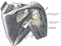
The suprascapular, axillary, and radial nerves. (Suprascapular labeled at upper left.)
Gray808.png
The right brachial plexus with its short branches, viewed from in front. (Suprascapular labeled at upper left.)
Details
From Upper trunk (C5–C6) of brachial plexus
Innervates Supraspinatus, infraspinatus
Identifiers
Latin nervus suprascapularis
TA98 A14.2.03.014
TA2 6411
FMA 37025
Anatomical terms of neuroanatomy

The suprascapular nerve is a mixed (sensory and motor) nerve that branches from the upper trunk of the brachial plexus. It is derived from the ventral rami of cervical nerves C5-C6. It provides motor innervation to the supraspinatus muscle, and the infraspinatus muscle.

Contents

Structure

Origin

The suprascapular nerve arises from the upper trunk of the brachial plexus which is formed by the union of the ventral rami of the cervical nerves C5-C6. [1]

Course and relations

After branching from the upper trunk, the nerve passes across the posterior triangle of the neck parallel to the inferior belly of the omohyoid muscle and deep to the trapezius muscle. It then runs along the superior border of the scapula through the suprascapular canal, [1] in which it enters via the suprascapular notch inferior to the superior transverse scapular ligament and enters the supraspinous fossa. [2] It then passes beneath the supraspinatus and curves around the lateral border of the spine of the scapula through spinogleniod notch to the infraspinous fossa.[ citation needed ]

Function

The suprascapular nerve is a mixed peripheral nerve containing motor and sensory components.

Motor innervation

Sensory innervation

In the supraspinous fossa it gives off two branches to the supraspinatus muscle and in the infraspinous fossa it gives off two branches to the infraspinatus muscle.

Clinical significance

Additional images

Related Research Articles

<span class="mw-page-title-main">Scapula</span> Bone that connects the humerus (upper arm bone) with the clavicle (collar bone)

The scapula, also known as the shoulder blade, is the bone that connects the humerus with the clavicle. Like their connected bones, the scapulae are paired, with each scapula on either side of the body being roughly a mirror image of the other. The name derives from the Classical Latin word for trowel or small shovel, which it was thought to resemble.

<span class="mw-page-title-main">Brachial plexus</span> Network of nerves

The brachial plexus is a network of nerves formed by the anterior rami of the lower four cervical nerves and first thoracic nerve. This plexus extends from the spinal cord, through the cervicoaxillary canal in the neck, over the first rib, and into the armpit, it supplies afferent and efferent nerve fibers to the chest, shoulder, arm, forearm, and hand.

<span class="mw-page-title-main">Spinal nerve</span> Nerve that carries signals between the spinal cord and the body

A spinal nerve is a mixed nerve, which carries motor, sensory, and autonomic signals between the spinal cord and the body. In the human body there are 31 pairs of spinal nerves, one on each side of the vertebral column. These are grouped into the corresponding cervical, thoracic, lumbar, sacral and coccygeal regions of the spine. There are eight pairs of cervical nerves, twelve pairs of thoracic nerves, five pairs of lumbar nerves, five pairs of sacral nerves, and one pair of coccygeal nerves. The spinal nerves are part of the peripheral nervous system.

<span class="mw-page-title-main">Deltoid muscle</span> Shoulder muscle

The deltoid muscle is the muscle forming the rounded contour of the human shoulder. It is also known as the 'common shoulder muscle', particularly in other animals such as the domestic cat. Anatomically, the deltoid muscle appears to be made up of three distinct sets of muscle fibers, namely the

  1. anterior or clavicular part
  2. posterior or scapular part
  3. intermediate or acromial part
<span class="mw-page-title-main">Upper limb</span> Consists of the arm, forearm, and hand

The upper limbs or upper extremities are the forelimbs of an upright-postured tetrapod vertebrate, extending from the scapulae and clavicles down to and including the digits, including all the musculatures and ligaments involved with the shoulder, elbow, wrist and knuckle joints. In humans, each upper limb is divided into the arm, forearm and hand, and is primarily used for climbing, lifting and manipulating objects.

<span class="mw-page-title-main">Supraspinatus muscle</span> Muscle of the upper back

The supraspinatus is a relatively small muscle of the upper back that runs from the supraspinous fossa superior portion of the scapula to the greater tubercle of the humerus. It is one of the four rotator cuff muscles and also abducts the arm at the shoulder. The spine of the scapula separates the supraspinatus muscle from the infraspinatus muscle, which originates below the spine.

<span class="mw-page-title-main">Infraspinatus muscle</span> Main external rotator of the shoulder

In human anatomy, the infraspinatus muscle is a thick triangular muscle, which occupies the chief part of the infraspinatous fossa. As one of the four muscles of the rotator cuff, the main function of the infraspinatus is to externally rotate the humerus and stabilize the shoulder joint.

<span class="mw-page-title-main">Shoulder joint</span> Synovial ball and socket joint in the shoulder

The shoulder joint is structurally classified as a synovial ball-and-socket joint and functionally as a diarthrosis and multiaxial joint. It involves an articulation between the glenoid fossa of the scapula and the head of the humerus. Due to the very loose joint capsule that gives a limited interface of the humerus and scapula, it is the most mobile joint of the human body.

<span class="mw-page-title-main">Teres major muscle</span> Muscle of the upper limb

The teres major muscle is a muscle of the upper limb. It attaches to the scapula and the humerus and is one of the seven scapulohumeral muscles. It is a thick but somewhat flattened muscle.

<span class="mw-page-title-main">Nerve plexus</span> Network of nerve fibres

A nerve plexus is a plexus of intersecting nerves. A nerve plexus is composed of afferent and efferent fibers that arise from the merging of the anterior rami of spinal nerves and blood vessels. There are five spinal nerve plexuses, except in the thoracic region, as well as other forms of autonomic plexuses, many of which are a part of the enteric nervous system. The nerves that arise from the plexuses have both sensory and motor functions. These functions include muscle contraction, the maintenance of body coordination and control, and the reaction to sensations such as heat, cold, pain, and pressure. There are several plexuses in the body, including:

<span class="mw-page-title-main">Lower subscapular nerve</span>

The lower subscapular nerve, also known as the inferior subscapular nerve, is the third branch of the posterior cord of the brachial plexus. It innervates the inferior portion of the subscapularis muscle and the teres major muscle.

<span class="mw-page-title-main">Upper subscapular nerve</span> Nerve of upper limb

The upper (superior) subscapular nerve is the first branch of the posterior cord of the brachial plexus. The upper subscapular nerve contains axons from the ventral rami of the C5 and C6 cervical spinal nerves. It innervates the superior portion of the subscapularis muscle. The inferior portion of the subscapularis is innervated by the lower subscapular nerve.

<span class="mw-page-title-main">Suprascapular artery</span> Artery of the neck

The suprascapular artery is a branch of the thyrocervical trunk on the neck.

<span class="mw-page-title-main">Deep branch of radial nerve</span>

The radial nerve divides into a superficial (sensory) and deep (motor) branch at the cubital fossa. The deep branch of the radial nerve winds to the back of the forearm around the lateral side of the radius between the two planes of fibers of the supinator, and is prolonged downward between the superficial and deep layers of muscles, to the middle of the forearm. The deep branch provides motor function to the muscles in the posterior compartment of the forearm, which is mostly the extensor muscles of the hand.

<span class="mw-page-title-main">Supraspinous fossa</span> Part of the shoulderblade

The supraspinous fossa of the posterior aspect of the scapula is smaller than the infraspinous fossa, concave, smooth, and broader at its vertebral than at its humeral end. Its medial two-thirds give origin to the Supraspinatus.

<span class="mw-page-title-main">Great scapular notch</span> Notch which serves to connect the supraspinous fossa and infraspinous fossa

The great scapular notch is a notch which serves to connect the supraspinous fossa and infraspinous fossa. It lies immediately medial to the attachment of the acromion to the lateral angle of the scapular spine.

<span class="mw-page-title-main">Ventral ramus of spinal nerve</span> Anterior division of a spinal nerve

The ventral ramus is the anterior division of a spinal nerve. The ventral rami supply the antero-lateral parts of the trunk and the limbs. They are mainly larger than the dorsal rami.

<span class="mw-page-title-main">Outline of human anatomy</span> Overview of and topical guide to human anatomy

The following outline is provided as an overview of and topical guide to human anatomy:

The upper (superior) trunk is part of the brachial plexus. It is formed by joining of the ventral rami of the fifth (C5) and sixth (C6) cervical nerves. The upper trunk divides into an anterior and posterior division.

<span class="mw-page-title-main">Suprascapular canal</span> Anatomical passage between two openings found on the upper dorsal aspect of the shoulder

The suprascapular canal is an anatomical passage between two openings found on the upper dorsal aspect of the shoulder. It is found bilaterally running on superior-lateral aspect of the dorsal surface of the scapula underneath the supraspinatus muscle.

References

PD-icon.svgThis article incorporates text in the public domain from page 932 of the 20th edition of Gray's Anatomy (1918)

  1. 1 2 3 Al-Redouan, Azzat; Holding, keiv; Kachlik, David (2020). ""Suprascapular canal": Anatomical and topographical description andits clinical implication in entrapment syndrome". Annals of Anatomy. 233: 151593. doi:10.1016/j.aanat.2020.151593. PMID   32898658.
  2. 1 2 3 4 5 Avery, BW; Pilon, FM; Barclay, JK (November 2002). "Anterior coracoscapular ligament and suprascapular nerve entrapment". Clinical Anatomy. 15 (6): 383–6. doi:10.1002/ca.10058. PMID   12373728. S2CID   9826767.