Acromioclavicular joint

Last updated
Acromioclavicular joint
Gray326.png
The left shoulder and acromioclavicular joint
Gray328.png
Glenoid cavity of the right scapula
Details
Identifiers
Latin articulatio acromioclavicularis
MeSH D000173
TA98 A03.5.03.001
TA2 1744
FMA 25898
Anatomical terminology

The acromioclavicular joint, or AC joint, is a joint at the top of the shoulder. It is the junction between the acromion (part of the scapula that forms the highest point of the shoulder) and the clavicle. [1] It is a plane synovial joint.

Contents

Structure

Ligaments

The joint is stabilized by three ligaments:
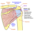
Superior Acromioclavicular Ligament This ligament is a quadrilateral band, covering the superior part of the articulation, and extending between the upper part of the lateral end of the clavicle and the adjoining part of the upper surface of the acromion.

It is composed of parallel fibers, which interlace with the aponeuroses of the Trapezius and Deltoideus; below, it is in contact with the articular disk when this is present.

Inferior Acromioclavicular Ligament This ligament is somewhat thinner than the preceding; it covers the under part of the articulation, and is attached to the adjoining surfaces of the two bones.

It is in relation, above, in rare cases with the articular disk; below, with the tendon of the Supraspinatus

The Coracoacromial Ligament is a strong triangular band, extending between the coracoid process and the acromion.

It is attached, by its apex, to the summit of the acromion just in front of the articular surface for the clavicle; and by its broad base to the whole length of the lateral border of the coracoid process.

This ligament, together with the coracoid process and the acromion, forms a vault for the protection of the head of the humerus.

It is in relation, above, with the clavicle and under surface of the Deltoideus; below, with the tendon of the Supraspinatus, a bursa being interposed.

Its lateral border is continuous with a dense lamina that passes beneath the Deltoideus upon the tendons of the Supraspinatus and Infraspinatus.

The ligament is sometimes described as consisting of two marginal bands and a thinner intervening portion, the two bands being attached respectively to the apex and the base of the coracoid process, and joining at the acromion.

When the Pectoralis minor is inserted, as occasionally is the case, into the capsule of the shoulder-joint instead of into the coracoid process, it passes between these two bands, and the intervening portion of the ligament is then deficient.

The Coracoclavicular Ligament serves to connect the clavicle with the coracoid process of the scapula.

It does not properly belong to the acromioclavicular joint articulation, but is usually described with it, since it forms a most efficient means of retaining the clavicle in contact with the acromion. It consists of two fasciculi, called the trapezoid ligament and conoid ligament.

These ligaments are in relation, in front, with the Subclavius and Deltoideus; behind, with the Trapezius.

Variation

An X-ray study of 100 shoulders in US soldiers found considerable variation in the size and shape of the joint. [2] The articular surfaces were notably different in size and form. On some they are separated by a meniscus attached to the superior acromioclavicular ligament. This meniscus may be a blade of fibrocartilage that extends nearly halfway into the joint or it may form a complete disc that divides the joint into two parts. In other joints no synovial joint is present with the joint being made by a pad of fibrous tissue attached to the outer end of the clavicle, and no articular cavity. [2]

Function

The acromioclavicular joint provides the ability to raise the arm above the head. This joint functions as a pivot point (although technically it is a gliding synovial joint), acting like a strut to help with movement of the scapula resulting in a greater degree of arm rotation.

Clinical significance

Injury

A common injury to the AC joint is dislocation, often called AC separation or shoulder separation. This is not the same as a "shoulder dislocation," which refers to dislocation of the glenohumeral joint.

Acromioclavicular joint dislocation is particularly common in collision sports such as ice hockey, football, Judo, rugby and Aussie rules, and is also a problem for those who participate in swimming, horseback riding, mountain biking, biking, snow skiing and skateboarding. The most common mechanism of injury is a fall on the tip of the shoulder or FOOSH (Fall On OutStretched Hand).

Acromioclavicular joint dislocations are graded from I to VI. Grading is based upon the degree of separation of the acromion from the clavicle with weight applied to the arm. Grade I is slight displacement of the joint, and a badly stretched or partially torn AC ligament. It has the normal separation of <4 mm. Grade II is a partial dislocation of the AC joint with a complete disruption tear of the AC joint and a partial disruption of coracoclavicular ligament. The AC gap is >5 mm. Grades I and II never require surgery and heal by themselves, though physical therapy may be required. Grade III is complete disruption of AC and CC ligaments. On plain film the inferior aspect of the clavicle will be above the superior aspect of the acromion. This can also be assessed with an MRI scan, which will also demonstrate disruption of the coracoclavicular ligaments (the degree depending on the severity of AC joint disruption) as well as tearing of the joint capsule. The joint will be very tender and swollen on examination. Grade III separations most often do not require surgery and shoulder function should return to normal after 16–20 weeks. However, there will be some physical deformity of the shoulder with a noticeable bump resulting from the dislocation of the clavicle. Grades IV-VI are complications on a 'standard' dislocation involving a displacement of the clavicle, and will almost always require surgery. Dynamic US is critical to detect mild (grade I) dislocations, especially because they may present as a normal joint in static images. Doppler US may also be used to increase diagnostic confidence to detect low-grade injuries because it depicts reparative process to injured ligament. [3]

Osteoarthritis

Osteoarthritis of the acromioclavicular joint is not uncommon. It may be caused by a prior trauma (secondary osteoarthritis) or occur as a chronic degenerative disorder often co-existing with subacromial impingement.

Additional images

See also

Related Research Articles

<span class="mw-page-title-main">Clavicle</span> Long bone that serves as a strut between the scapula and the sternum

The clavicle, or collarbone, is a slender, S-shaped long bone approximately 6 inches (15 cm) long that serves as a strut between the shoulder blade and the sternum (breastbone). There are two clavicles, one on the left and one on the right. The clavicle is the only long bone in the body that lies horizontally. Together with the shoulder blade, it makes up the shoulder girdle. It is a palpable bone and, in people who have less fat in this region, the location of the bone is clearly visible. It receives its name from Latin clavicula 'little key' because the bone rotates along its axis like a key when the shoulder is abducted. The clavicle is the most commonly fractured bone. It can easily be fractured by impacts to the shoulder from the force of falling on outstretched arms or by a direct hit.

<span class="mw-page-title-main">Acromion</span> Bony process on the scapula (shoulder blade)

In human anatomy, the acromion is a bony process on the scapula. Together with the coracoid process it extends laterally over the shoulder joint. The acromion is a continuation of the scapular spine, and hooks over anteriorly. It articulates with the clavicle to form the acromioclavicular joint.

<span class="mw-page-title-main">Scapula</span> Bone that connects the humerus (upper arm bone) with the clavicle (collar bone)

The scapula, also known as the shoulder blade, is the bone that connects the humerus with the clavicle. Like their connected bones, the scapulae are paired, with each scapula on either side of the body being roughly a mirror image of the other. The name derives from the Classical Latin word for trowel or small shovel, which it was thought to resemble.

<span class="mw-page-title-main">Humerus</span> Long bone of the upper arm

The humerus is a long bone in the arm that runs from the shoulder to the elbow. It connects the scapula and the two bones of the lower arm, the radius and ulna, and consists of three sections. The humeral upper extremity consists of a rounded head, a narrow neck, and two short processes. The body is cylindrical in its upper portion, and more prismatic below. The lower extremity consists of 2 epicondyles, 2 processes, and 3 fossae. As well as its true anatomical neck, the constriction below the greater and lesser tubercles of the humerus is referred to as its surgical neck due to its tendency to fracture, thus often becoming the focus of surgeons.

<span class="mw-page-title-main">Coracoid process</span> Small hook-like structure on the lateral edge of the superior anterior portion of the scapula

The coracoid process is a small hook-like structure on the lateral edge of the superior anterior portion of the scapula. Pointing laterally forward, it, together with the acromion, serves to stabilize the shoulder joint. It is palpable in the deltopectoral groove between the deltoid and pectoralis major muscles.

<span class="mw-page-title-main">Shoulder problem</span> Medical condition

Shoulder problems including pain, are one of the more common reasons for physician visits for musculoskeletal symptoms. The shoulder is the most movable joint in the body. However, it is an unstable joint because of the range of motion allowed. This instability increases the likelihood of joint injury, often leading to a degenerative process in which tissues break down and no longer function well.

<span class="mw-page-title-main">Shoulder</span> Part of the body

The human shoulder is made up of three bones: the clavicle (collarbone), the scapula, and the humerus as well as associated muscles, ligaments and tendons.

<span class="mw-page-title-main">Shoulder joint</span> Synovial ball and socket joint in the shoulder

The shoulder joint is structurally classified as a synovial ball-and-socket joint and functionally as a diarthrosis and multiaxial joint. It involves an articulation between the glenoid fossa of the scapula and the head of the humerus. Due to the very loose joint capsule that gives a limited interface of the humerus and scapula, it is the most mobile joint of the human body.

<span class="mw-page-title-main">Shoulder girdle</span> Set of bones which connects the arm to the axial skeleton on each side

The shoulder girdle or pectoral girdle is the set of bones in the appendicular skeleton which connects to the arm on each side. In humans it consists of the clavicle and scapula; in those species with three bones in the shoulder, it consists of the clavicle, scapula, and coracoid. Some mammalian species have only the scapula.

<span class="mw-page-title-main">Coracoclavicular ligament</span> Ligament of the shoulder

The coracoclavicular ligament is a ligament of the shoulder. It connects the clavicle to the coracoid process of the scapula.

<span class="mw-page-title-main">Sternoclavicular joint</span> Joint between the manubrium of the sternum and the clavicle bone.

The sternoclavicular joint or sternoclavicular articulation is a synovial saddle joint between the manubrium of the sternum, and the clavicle, and the first costal cartilage. The joint possesses a joint capsule, and an articular disc, and is reinforced by multiple ligaments.

<span class="mw-page-title-main">Glenoid fossa</span> Part of the shoulder

The glenoid fossa of the scapula or the glenoid cavity is a bone part of the shoulder. The word glenoid is pronounced or and is from Greek: gléne, "socket", reflecting the shoulder joint's ball-and-socket form. It is a shallow, pyriform articular surface, which is located on the lateral angle of the scapula. It is directed laterally and forward and articulates with the head of the humerus; it is broader below than above and its vertical diameter is the longest.

<span class="mw-page-title-main">Acromioclavicular ligament</span>

The acromioclavicular ligament is part of the acromioclavicular joint. It is divided into two parts: superior and inferior.

<span class="mw-page-title-main">Coracoacromial ligament</span> Ligament between the coracoid process and the acromion of the scapula

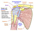
The coracoacromial ligament is a strong triangular ligament between the coracoid process and the acromion. It protects the head of the humerus. Its acromial attachment may be repositioned to the clavicle during reconstructive surgery of the acromioclavicular joint.

<span class="mw-page-title-main">Separated shoulder</span> Medical condition

A separated shoulder, also known as acromioclavicular joint injury, is a common injury to the acromioclavicular joint. The AC joint is located at the outer end of the clavicle where it attaches to the acromion of the scapula. Symptoms include non-radiating pain which may make it difficult to move the shoulder. The presence of swelling or bruising and a deformity in the shoulder is also common depending on how severe the dislocation is.

Shoulder surgery is a means of treating injured shoulders. Many surgeries have been developed to repair the muscles, connective tissue, or damaged joints that can arise from traumatic or overuse injuries to the shoulder.

<span class="mw-page-title-main">Weaver–Dunn procedure</span>

The Weaver–Dunn procedure is a generally successful type of surgery involved in the treatment of severe separated shoulders developed by James K. Weaver, M.D., Albuquerque, New Mexico, and Harold K. Dunn, M.D., Salt Lake City, Utah, in the early 1970s.

<span class="mw-page-title-main">Axillary joints</span>

The axillary joints are two joints in the axillary region of the body, and include the shoulder joint and the acromioclavicular joint.

<span class="mw-page-title-main">Shoulder impingement syndrome</span> Medical condition

Shoulder impingement syndrome is a syndrome involving tendonitis of the rotator cuff muscles as they pass through the subacromial space, the passage beneath the acromion. It is particularly associated with tendonitis of the supraspinatus muscle. This can result in pain, weakness, and loss of movement at the shoulder.

<span class="mw-page-title-main">Clavicular facet of scapula</span>

Clavicular facet of scapula is small oval facet on the medial border of the acromion for articulation with the acromial facet on the lateral end of the clavicle. Also called Clavicular articular facet of acromion.

References

  1. SimonMoyes.co.uk. "What is Acromioclavicular Joint Osteoarthritis?". Archived from the original on 2011-05-06.{{cite journal}}: Cite journal requires |journal= (help)
  2. 1 2 URIST, M. R. (1946). COMPLETE DISLOCATIONS OF THE ACROMIOCLAVICULAR JOINT: The Nature of the Traumatic Lesion and Effective Methods of Treatment with an Analysis of Forty-One Cases. Archived 2011-01-27 at the Wayback Machine J. Bone Joint Surg. Am.28: 813 - 837.
  3. Arend CF. Ultrasound of the Shoulder. Master Medical Books, 2013