The lesser omentum is the double layer of peritoneum that extends from the liver to the lesser curvature of the stomach and the first part of the duodenum.

The olecranon from the Greekolene meaning elbow and kranon meaning head is the large, thick, curved bony eminence of the ulna, a long bone in the forearm that projects behind the elbow. It forms the most pointed portion of the elbow and is opposite to the cubital fossa or elbow pit. The olecranon serves as a lever for the extensor muscles that straighten the elbow joint.

The talus, talus bone, astragalus, or ankle bone is one of the group of foot bones known as the tarsus. The tarsus forms the lower part of the ankle joint. It transmits the entire weight of the body from the lower legs to the foot.
The semimembranosus is the most medial of the three hamstring muscles. It is so named because it has a flat tendon of origin. It lies posteromedially in the thigh, deep to the semitendinosus.

The sacrotuberous ligament is situated at the lower and back part of the pelvis. It is flat, and triangular in form; narrower in the middle than at the ends.

The falciform ligament is a ligament that attaches the liver to the front body wall, and separates the liver into the left medial lobe and right lateral lobe. The falciform ligament, from Latin 'sickle-shaped', is a broad and thin fold of peritoneum, its base being directed downward and backward and its apex upward and forward. The falciform ligament droops down from the hilum of the liver.

The suspensory ligament of the ovary, also infundibulopelvic ligament, is a fold of peritoneum that extends out from the ovary to the wall of the pelvis.

The deep cervical fascia lies under cover of the platysma, and invests the muscles of the neck; it also forms sheaths for the carotid vessels, and for the structures situated in front of the vertebral column. Its attachment to the hyoid bone prevents the formation of a dewlap.

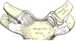
The coracoclavicular ligament serves to connect the clavicle with the coracoid process of the scapula.

The sternoclavicular joint or sternoclavicular articulation is the joint between the manubrium of the sternum and the clavicle bone. It is structurally classed as a synovial saddle joint and functionally classed as a diarthrosis and multiaxial joint. It is composed of two portions separated by an articular disc of fibrocartilage. The bone areas entering into its formation are the sternal end of the clavicle, the upper and lateral part of the sternum,, and the cartilage of the first rib, visible from the outside as the suprasternal notch. The articular surface of the clavicle is much larger than that of the sternum, and is invested with a layer of cartilage, which is considerably thicker than that on the sternum.

The iliolumbar ligament is a strong ligament passing from the tip of the transverse process of the fifth lumbar vertebra to the posterior part of the inner lip of the iliac crest.
The acromioclavicular ligament is part of the acromioclavicular joint. It is divided into two parts: superior and inferior.
The coracohumeral ligament is a broad ligament which strengthens the upper part of the capsule of the shoulder joint.
In human anatomy, the glenohumeral ligaments (GHL) are three ligaments on the anterior side of the glenohumeral joint. Reinforcing the anterior glenohumeral joint capsule, the superior, middle, and inferior glenohumeral ligaments play different roles in the stability of the head of the humerus depending on arm position and degree of rotation.

The oblique popliteal ligament is a broad, flat, fibrous band, formed of fasciculi separated from one another by apertures for the passage of vessels and nerves.
The anterior sternoclavicular ligament is a broad band of fibers, covering the anterior (front) surface of the joint between the sternum and clavicle.

The coronary ligament of the liver refers to parts of the peritoneal reflections that hold the liver to the inferior surface of the diaphragm.
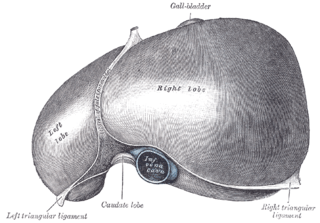
The left triangular ligament is a fold of some considerable size, which connects the posterior part of the upper surface of the left lobe of the liver to the diaphragm; its anterior layer is continuous with the left layer of the falciform ligament.

The anatomical neck of the humerus is obliquely directed, forming an obtuse angle with the body of the humerus. It represents the fused epiphyseal plate.

The root of the penis is triradiate in form, consisting of the diverging crura, one on either side, and the median urethral bulb.