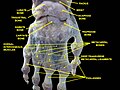
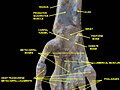
In human anatomy, the wrist is variously defined as (1) the carpus or carpal bones, the complex of eight bones forming the proximal skeletal segment of the hand; (2) the wrist joint or radiocarpal joint, the joint between the radius and the carpus and; (3) the anatomical region surrounding the carpus including the distal parts of the bones of the forearm and the proximal parts of the metacarpus or five metacarpal bones and the series of joints between these bones, thus referred to as wrist joints. This region also includes the carpal tunnel, the anatomical snuff box, bracelet lines, the flexor retinaculum, and the extensor retinaculum.

The lumbricals are intrinsic muscles of the hand that flex the metacarpophalangeal joints, and extend the interphalangeal joints.
The flexor digitorum profundus is a muscle in the forearm of humans that flexes the fingers. It is considered an extrinsic hand muscle because it acts on the hand while its muscle belly is located in the forearm.

In human anatomy, the ulnar nerve is a nerve that runs near the ulna bone. The ulnar collateral ligament of elbow joint is in relation with the ulnar nerve. The nerve is the largest in the human body unprotected by muscle or bone, so injury is common. This nerve is directly connected to the little finger, and the adjacent half of the ring finger, innervating the palmar aspect of these fingers, including both front and back of the tips, perhaps as far back as the fingernail beds.
The trapezium bone is a carpal bone in the hand. It forms the radial border of the carpal tunnel.

In human anatomy, the metacarpal bones or metacarpus, also known as the "palm bones", are the appendicular bones that form the intermediate part of the hand between the phalanges (fingers) and the carpal bones, which articulate with the forearm. The metacarpal bones are homologous to the metatarsal bones in the foot.

The capitate bone is a bone in the human wrist found in the center of the carpal bone region, located at the distal end of the radius and ulna bones. It articulates with the third metacarpal bone and forms the third carpometacarpal joint. The capitate bone is the largest of the carpal bones in the human hand. It presents, above, a rounded portion or head, which is received into the concavity formed by the scaphoid and lunate bones; a constricted portion or neck; and below this, the body. The bone is also found in many other mammals, and is homologous with the "third distal carpal" of reptiles and amphibians.

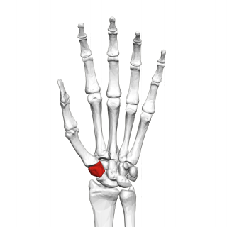
The hamate bone, or unciform bone, Latin os hamatum and occasionally abbreviated as just hamatum, is a bone in the human wrist readily distinguishable by its wedge shape and a hook-like process ("hamulus") projecting from its palmar surface.

The extensor digitorum muscle is a muscle of the posterior forearm present in humans and other animals. It extends the medial four digits of the hand. Extensor digitorum is innervated by the posterior interosseous nerve, which is a branch of the radial nerve.
In human anatomy, the extensor pollicis longus muscle (EPL) is a skeletal muscle located dorsally on the forearm. It is much larger than the extensor pollicis brevis, the origin of which it partly covers and acts to stretch the thumb together with this muscle.

The flexor pollicis brevis is a muscle in the hand that flexes the thumb. It is one of three thenar muscles. It has both a superficial part and a deep part.

In human anatomy, the palmar or volar interossei are four muscles, one on the thumb that is occasionally missing, and three small, unipennate, central muscles in the hand that lie between the metacarpal bones and are attached to the index, ring, and little fingers. They are smaller than the dorsal interossei of the hand.

The carpometacarpal (CMC) joints are five joints in the wrist that articulate the distal row of carpal bones and the proximal bases of the five metacarpal bones.

In human anatomy, the dorsal interossei (DI) are four muscles in the back of the hand that act to abduct (spread) the index, middle, and ring fingers away from hand's midline and assist in flexion at the metacarpophalangeal joints and extension at the interphalangeal joints of the index, middle and ring fingers.

The patellar tendon is the distal portion of the common tendon of the quadriceps femoris, which is continued from the patella to the tibial tuberosity. It is also sometimes called the patellar ligament as it forms a bone to bone connection when the patella is fully ossified.

The extensor retinaculum is a thickened portion of the antebrachial fascia that holds the tendons of the extensor muscles in place. It is located on the back of the forearm, just proximal to the hand. It is continuous with the palmar carpal ligament.

In human anatomy, the annular ligaments of the fingers, often referred to as A pulleys, are the annular part of the fibrous sheathes of the fingers. Four or five such annular pulleys, together with three cruciate pulleys, form a fibro-osseous tunnel on the palmar aspect of the hand through which passes the deep and superficial flexor tendons. The annular and cruciate ligaments serve to govern the flexor mechanism of the hand and wrist, providing critical constraints to the flexor tendons to prevent bowstringing upon contraction and excursion of extrinsic flexor musculo-tendinous units.

In the human hand, palmar or volar plates are found in the metacarpophalangeal (MCP) and interphalangeal (IP) joints, where they reinforce the joint capsules, enhance joint stability, and limit hyperextension. The plates of the MCP and IP joints are structurally and functionally similar, except that in the MCP joints they are interconnected by a deep transverse ligament. In the MCP joints, they also indirectly provide stability to the longitudinal palmar arches of the hand. The volar plate of the thumb MCP joint has a transverse longitudinal rectangular shape, shorter than those in the fingers.

A hand is a prehensile, multi-fingered appendage located at the end of the forearm or forelimb of primates such as humans, chimpanzees, monkeys, and lemurs. A few other vertebrates such as the koala are often described as having "hands" instead of paws on their front limbs. The raccoon is usually described as having "hands" though opposable thumbs are lacking.

Trapeziometacarpal osteoarthritis (TMC OA) is, also known as osteoarthritis at the base of the thumb, thumb carpometacarpal osteoarthritis, basilar (or basal) joint arthritis, or as rhizarthrosis. This joint is formed by the trapezium bone of the wrist and the metacarpal bone of the thumb. This is one of the joints where most humans develop osteoarthritis with age. Osteoarthritis is age-related loss of the smooth surface of the bone where it moves against another bone (cartilage of the joint). In reaction to the loss of cartilage, the bones thicken at the joint surface, resulting in subchondral sclerosis. Also, bony outgrowths, called osteophytes (also known as “bone spurs”), are formed at the joint margins.