Carpal tunnel

Last updated
Carpal tunnel
815 The Carpal Tunnel.jpg
The carpal tunnel
Details
Identifiers
Latin canalis carpi
TA98 A03.5.11.201
TA2 2551
FMA 42352
Anatomical terminology

In the human body, the carpal tunnel or carpal canal is a flattened body cavity on the flexor (palmar/volar) side of the wrist, bounded by the carpal bones and flexor retinaculum. It forms the passageway that transmits the median nerve and the tendons of the extrinsic flexor muscles of the hand from the forearm to the hand. [1] There are described cases of the anatomical variant median artery occurrence.

Contents

When swelling or degeneration occurs in the tendons and sheaths of any of the nine flexor muscles (flexor pollicis longus, four flexor digitorum profundus and four flexor digitorum superficialis) passing through the carpal tunnel, the canal can narrow and compress/entrap the median nerve, resulting in a compression neuropathy known as carpal tunnel syndrome (CTS). [2] [3] If untreated, neuropraxia, parasthesia and muscle atrophy (especially of the thenar muscles) can occur. The condition often require surgical division of the retinaculum to relieve the pressure upon the nerve.

Structure

Carpal tunnel as seen on MRI Wrist MRI T1FSE PDfrFSE STIR 14.jpg
Carpal tunnel as seen on MRI

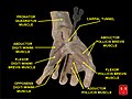
The carpal bones that make up the wrist form an arch which is convex on the dorsal side of the hand and concave on the palmar side. The groove on the palmar side, the sulcus carpi, is covered by the flexor retinaculum, a rigid band of fibrous tissue, thus forming the carpal tunnel. On the side of the radius, the flexor retinaculum is attached to the scaphoid bone, more precisely its tubercle, as well as the ridge of trapezium. On the ulnar side, it is attached to the pisiform and hook of hamate. [4]

The narrowest section of the tunnel is located a centimetre beyond the mid-line of the distal row of carpal bones where the sectional area is limited to 1.6 cm2. [2]

The tendons of the flexor digitorum superficialis and profundus pass through a common ulnar sheath, while the tendon of the flexor pollicis longus passes through a separate radial sheath. The mesotendon shared by these tendons is attached to the radial and palmar walls of the carpal tunnel. [4]

Superficial to the carpal tunnel and the flexor retinaculum, the ulnar artery and ulnar nerve pass through the ulnar tunnel/Guyon's canal. [4]

Function

Transverse section across the wrist and carpals; the palm is at the top (carpal tunnel not labeled but visible at centre) Carpal-Tunnel.svg
Transverse section across the wrist and carpals; the palm is at the top (carpal tunnel not labeled but visible at centre)

Ten structures pass through the carpal tunnel, most of them flexor tendons [2] (not the muscles themselves):

The flexor carpi radialis (one tendon) is often incorrectly stated to travel within the carpal tunnel. More precisely, it travels within the fibers of the flexor retinaculum which forms the roof of the carpal tunnel, rather than running inside the tunnel itself.

Effect of wrist movements

Movements in the wrist affect the shape and width of the carpal tunnel. The width decreases considerably during the normal range of motion in the wrist; and because the carpal bones move in relation to each other with every motion of the hand, the bony walls of the tunnel are not rigid. Both flexion and extension increase compression in the carpal tunnel.

Clinical significance

A 3D medical animation still of carpal tunnel syndrome Carpal Tunnel Syndrome.jpg
A 3D medical animation still of carpal tunnel syndrome

Carpal tunnel syndrome is characterized by tingling, burning and pain (needle, pin) through the course of median nerve particularly over the outer fingers and radiating up the arm, that is caused by compression of the carpal tunnel contents. It is associated with repetitive use, rheumatoid arthritis, and a number of other states. It can be detected using Tinel's sign and the Phalen maneuver. It may be treated non-surgically by splinting or corticosteroid injection. Definitive management often requires surgical division of the flexor retinaculum, the aponeuroses which forms the roof of the carpal tunnel. Symptoms of the syndrome can sometimes be caused by tight muscles in the neck and shoulder region.

Additional images

See also

Related Research Articles

<span class="mw-page-title-main">Carpal bones</span> Eight small bones that make up the wrist (or carpus) that connects the hand to the forearm

The carpal bones are the eight small bones that make up the wrist (carpus) that connects the hand to the forearm. The term "carpus" and "carpal" is derived from the Latin carpus and the Greek καρπός (karpós), meaning "wrist". In human anatomy, the main role of the carpal bones is to articulate with the radial and ulnar heads to form a highly mobile condyloid joint, to provide attachments for thenar and hypothenar muscles, and to form part of the rigid carpal tunnel which allows the median nerve and tendons of the anterior forearm muscles to be transmitted to the hand and fingers.

<span class="mw-page-title-main">Median nerve</span> Nerve of the upper limb

The median nerve is a nerve in humans and other animals in the upper limb. It is one of the five main nerves originating from the brachial plexus.

<span class="mw-page-title-main">Wrist</span> Part of the arm between the lower arm and the hand

In human anatomy, the wrist is variously defined as (1) the carpus or carpal bones, the complex of eight bones forming the proximal skeletal segment of the hand; (2) the wrist joint or radiocarpal joint, the joint between the radius and the carpus and; (3) the anatomical region surrounding the carpus including the distal parts of the bones of the forearm and the proximal parts of the metacarpus or five metacarpal bones and the series of joints between these bones, thus referred to as wrist joints. This region also includes the carpal tunnel, the anatomical snuff box, bracelet lines, the flexor retinaculum, and the extensor retinaculum.

The flexor digitorum profundus is a muscle in the forearm of humans that flexes the fingers. It is considered an extrinsic hand muscle because it acts on the hand while its muscle belly is located in the forearm.

<span class="mw-page-title-main">Flexor digitorum superficialis muscle</span>

Flexor digitorum superficialis is an extrinsic flexor muscle of the fingers at the proximal interphalangeal joints.

<span class="mw-page-title-main">Ulnar nerve</span> Nerve which runs near the ulna bone

In human anatomy, the ulnar nerve is a nerve that runs near the ulna bone. The ulnar collateral ligament of elbow joint is in relation with the ulnar nerve. The nerve is the largest in the human body unprotected by muscle or bone, so injury is common. This nerve is directly connected to the little finger, and the adjacent half of the ring finger, innervating the palmar aspect of these fingers, including both front and back of the tips, perhaps as far back as the fingernail beds.

<span class="mw-page-title-main">Upper limb</span> Consists of the arm, forearm, and hand

The upper limbs or upper extremities are the forelimbs of an upright-postured tetrapod vertebrate, extending from the scapulae and clavicles down to and including the digits, including all the musculatures and ligaments involved with the shoulder, elbow, wrist and knuckle joints. In humans, each upper limb is divided into the arm, forearm and hand, and is primarily used for climbing, lifting and manipulating objects.

<span class="mw-page-title-main">Ulnar artery</span> Artery of the forearm

The ulnar artery is the main blood vessel, with oxygenated blood, of the medial aspects of the forearm. It arises from the brachial artery and terminates in the superficial palmar arch, which joins with the superficial branch of the radial artery. It is palpable on the anterior and medial aspect of the wrist.

<span class="mw-page-title-main">Flexor pollicis brevis muscle</span> Muscle in the thenar compartment

The flexor pollicis brevis is a muscle in the hand that flexes the thumb. It is one of three thenar muscles. It has both a superficial part and a deep part.

The flexor pollicis longus is a muscle in the forearm and hand that flexes the thumb. It lies in the same plane as the flexor digitorum profundus. This muscle is unique to humans, being either rudimentary or absent in other primates. A meta-analysis indicated accessory flexor pollicis longus is present in around 48% of the population.

<span class="mw-page-title-main">Abductor digiti minimi muscle of hand</span> Muscle in the hypothenar compartment

In human anatomy, the abductor digiti minimi is a skeletal muscle situated on the ulnar border of the palm of the hand. It forms the ulnar border of the palm and its spindle-like shape defines the hypothenar eminence of the palm together with the skin, connective tissue, and fat surrounding it. Its main function is to pull the little finger away from the other fingers.

<span class="mw-page-title-main">Flexor retinaculum of the hand</span> Thickened fascia over the carpal tunnel

The flexor retinaculum is a fibrous band on the palmar side of the hand near the wrist. It arches over the carpal bones of the hands, covering them and forming the carpal tunnel.

<span class="mw-page-title-main">Anterior interosseous artery</span>

The anterior interosseous artery is an artery in the forearm. It is a branch of the common interosseous artery.

<span class="mw-page-title-main">Anterior interosseous nerve</span>

The anterior interosseous nerve is a branch of the median nerve that supplies the deep muscles on the anterior of the forearm, except the ulnar (medial) half of the flexor digitorum profundus. Its nerve roots come from C8 and T1.

<span class="mw-page-title-main">Common flexor sheath of hand</span>

The common synovial sheath for the flexor tendons or the ulnar bursa is a synovial sheath in the carpal tunnel of the human hand.

<span class="mw-page-title-main">Pronator teres syndrome</span> Medical condition

Pronator teres syndrome is a compression neuropathy of the median nerve at the elbow. It is rare compared to compression at the wrist or isolated injury of the anterior interosseous branch of the median nerve.

<span class="mw-page-title-main">Median nerve palsy</span> Medical condition

Injuries to the arm, forearm or wrist area can lead to various nerve disorders. One such disorder is median nerve palsy. The median nerve controls the majority of the muscles in the forearm. It controls abduction of the thumb, flexion of hand at wrist, flexion of digital phalanx of the fingers, is the sensory nerve for the first three fingers, etc. Because of this major role of the median nerve, it is also called the eye of the hand. If the median nerve is damaged, the ability to abduct and oppose the thumb may be lost due to paralysis of the thenar muscles. Various other symptoms can occur which may be repaired through surgery and tendon transfers. Tendon transfers have been very successful in restoring motor function and improving functional outcomes in patients with median nerve palsy.

<span class="mw-page-title-main">Extrinsic extensor muscles of the hand</span>

The extrinsic extensor muscles of the hand are located in the back of the forearm and have long tendons connecting them to bones in the hand, where they exert their action. Extrinsic denotes their location outside the hand. Extensor denotes their action which is to extend, or open flat, joints in the hand. They include the extensor carpi radialis longus (ECRL), extensor carpi radialis brevis (ECRB), extensor digitorum (ED), extensor digiti minimi (EDM), extensor carpi ulnaris (ECU), abductor pollicis longus (APL), extensor pollicis brevis (EPB), extensor pollicis longus (EPL), and extensor indicis (EI).

<span class="mw-page-title-main">Muscles of the thumb</span>

The muscles of the thumb are nine skeletal muscles located in the hand and forearm. The muscles allow for flexion, extension, adduction, abduction and opposition of the thumb. The muscles acting on the thumb can be divided into two groups: The extrinsic hand muscles, with their muscle bellies located in the forearm, and the intrinsic hand muscles, with their muscles bellies located in the hand proper.

<span class="mw-page-title-main">Palmaris profundus muscle</span>

Palmaris profundus is a rare anatomical variant in the anterior compartment of forearm. It was first described in 1908. It is usually found incidentally in cadaveric dissection or surgery.

References

  1. 1 2 Schmidt, Hans-Martin; Lanz, Ulrich (2003). Surgical anatomy of the hand. Thieme. p. 29. ISBN   1-58890-007-X.
  2. 1 2 3 Thieme Atlas of Anatomy: General Anatomy and Musculoskeletal System. Thieme. 2006. pp. 248–249. ISBN   1-58890-419-9.
  3. "Carpal Tunnel Syndrome Fact Sheet | National Institute of Neurological Disorders and Stroke". www.ninds.nih.gov. Retrieved 2021-05-27.
  4. 1 2 3 Thieme Atlas of Anatomy: General Anatomy and Musculoskeletal System. Thieme. 2006. p. 354. ISBN   1-58890-419-9.