In human anatomy, the wrist is variously defined as (1) the carpus or carpal bones, the complex of eight bones forming the proximal skeletal segment of the hand;[1][2] (2) the wrist joint or radiocarpal joint, the joint between the radius and the carpus[2] and; (3) the anatomical region surrounding the carpus including the distal parts of the bones of the forearm and the proximal parts of the metacarpus or five metacarpal bones and the series of joints between these bones, thus referred to as wrist joints.[3][4] This region also includes the carpal tunnel, the anatomical snuff box, bracelet lines, the flexor retinaculum, and the extensor retinaculum.
As a consequence of these various definitions, fractures to the carpal bones are referred to as carpal fractures, while fractures such as distal radius fracture are often considered fractures to the wrist.
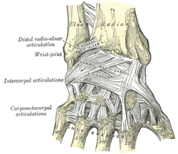
The radiocarpal (wrist) joint is an ellipsoid joint formed by the radius and the articular disc proximally and the proximal row of carpal bones distally. The carpal bones on the ulnar side only make intermittent contact with the proximal side — the triquetrum only makes contact during ulnar abduction. The capsule, lax and un-branched, is thin on the dorsal side and can contain synovial folds. The capsule is continuous with the midcarpal joint and strengthened by numerous ligaments, including the palmar and dorsal radiocarpal ligaments, and the ulnar and radial collateral ligaments. [6]
The parts forming the radiocarpal joint are the lower end of the radius and under surface of the articular disk above; and the scaphoid, lunate, and triquetral bones below. The articular surface of the radius and the undersurface of the articular disk form together with a transversely elliptical concave surface, the receiving cavity. The superior articular surfaces of the scaphoid, lunate, and triquetrum form a smooth convex surface, the condyle, which is received into the concavity.[7]
The earliest carpal bones to ossify are capitate bone and hamate bone in the first six months of an infant life.[9]
Articulations
The radiocarpal, intercarpal, midcarpal, carpometacarpal, and intermetacarpal joints often intercommunicate through a common synovial cavity. [10]
Articular surfaces
It has two articular surfaces named, proximal and distal articular surfaces respectively. The proximal articular surface is made up of the lower end of the radius and a triangular articular disc of the inferior radio-ulnar joint. On the other hand, the distal articular surface is made up of proximal surfaces of the scaphoid, triquetral and lunate bones.[11]
Micro-radiography of 8-weeks human embryo hand
Function
Movement
The extrinsic hand muscles are located in the forearm where their bellies form the proximal fleshy roundness. When contracted, most of the tendons of these muscles are prevented from standing up like taut bowstrings around the wrist by passing under the flexor retinaculum on the palmar side and the extensor retinaculum on the dorsal side. On the palmar side the carpal bones form the carpal tunnel,[12] through which some of the flexor tendons pass in tendon sheaths that enable them to slide back and forth through the narrow passageway (see carpal tunnel syndrome).[13]
Starting from the mid-position of the hand, the movements permitted in the wrist proper are (muscles in order of importance):[14][15]
Magnetic resonance imaging (MRI) of radial abduction (rightwards in image) and ulnar adduction (leftwards in image)
Marginal movements: radial deviation (abduction, movement towards the thumb) and ulnar deviation (adduction, movement towards the little finger). These movements take place about a dorsopalmar axis (back to front) at the radiocarpal and midcarpal joints passing through the capitate bone.
Movements in the plane of the hand: flexion (palmar flexion, tilting towards the palm) and extension (dorsiflexion, tilting towards the back of the hand). These movements take place through a transverse axis passing through the capitate bone. Palmar flexion is the most powerful of these movements because the flexors, especially the finger flexors, are considerably stronger than the extensors.
However, movements at the wrist can not be properly described without including movements in the distal radioulnar joint in which the rotary actions of supination and pronation occur and this joint is therefore normally regarded as part of the wrist.[17]
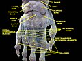
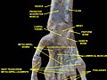
Wrist joint. Deep dissection. Anterior, palmar, view.
Wrist joint. Deep dissection. Anterior, palmar, view.
References
↑ Behnke 2006, p.76 "The wrist contains eight bones, roughly aligned in two rows, known as the carpal bones."
1 2 Moore KL, Agur AM (2006). Essential clinical anatomy. Lippincott Williams & Wilkins. p.485. ISBN0-7817-6274-X. The wrist (carpus), the proximal segment of the hand, is a complex of eight carpal bones. The carpus articulates proximally with the forearm at the wrist joint and distally with the five metacarpals. The joints formed by the carpus include the wrist (the radiocarpal joint), intercarpal, carpometacarpal, and intermetacarpal joints. Augmenting movement at the wrist joint, the rows of carpals glide on each other[...]
↑ Behnke 2006, p.77 "With the large number of bones composing the wrist (ulna, radius, eight carpas, and five metacarpals), it makes sense that there are many, many joints that make up the structure known as the wrist."
↑ Baratz M, Watson AD, Imbriglia JE (1999). Orthopaedic surgery: the essentials. Thieme. p.391. ISBN0-86577-779-9. The wrist joint is composed of not only the radiocarpal and distal radioulnar joints but also the intercarpal articulations.
↑ Rea P (2016-01-01). "Chapter 3 - Neck". In Rea P (ed.). Essential Clinically Applied Anatomy of the Peripheral Nervous System in the Head and Neck. Academic Press. pp.131–183. doi:10.1016/b978-0-12-803633-4.00003-x. ISBN978-0-12-803633-4.
↑ Saladin KS (2003). Anatomy & Physiology: The Unity of Form and Function (3rded.). McGraw-Hill. pp.361, 365.
1 2 3 4 5 Lalani I, Argoff CE (2008-01-01). "Chapter 10 - History and Physical Examination of the Pain Patient". In Benzon HT, Rathmell JP, Wu CL, Turk DC (eds.). Raj's Practical Management of Pain (Fourthed.). Philadelphia: Mosby. pp.177–188. doi:10.1016/B978-032304184-3.50013-3. ISBN978-0-323-04184-3.
This page is based on this Wikipedia article Text is available under the CC BY-SA 4.0 license; additional terms may apply. Images, videos and audio are available under their respective licenses.