Capitate bone

Last updated

Capitate bone
Capitate bone (left hand) 01 palmar view.png
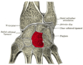
Left hand anterior view (palmar view). Capitate-bone shown in red.
Gray227 retouched.png
The left capitate bone.
Left: ulnar surface (little-finger-side surface).
Right: radial surface (thumb-side surface)
Details
Pronunciation /ˈkæpɪtt/
Part of Carpal bones of the hand
Identifiers
Latin os capitatum; os magnum
MeSH D051224
TA98 A02.4.08.011
TA2 1258
FMA 23727
Anatomical terms of bone

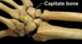
The capitate bone is a bone in the human wrist found in the center of the carpal bone region, located at the distal end of the radius and ulna bones. It articulates with the third metacarpal bone (the middle finger) and forms the third carpometacarpal joint. The capitate bone is the largest of the carpal bones in the human hand. It presents, above, a rounded portion or head, which is received into the concavity formed by the scaphoid and lunate bones; a constricted portion or neck; and below this, the body. [1] The bone is also found in many other mammals, and is homologous with the "third distal carpal" of reptiles and amphibians.

Contents

Structure

The capitate is the largest carpal bone found within the hand. [2] The capitate is found within the distal row of carpal bones. The capitate lies directly adjacent to the metacarpal of the ring finger on its distal surface, has the hamate on its ulnar surface and trapezoid on its radial surface, and abuts the lunate and scaphoid proximally. [3] :708–709

Surfaces

The proximal surface is round, smooth, and articulates with the lunate bone. [1]

The distal surface is divided by two ridges into three facets, for articulation with the second, third, and fourth metacarpal bones, that for the third being the largest. [1]

The dorsal surface is broad and rough. [1]

The palmar surface is narrow, rounded, and rough, for the attachment of ligaments and a part of the adductor pollicis muscle. [1]

The lateral surface articulates with the lesser multangular by a small facet at its anterior inferior angle, behind which is a rough depression for the attachment of an interosseous ligament. Above this is a deep, rough groove, forming part of the neck, and serving for the attachment of ligaments; it is bounded superiorly by a smooth, convex surface, for articulation with the scaphoid bone. [1]

The medial surface articulates with the hamate bone by a smooth, concave, oblong facet, which occupies its posterior and superior parts; it is rough in front, for the attachment of an interosseous ligament. [1]

Variation

The capitate bone variably articulates with the metacarpal of the index finger. However, its normal articulation is with the middle finger. [2]

Development

The ossification of capitate starts at 1 – 5 months. [4]

Function

The carpal bones function as a unit to provide a bony superstructure for the hand. [3] :708 They allow movements of the wrist from side to side (medial to lateral) as well as up and down (anterior to posterior). H. A. Harris wrote in the British Medical Journal in 1944 that "the strength of construction of the hand in a man is concentrated in the radius, thumb, and index and middle fingers." Therefore, the capitate is larger to support the strength and stress that the middle finger undergoes.

Clinical significance

A capitate fracture accounts for 1.3% of all wrist fractures. Isolated fractures of the capitate comprise only 0.3% and are often non-displaced. This is since the capitate is at the centre of the carpal region and is therefore quite well protected. Capitate fractures occur together with fractures of another carpal bone, the scaphoid. [5]

Various mechanisms for fractures of the capitate have been postulated. Adler et al. described three mechanisms—the first is direct trauma to the dorsal surface of the bone, the second is fall on the palm with the wrist in forced extension and the third is fall on the forcefully flexed hand; the second being the most frequent and the third rarest. [5]

In the case of an acute capitate fracture where there is X-ray evidence of excellent alignment of the fracture fragments, the attending doctor will immobilise the wrist in a plaster or lightweight wrist brace. Once the cast has been removed, the patient begins physiotherapy to regain the range of movement of the wrist joint and strength in the muscles involved.

If X-rays show that the capitate fracture fragments are out of alignment, surgery is indicated. A surgeon can use small compression screws or K-wires to unite the two pieces of bone. The headless compression screw has advantage over the K-wire as it provides compression across the fracture site and allows early motion. It may be the case that the ligament between the сapitate and the scaphoid bone is also injured; if so, this would be repaired at the same time. [6]

Because the capitate has a poor blood supply there are sometimes complications with the healing process. This may manifest itself as a diffuse ache in the wrist upon activity, and can persist for many months. This is due to a breakdown of the capitate caused by the lack of blood supply and healing (avascular necrosis). Nonunion has been reported as the most common complication; 19.6% to 56% in isolated capitate fractures. Early diagnosis is key to preventing this.

Etymology

The name of the bone derives from Latin : capitātus 'having a head', from Latin : capit- 'head'. [7]

Additional images

See also

Related Research Articles

<span class="mw-page-title-main">Carpal bones</span> Eight small bones that make up the wrist (or carpus) that connects the hand to the forearm

The carpal bones are the eight small bones that make up the wrist (carpus) that connects the hand to the forearm. The term "carpus" and "carpal" is derived from the Latin carpus and the Greek καρπός (karpós), meaning "wrist". In human anatomy, the main role of the carpal bones is to articulate with the radial and ulnar heads to form a highly mobile condyloid joint, to provide attachments for thenar and hypothenar muscles, and to form part of the rigid carpal tunnel which allows the median nerve and tendons of the anterior forearm muscles to be transmitted to the hand and fingers.

<span class="mw-page-title-main">Wrist</span> Part of the arm between the lower arm and the hand

In human anatomy, the wrist is variously defined as (1) the carpus or carpal bones, the complex of eight bones forming the proximal skeletal segment of the hand; (2) the wrist joint or radiocarpal joint, the joint between the radius and the carpus and; (3) the anatomical region surrounding the carpus including the distal parts of the bones of the forearm and the proximal parts of the metacarpus or five metacarpal bones and the series of joints between these bones, thus referred to as wrist joints. This region also includes the carpal tunnel, the anatomical snuff box, bracelet lines, the flexor retinaculum, and the extensor retinaculum.

<span class="mw-page-title-main">Anatomical snuffbox</span> Indent on back of hand between tendons

The anatomical snuff box or snuffbox or foveola radialis is a triangular deepening on the radial, dorsal aspect of the hand—at the level of the carpal bones, specifically, the scaphoid and trapezium bones forming the floor. The name originates from the use of this surface for placing and then sniffing powdered tobacco, or "snuff." It is sometimes referred to by its French name tabatière.

<span class="mw-page-title-main">Trapezoid bone</span> Carpal (wrist) bone

The trapezoid bone is a carpal bone in tetrapods, including humans. It is the smallest bone in the distal row of carpal bones that give structure to the palm of the hand. It may be known by its wedge-shaped form, the broad end of the wedge constituting the dorsal, the narrow end the palmar surface; and by its having four articular facets touching each other, and separated by sharp edges. It is homologous with the "second distal carpal" of reptiles and amphibians.

<span class="mw-page-title-main">Trapezium (bone)</span> Bone of the wrist

The trapezium bone is a carpal bone in the hand. It forms the radial border of the carpal tunnel.

<span class="mw-page-title-main">Metacarpal bones</span> Bones of hand

In human anatomy, the metacarpal bones or metacarpus, also known as the "palm bones", are the appendicular bones that form the intermediate part of the hand between the phalanges (fingers) and the carpal bones, which articulate with the forearm. The metacarpal bones are homologous to the metatarsal bones in the foot.

<span class="mw-page-title-main">Scaphoid bone</span> Bone of the carpus

The scaphoid bone is one of the carpal bones of the wrist. It is situated between the hand and forearm on the thumb side of the wrist. It forms the radial border of the carpal tunnel. The scaphoid bone is the largest bone of the proximal row of wrist bones, its long axis being from above downward, lateralward, and forward. It is approximately the size and shape of a medium cashew nut.

<span class="mw-page-title-main">Hamate bone</span> Carpal bone in the wrist

The hamate bone, or unciform bone, Latin os hamatum and occasionally abbreviated as just hamatum, is a bone in the human wrist readily distinguishable by its wedge shape and a hook-like process ("hamulus") projecting from its palmar surface.

<span class="mw-page-title-main">Lunate bone</span> Carpal bone in the human hand

The lunate bone is a carpal bone in the human hand. It is distinguished by its deep concavity and crescentic outline. It is situated in the center of the proximal row carpal bones, which lie between the ulna and radius and the hand. The lunate carpal bone is situated between the lateral scaphoid bone and medial triquetral bone.

<span class="mw-page-title-main">Radius (bone)</span> One of the two long bones of the forearm

The radius or radial bone is one of the two large bones of the forearm, the other being the ulna. It extends from the lateral side of the elbow to the thumb side of the wrist and runs parallel to the ulna. The ulna is longer than the radius, but the radius is thicker. The radius is a long bone, prism-shaped and slightly curved longitudinally.

<span class="mw-page-title-main">Triquetral bone</span> Bone in the wrist

The triquetral bone is located in the wrist on the medial side of the proximal row of the carpus between the lunate and pisiform bones. It is on the ulnar side of the hand, but does not directly articulate with the ulna. Instead, it is connected to and articulates with the ulna through the Triangular fibrocartilage disc and ligament, which forms part of the ulnocarpal joint capsule. It connects with the pisiform, hamate, and lunate bones. It is the 2nd most commonly fractured carpal bone.

<span class="mw-page-title-main">Upper limb</span> Consists of the arm, forearm, and hand

The upper limbs or upper extremities are the forelimbs of an upright-postured tetrapod vertebrate, extending from the scapulae and clavicles down to and including the digits, including all the musculatures and ligaments involved with the shoulder, elbow, wrist and knuckle joints. In humans, each upper limb is divided into the arm, forearm and hand, and is primarily used for climbing, lifting and manipulating objects.

<span class="mw-page-title-main">Palmar interossei muscles</span> Muscles between the metacarpals

In human anatomy, the palmar or volar interossei are four muscles, one on the thumb that is occasionally missing, and three small, unipennate, central muscles in the hand that lie between the metacarpal bones and are attached to the index, ring, and little fingers. They are smaller than the dorsal interossei of the hand.

<span class="mw-page-title-main">Carpometacarpal joint</span>

The carpometacarpal (CMC) joints are five joints in the wrist that articulate the distal row of carpal bones and the proximal bases of the five metacarpal bones.

<span class="mw-page-title-main">First metacarpal bone</span>

The first metacarpal bone or the metacarpal bone of the thumb is the first bone proximal to the thumb. It is connected to the trapezium of the carpus at the first carpometacarpal joint and to the proximal thumb phalanx at the first metacarpophalangeal joint.

<span class="mw-page-title-main">Intercarpal joints</span> Joints of the carpal bones of the wrist

The intercarpal joints can be subdivided into three sets of joints : Those of the proximal row of carpal bones, those of the distal row of carpal bones, and those of the two rows with each other.

<span class="mw-page-title-main">Midcarpal joint</span>

The midcarpal joint is formed by the scaphoid, lunate, and triquetral bones in the proximal row, and the trapezium, trapezoid, capitate, and hamate bones in the distal row. The distal pole of the scaphoid articulates with two trapezial bones as a gliding type of joint. The proximal end of the scaphoid combines with the lunate and triquetrum to form a deep concavity that articulates with the convexity of the combined capitate and hamate in a form of diarthrodial, almost condyloid joint.

<span class="mw-page-title-main">Hand</span> Extremity at the end of an arm or forelimb

A hand is a prehensile, multi-fingered appendage located at the end of the forearm or forelimb of primates such as humans, chimpanzees, monkeys, and lemurs. A few other vertebrates such as the koala are often described as having "hands" instead of paws on their front limbs. The raccoon is usually described as having "hands" though opposable thumbs are lacking.

<span class="mw-page-title-main">Wrist osteoarthritis</span> Medical condition

Wrist osteoarthritis is gradual loss of articular cartilage and hypertrophic bone changes (osteophytes). While in many joints this is part of normal aging (senescence), in the wrist osteoarthritis usually occurs over years to decades after scapholunate interosseous ligament rupture or an unhealed fracture of the scaphoid. Characteristic symptoms including pain, deformity and stiffness. Pain intensity and incapability are notably variable and do not correspond with arthritis severity on radiographs.

<span class="mw-page-title-main">Muscles of the thumb</span>

The muscles of the thumb are nine skeletal muscles located in the hand and forearm. The muscles allow for flexion, extension, adduction, abduction and opposition of the thumb. The muscles acting on the thumb can be divided into two groups: The extrinsic hand muscles, with their muscle bellies located in the forearm, and the intrinsic hand muscles, with their muscles bellies located in the hand proper.

References

PD-icon.svgThis article incorporates text in the public domain from page 226 of the 20th edition of Gray's Anatomy (1918)

  1. 1 2 3 4 5 6 7 Gray's Anatomy (1918). See infobox.
  2. 1 2 Eathorne, SW (March 2005). "The wrist: clinical anatomy and physical examination--an update". Primary Care. 32 (1): 17–33. doi:10.1016/j.pop.2004.11.009. PMID   15831311.
  3. 1 2 Drake, Richard L.; Vogl, Wayne; Tibbitts, Adam W.M. Mitchell; illustrations by Richard; Richardson, Paul (2005). Gray's anatomy for students. Philadelphia: Elsevier/Churchill Livingstone. ISBN   978-0-8089-2306-0.
  4. Balachandran, Ajay; Kartha, Moumitha; Krishna, Anooj; Thomas, Jerry; K, Prathilash; TN, Prem; GK, Libu; B, Krishnan; John, Liza (2014). "A Study of Ossification of Capitate, Hamate, Triquetral & Lunate in Forensic Age Estimation". Indian Journal of Forensic Medicine & Toxicology. 8 (2): 218–224. doi:10.5958/0973-9130.2014.00720.8. ISSN   0973-9130 . Retrieved 18 August 2014.
  5. 1 2 Sabat, D; Arora, S; Dhal, A (2011). "Isolated capitate fracture with dorsal dislocation of proximal pole: a case report". Hand (N Y). 6 (3): 333–6. doi:10.1007/s11552-011-9337-5. PMC   3153623 . PMID   22942861.
  6. "Carpal Fractures Treatment & Management: Approach Considerations, Surgical Therapy, Postoperative Care". 6 October 2021.
  7. Harper, Douglas. "Capitate". Online Etymology Dictionary. Retrieved 5 January 2014.

Sources