
The humerus is a long bone in the arm that runs from the shoulder to the elbow. It connects the scapula and the two bones of the lower arm, the radius and ulna, and consists of three sections. The humeral upper extremity consists of a rounded head, a narrow neck, and two short processes. The body is cylindrical in its upper portion, and more prismatic below. The lower extremity consists of 2 epicondyles, 2 processes, and 3 fossae. As well as its true anatomical neck, the constriction below the greater and lesser tubercles of the humerus is referred to as its surgical neck due to its tendency to fracture, thus often becoming the focus of surgeons.

The brachioradialis is a muscle of the forearm that flexes the forearm at the elbow. It is also capable of both pronation and supination, depending on the position of the forearm. It is attached to the distal styloid process of the radius by way of the brachioradialis tendon, and to the lateral supracondylar ridge of the humerus.

The inguinal canal is a passage in the anterior abdominal wall on each side of the body, which in males, convey the spermatic cords and in females, the round ligament of the uterus. The inguinal canals are larger and more prominent in males.

Pectoralis minor muscle is a thin, triangular muscle, situated at the upper part of the chest, beneath the pectoralis major in the human body. It arises from ribs III-V; it inserts onto the coracoid process of the scapula. It is innervated by the medial pectoral nerve. Its function is to stabilise the scapula by holding it fast in position against the chest wall.

The internal pudendal artery is one of the three pudendal arteries. It branches off the internal iliac artery, and provides blood to the external genitalia.

The rectus abdominis muscle, also known as the "abdominal muscle" or simply the "abs", is a pair of segmented skeletal muscle on the ventral aspect of a person's abdomen. The paired muscle is separated at the midline by a band of dense connective tissue called the linea alba, and the connective tissue defining each lateral margin of the rectus abdominus is the linea semilunaris. The muscle extends from the pubic symphysis, pubic crest and pubic tubercle inferiorly, to the xiphoid process and costal cartilages of the 5th–7th ribs superiorly.

The pectineus muscle is a flat, quadrangular muscle, situated at the anterior (front) part of the upper and medial (inner) aspect of the thigh. The pectineus muscle is the most anterior adductor of the hip. The muscle's primary action is hip flexion; it also produces adduction and internal rotation of the hip.

The inguinal ligament, also known as Poupart's ligament or groin ligament, is a band running from the pubic tubercle to the anterior superior iliac spine. It forms the base of the inguinal canal through which an indirect inguinal hernia may develop.

The transverse abdominal muscle (TVA), also known as the transverse abdominis, transversalis muscle and transversus abdominis muscle, is a muscle layer of the anterior and lateral abdominal wall, deep to the internal oblique muscle. It is thought by most fitness instructors to be a significant component of the core.

The external iliac arteries are two major arteries which bifurcate off the common iliac arteries anterior to the sacroiliac joint of the pelvis.

The lesser omentum is the double layer of peritoneum that extends from the liver to the lesser curvature of the stomach, and to the first part of the duodenum. The lesser omentum is usually divided into these two connecting parts: the hepatogastric ligament, and the hepatoduodenal ligament.

The sacrotuberous ligament is situated at the lower and back part of the pelvis. It is flat, and triangular in form; narrower in the middle than at the ends.

The bicipital groove is a deep groove on the humerus that separates the greater tubercle from the lesser tubercle. It allows for the long tendon of the biceps brachii muscle to pass.

The petrotympanic fissure is a fissure in the temporal bone that runs from the temporomandibular joint to the tympanic cavity.
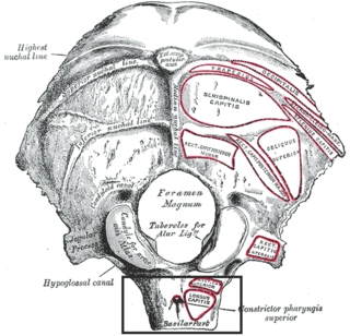
The pharyngeal tubercle is a part of the occipital bone of the head and neck. It is located on the lower surface of the basilar part of occipital bone. It is the site of attachment of the pharyngeal raphe.

The lateral condyle is the lateral portion of the upper extremity of tibia.

The fascia of Camper is a thick superficial layer of the anterior abdominal wall.

The greater tubercle of the humerus is the outward part the upper end of that bone, adjacent to the large rounded prominence of the humerus head. It provides attachment points for the supraspinatus, infraspinatus, and teres minor muscles, three of the four muscles of the rotator cuff, a muscle group that stabilizes the shoulder joint. In doing so the tubercle acts as a location for the transfer of forces from the rotator cuff muscles to the humerus.
The coracohumeral ligament is a broad ligament of the shoulder. It attaches to the coracoid process at one end, and to the greater and lesser tubercles of the humerus at the other. It strengthens the upper part of the joint capsule of the shoulder joint.
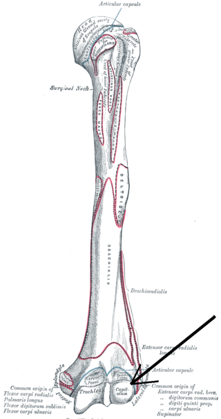
In human anatomy of the arm, the capitulum of the humerus is a smooth, rounded eminence on the lateral portion of the distal articular surface of the humerus. It articulates with the cup-shaped depression on the head of the radius, and is limited to the front and lower part of the bone.


