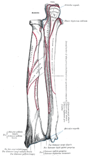
The ulna is a long bone found in the forearm that stretches from the elbow to the smallest finger, and when in anatomical position, is found on the medial side of the forearm. It runs parallel to the radius, the other long bone in the forearm. The ulna is usually slightly longer than the radius, but the radius is thicker. Therefore the radius is considered to be the larger of the two.

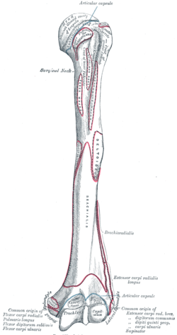
The humerus is a long bone in the arm that runs from the shoulder to the elbow. It connects the scapula and the two bones of the lower arm, the radius and ulna, and consists of three sections. The humeral upper extremity consists of a rounded head, a narrow neck, and two short processes. The body is cylindrical in its upper portion, and more prismatic below. The lower extremity consists of 2 epicondyles, 2 processes, and 3 fossae. As well as its true anatomical neck, the constriction below the greater and lesser tubercles of the humerus is referred to as its surgical neck due to its tendency to fracture, thus often becoming the focus of surgeons.

The brachioradialis is a muscle of the forearm that flexes the forearm at the elbow. It is also capable of both pronation and supination, depending on the position of the forearm. It is attached to the distal styloid process of the radius by way of the brachioradialis tendon, and to the lateral supracondylar ridge of the humerus.

The brachial artery is the major blood vessel of the (upper) arm. It is the continuation of the axillary artery beyond the lower margin of teres major muscle. It continues down the ventral surface of the arm until it reaches the cubital fossa at the elbow. It then divides into the radial and ulnar arteries which run down the forearm. In some individuals, the bifurcation occurs much earlier and the ulnar and radial arteries extend through the upper arm. The pulse of the brachial artery is palpable on the anterior aspect of the elbow, medial to the tendon of the biceps, and, with the use of a stethoscope and sphygmomanometer often used to measure the blood pressure.

The median nerve is a nerve in humans and other animals in the upper limb. It is one of the five main nerves originating from the brachial plexus.

The brachialis is a muscle in the upper arm that flexes the elbow joint. It lies deeper than the biceps brachii, and makes up part of the floor of the region known as the cubital fossa. The brachialis is the prime mover of elbow flexion. While the biceps brachii appears as a large anterior bulge on the arm and commands considerable interest among body builders, the brachialis underlying it actually generates about 50% more power and is thus the prime mover of elbow flexion.

In human anatomy, the ulnar nerve is a nerve that runs near the ulna bone. The ulnar collateral ligament of elbow joint is in relation with the ulnar nerve. The nerve is the largest in the human body unprotected by muscle or bone, so injury is common. This nerve is directly connected to the little finger, and the adjacent half of the ring finger, innervating the palmar aspect of these fingers, including both front and back of the tips, perhaps as far back as the fingernail beds.

The ulnar collateral ligament is a thick triangular band at the medial aspect of the elbow uniting the distal aspect of the humerus to the proximal aspect of the ulna.
The cubital fossa,chelidon, or elbow pit is the triangular area on the anterior view of the elbow of a human or other hominid animal. It lies anteriorly to the elbow when in standard anatomical position.

The ulnar artery is the main blood vessel, with oxygenated blood, of the medial aspects of the forearm. It arises from the brachial artery and terminates in the superficial palmar arch, which joins with the superficial branch of the radial artery. It is palpable on the anterior and medial aspect of the wrist.

The olecranon from the Greekolene meaning elbow and kranon meaning head is the large, thick, curved bony eminence of the ulna, a long bone in the forearm that projects behind the elbow. It forms the most pointed portion of the elbow and is opposite to the cubital fossa or elbow pit. The olecranon serves as a lever for the extensor muscles that straighten the elbow joint.
The posterior interosseous artery is an artery of the forearm.
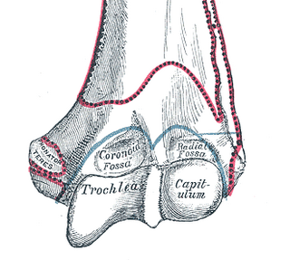
In the human arm, the humeral trochlea is the medial portion of the articular surface of the elbow joint which articulates with the trochlear notch on the ulna in the forearm.

The lower extremity of the humerus is flattened from before backward, and curved slightly forward; it ends below in a broad, articular surface, which is divided into two parts by a slight ridge.

The medial epicondyle of the humerus is an epicondyle of the humerus bone of the upper arm in humans. It is larger and more prominent than the lateral epicondyle and is directed slightly more posteriorly in the anatomical position. In birds, where the arm is somewhat rotated compared to other tetrapods, it is called the ventral epicondyle of the humerus. In comparative anatomy, the more neutral term entepicondyle is used.

The olecranon fossa is a deep triangular depression on the posterior side of the humerus, superior to the trochlea, in which the summit of the olecranon is received during extension of the forearm.
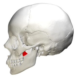
In human anatomy, the mandible's coronoid process is a thin, triangular eminence, which is flattened from side to side and varies in shape and size. Its anterior border is convex and is continuous below with the anterior border of the ramus. Its posterior border is concave and forms the anterior boundary of the mandibular notch. The lateral surface is smooth, and affords insertion to the temporalis and masseter muscles. Its medial surface gives insertion to the temporalis, and presents a ridge which begins near the apex of the process and runs downward and forward to the inner side of the last molar tooth.
The Ulna's coronoid process is a triangular eminence projecting forward from the anterior proximal portion of the ulna.
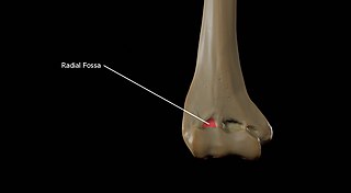
The radial fossa is a slight depression found on the humerus above the front part of the capitulum. It receives the anterior border of the head of the radius when the forearm is flexed.
The head of the radius has a cylindrical form, and on its upper surface is a shallow cup or fovea for articulation with the capitulum of the humerus. The circumference of the head is smooth; it is broad medially where it articulates with the radial notch of the ulna, narrow in the rest of its extent, which is embraced by the annular ligament.