| Hamate bone | |
|---|---|
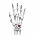
 Left hand anterior view (palmar view). Hamate bone shown in red. | |
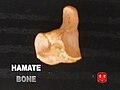
 The left hamate bone | |
| Details | |
| Pronunciation | /ˈheɪmət/ |
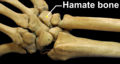
| Articulations | Articulates with five bones: the lunate proximally the fourth and fifth metacarpals distally the triangular medially the capitate laterally |
| Identifiers | |
| Latin | os hamatum |
| MeSH | D051225 |
| TA98 | A02.4.08.012 |
| TA2 | 1259 |
| FMA | 23730 |
| Anatomical terms of bone | |
The hamate bone (from Latin hamatus, "hooked"), or unciform bone (from Latin uncus , "hook"), Latin os hamatum and occasionally abbreviated as just hamatum, [1] [2] [3] is a bone in the human wrist readily distinguishable by its wedge shape and a hook-like process ("hamulus") projecting from its palmar surface.