
The rib cage is an endoskeletal enclosure in the thorax of most vertebrate animals that comprises the ribs, vertebral column and sternum, which protects vital organs such as the heart, lungs and great vessels. The circumferential enclosure formed by left and right rib cages, together known as the thoracic cage, is a semi-rigid bony and cartilaginous structure which surrounds the thoracic cavity and supports the shoulder girdles to form the core part of the axial skeleton.

The femur, or thigh bone is the only bone in the thigh. The thigh is the region of the lower limb between the hip and the knee. In many four-legged animals the femur is the upper bone of the hindleg.

The scapula, also known as the shoulder blade, is the bone that connects the humerus with the clavicle. Like their connected bones, the scapulae are paired, with each scapula on either side of the body being roughly a mirror image of the other. The name derives from the Classical Latin word for trowel or small shovel, which it was thought to resemble.

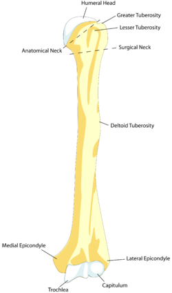
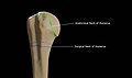
The humerus is a long bone in the arm that runs from the shoulder to the elbow. It connects the scapula and the two bones of the lower arm, the radius and ulna, and consists of three sections. The humeral upper extremity consists of a rounded head, a narrow neck, and two short processes. The body is cylindrical in its upper portion, and more prismatic below. The lower extremity consists of 2 epicondyles, 2 processes, and 3 fossae. As well as its true anatomical neck, the constriction below the greater and lesser tubercles of the humerus is referred to as its surgical neck due to its tendency to fracture, thus often becoming the focus of surgeons.

The coracoid process is a small hook-like structure on the lateral edge of the superior anterior portion of the scapula. Pointing laterally forward, it, together with the acromion, serves to stabilize the shoulder joint. It is palpable in the deltopectoral groove between the deltoid and pectoralis major muscles.

The human shoulder is made up of three bones: the clavicle (collarbone), the scapula, and the humerus as well as associated muscles, ligaments and tendons.

An epiphysis is one of the rounded ends or tips of a long bone that ossify from a secondary center of ossification. Between the epiphysis and diaphysis lies the metaphysis, including the epiphyseal plate. At the joint, the epiphysis is covered with articular cartilage; below that covering is a zone similar to the epiphyseal plate, known as subchondral bone.

The shoulder joint is structurally classified as a synovial ball-and-socket joint and functionally as a diarthrosis and multiaxial joint. It involves an articulation between the glenoid fossa of the scapula and the head of the humerus. Due to the very loose joint capsule that gives a limited interface of the humerus and scapula, it is the most mobile joint of the human body.

The subscapularis is a large triangular muscle which fills the subscapular fossa and inserts into the lesser tubercle of the humerus and the front of the capsule of the shoulder-joint.

Dog anatomy comprises the anatomical studies of the visible parts of the body of a domestic dog. Details of structures vary tremendously from breed to breed, more than in any other animal species, wild or domesticated, as dogs are highly variable in height and weight. The smallest known adult dog was a Yorkshire Terrier that stood only 6.3 cm (2.5 in) at the shoulder, 9.5 cm (3.7 in) in length along the head and body, and weighed only 113 grams (4.0 oz). The heaviest dog was an English Mastiff named Zorba which weighed 314 pounds (142 kg). The tallest known adult dog is a Great Dane that stands 106.7 cm (42.0 in) at the shoulder.
The shoulder girdle or pectoral girdle is the set of bones in the appendicular skeleton which connects to the arm on each side. In humans it consists of the clavicle and scapula; in those species with three bones in the shoulder, it consists of the clavicle, scapula, and coracoid. Some mammalian species have only the scapula.

The upper extremity, proximal extremity or superior epiphysis of the femur is the part of the femur closest to the pelvic bone and the trunk. It contains the following structures:

The lesser tubercle of the humerus, although smaller, is more prominent than the greater tubercle: it is situated in front, and is directed medially and anteriorly.

The greater tubercle of the humerus is the outward part the upper end of that bone, adjacent to the large rounded prominence of the humerus head. It provides attachment points for the supraspinatus, infraspinatus, and teres minor muscles, three of the four muscles of the rotator cuff, a muscle group that stabilizes the shoulder joint. In doing so the tubercle acts as a location for the transfer of forces from the rotator cuff muscles to the humerus.
The coracohumeral ligament is a broad ligament of the shoulder. It attaches to the coracoid process at one end, and to the greater and lesser tubercles of the humerus at the other. It strengthens the upper part of the joint capsule of the shoulder joint.
In human anatomy, the glenohumeral ligaments (GHL) are three ligaments on the anterior side of the glenohumeral joint. Reinforcing the anterior glenohumeral joint capsule, the superior, middle, and inferior glenohumeral ligaments play different roles in the stability of the head of the humerus depending on arm position and degree of rotation.

The fascial compartments of arm refers to the specific anatomical term of the compartments within the upper segment of the upper limb of the body. The upper limb is divided into two segments, the arm and the forearm. Each of these segments is further divided into two compartments which are formed by deep fascia – tough connective tissue septa (walls). Each compartment encloses specific muscles and nerves.

The surgical neck of the humerus is a bony constriction at the proximal end of shaft of humerus. It is situated distal to the greater tubercle and lesser tubercle, and proximal to the deltoid tuberosity.

The following outline is provided as an overview of and topical guide to human anatomy:
Humeral avulsion of the glenohumeral ligament (HAGL) is defined as an avulsion of the inferior glenohumeral ligament from the anatomic neck of the humerus. In other words, it occurs when we have disruption of the ligaments that join the humerus to the glenoid. HAGL tends to occur in 7.5-9.3% of cases of anterior shoulder instability. Making it an uncommon cause of anterior shoulder instability. Avulsion of this ligamentous complex may occur in three sites: glenoid insertion (40%), the midsubstance (35%) and the humeral insertion (25%). Bony humeral avulsion of the glenohumeral ligament (BHAGL) refers when we have HAGL with bony fracture.
