Ankle

Last updated
Ankle
Ankle.jpg
Human ankle
Ankle en.svg
Lateral view of the human ankle
Details
Identifiers
Latin tarsus
MeSH D000842
TA98 A01.1.00.041
TA2 165
FMA 9665
Anatomical terminology

The ankle, the talocrural region [1] or the jumping bone (informal) is the area where the foot and the leg meet. [2] The ankle includes three joints: the ankle joint proper or talocrural joint, the subtalar joint, and the inferior tibiofibular joint. [3] [4] [5] The movements produced at this joint are dorsiflexion and plantarflexion of the foot. In common usage, the term ankle refers exclusively to the ankle region. In medical terminology, "ankle" (without qualifiers) can refer broadly to the region or specifically to the talocrural joint. [1] [6]

Contents

The main bones of the ankle region are the talus (in the foot), the tibia, and fibula (both in the leg). The talocrural joint is a synovial hinge joint that connects the distal ends of the tibia and fibula in the lower limb with the proximal end of the talus. [7] The articulation between the tibia and the talus bears more weight than that between the smaller fibula and the talus.

Structure

Region

The ankle region is found at the junction of the leg and the foot. It extends downwards (distally) from the narrowest point of the lower leg and includes the parts of the foot closer to the body (proximal) to the heel and upper surface (dorsum) of the foot. [8] :768

Ankle joint

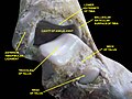
The talocrural joint is the only mortise and tenon joint in the human body, [9] :1418 the term likening the skeletal structure to the woodworking joint of the same name. The bony architecture of the ankle consists of three bones: the tibia, the fibula, and the talus. The articular surface of the tibia may be referred to as the plafond (French for "ceiling"). [10] The medial malleolus is a bony process extending distally off the medial tibia. The distal-most aspect of the fibula is called the lateral malleolus. Together, the malleoli, along with their supporting ligaments, stabilize the talus underneath the tibia.

Because the motion of the subtalar joint provides a significant contribution to positioning the foot, some authors will describe it as the lower ankle joint, and call the talocrural joint the upper ankle joint. [11] Dorsiflexion and Plantarflexion are the movements that take place in the ankle joint. When the foot is plantar flexed, the ankle joint also allows some movements of side to side gliding, rotation, adduction, and abduction. [12]

The bony arch formed by the tibial plafond and the two malleoli is referred to as the ankle "mortise" (or talar mortise). The mortise is a rectangular socket. [1] The ankle is composed of three joints: the talocrural joint (also called talotibial joint, tibiotalar joint, talar mortise, talar joint), the subtalar joint (also called talocalcaneal), and the Inferior tibiofibular joint. [3] [4] [5] The joint surface of all bones in the ankle is covered with articular cartilage.

The distances between the bones in the ankle are as follows: [13]

Decreased distances indicate osteoarthritis.

Ligaments

Ligaments of ankle and feet 919 Ankle Feet Joints.jpg
Ligaments of ankle and feet

The ankle joint is bound by the strong deltoid ligament and three lateral ligaments: the anterior talofibular ligament, the posterior talofibular ligament, and the calcaneofibular ligament.

Though it does not span the ankle joint itself, the syndesmotic ligament makes an important contribution to the stability of the ankle. This ligament spans the syndesmosis, i.e. the articulation between the medial aspect of the distal fibula and the lateral aspect of the distal tibia. An isolated injury to this ligament is often called a high ankle sprain.

The bony architecture of the ankle joint is most stable in dorsiflexion. [14] Thus, a sprained ankle is more likely to occur when the ankle is plantar-flexed, as ligamentous support is more important in this position. The classic ankle sprain involves the anterior talofibular ligament (ATFL), which is also the most commonly injured ligament during inversion sprains. Another ligament that can be injured in a severe ankle sprain is the calcaneofibular ligament.

Retinacula, tendons and their synovial sheaths, vessels, and nerves

A number of tendons pass through the ankle region. Bands of connective tissue called retinacula (singular: retinaculum) allow the tendons to exert force across the angle between the leg and foot without lifting away from the angle, a process called bowstringing. [11] The superior extensor retinaculum of foot extends between the anterior (forward) surfaces of the tibia and fibula near their lower (distal) ends. It contains the anterior tibial artery and vein and the tendons of the tibialis anterior muscle within its tendon sheath and the unsheathed tendons of extensor hallucis longus and extensor digitorum longus muscles. The deep peroneal nerve passes under the retinaculum while the superficial peroneal nerve is outside of it. The inferior extensor retinaculum of foot is a Y-shaped structure. Its lateral attachment is on the calcaneus, and the band travels towards the anterior tibia where it is attached and blends with the superior extensor retinaculum. Along with that course, the band divides and another segment attaches to the plantar aponeurosis. The tendons which pass through the superior extensor retinaculum are all sheathed along their paths through the inferior extensor retinaculum and the tendon of the fibularis tertius muscle is also contained within the retinaculum.

The flexor retinaculum of foot extends from the medial malleolus to the medical process of the calcaneus, and the following structures in order from medial to lateral: the tendon of the tibialis posterior muscle, the tendon of the flexor digitorum longus muscle, the posterior tibial artery and vein, the tibial nerve, and the tendon of the flexor hallucis longus muscle.

The fibular retinacula hold the tendons of the fibularis longus and fibularis brevis along the lateral aspect of the ankle region. The superior fibular retinaculum extends from the deep transverse fascia of the leg and lateral malleolus to calcaneus. The inferior fibular retinaculum is a continuous extension from the inferior extensor retinaculum to the calcaneus. [9] :1418–9

Mechanoreceptors

Mechanoreceptors of the ankle send proprioceptive sensory input to the central nervous system (CNS). [15] Muscle spindles are thought to be the main type of mechanoreceptor responsible for proprioceptive attributes from the ankle. [16] The muscle spindle gives feedback to the CNS system on the current length of the muscle it innervates and to any change in length that occurs.

It was hypothesized that muscle spindle feedback from the ankle dorsiflexors played the most substantial role in proprioception relative to other muscular receptors that cross at the ankle joint. However, due to the multi-planar range of motion at the ankle joint there is not one group of muscles that is responsible for this. [17] This helps to explain the relationship between the ankle and balance.

In 2011, a relationship between proprioception of the ankle and balance performance was seen in the CNS. This was done by using a fMRI machine in order to see the changes in brain activity when the receptors of the ankle are stimulated. [18] This implicates the ankle directly with the ability to balance. Further research is needed in order to see to what extent does the ankle affect balance.

Function

Historically, the role of the ankle in locomotion has been discussed by Aristotle and Leonardo da Vinci. There is no question that ankle push-off is a significant force in human gait, but how much energy is used in leg swing as opposed to advancing the whole-body center of mass is not clear. [19]

Clinical significance

A diagram illustrating varying severity of ankle sprain Ankle sprain -- Smart-Servier (cropped).jpg
A diagram illustrating varying severity of ankle sprain

Traumatic injury

Of all major joints, the ankle is the most commonly injured. If the outside surface of the foot is twisted under the leg during weight bearing, the lateral ligament, especially the anterior talofibular portion, is subject to tearing (a sprain) as it is weaker than the medial ligament and it resists inward rotation of the talocrural joint. [8] :825

Fractures

Fracture of both sides of the ankle with dislocation as seen on anteroposterior X-ray. (1) fibula, (2) tibia, (arrow) medial malleolus, (arrowhead) lateral malleolus Bimalleolar fracture legend.jpg
Fracture of both sides of the ankle with dislocation as seen on anteroposterior X-ray. (1) fibula, (2) tibia, (arrow) medial malleolus, (arrowhead) lateral malleolus

An ankle fracture is a break of one or more of the bones that make up the ankle joint. [20] Symptoms may include pain, swelling, bruising, and an inability to walk on the injured leg. [20] Complications may include an associated high ankle sprain, compartment syndrome, stiffness, malunion, and post-traumatic arthritis. [20] [21]

Ankle fractures may result from excessive stress on the joint such as from rolling an ankle or from blunt trauma. [20] [21] Types of ankle fractures include lateral malleolus, medial malleolus, posterior malleolus, bimalleolar, and trimalleolar fractures. [20] The Ottawa ankle rule can help determine the need for X-rays. [21] Special X-ray views called stress views help determine whether an ankle fracture is unstable.

Treatment depends on the fracture type. Ankle stability largely dictates non-operative vs. operative treatment. Non-operative treatment includes splinting or casting while operative treatment includes fixing the fracture with metal implants through an open reduction internal fixation (ORIF). [20] Significant recovery generally occurs within four months while completely recovery usually takes up to one year. [20]

Ankle fractures are common, occurring in over 1.8 per 1000 adults and 1 per 1000 children per year. [21] [22] In North America this figure increases to more than 14 in ever 10,000 patients admitted to the Emergency Room. [23] They occur most commonly in young males and older females. [21]

Imaging

The initial evaluation of suspected ankle pathology is usually by projectional radiography ("X-ray").

Tibiotalar surface angle (TTS) X-ray of frontal tibiotalar surface angle (TTS).jpg
Tibiotalar surface angle (TTS)

Varus or valgus deformity, if suspected, can be measured with the frontal tibiotalar surface angle (TTS), formed by the mid-longitudinal tibial axis (such as through a line bisecting the tibia at 8 and 13 cm above the tibial plafond) and the talar surface. [24] An angle of less than 84 degrees is regarded as talipes varus, and an angle of more than 94 degrees is regarded as talipes valgus. [25]

For ligamentous injury, there are three main landmarks on X-rays: The first is the tibiofibular clear space, the horizontal distance from the lateral border of the posterior tibial malleolus to the medial border of the fibula, with greater than 5 mm being abnormal. The second is tibiofibular overlap, the horizontal distance between the medial border of the fibula and the lateral border of the anterior tibial prominence, with less than 10 mm being abnormal. The final measurement is the medial clear space, the distance between the lateral aspect of the medial malleolus and the medial border of the talus at the level of the talar dome, with a measurement greater than 4 mm being abnormal. Loss of any of these normal anatomic spaces can indirectly reflect ligamentous injury or occult fracture, and can be followed by MRI or CT. [26]

Abnormalities

Clubfoot or talipes equinovarus, which occurs in one to two of every 1,000 live births, involves multiple abnormalities of the foot. [27] Equinus refers to the downard deflection of the ankle, and is named for the walking on the toes in the manner of a horse. [28] This does not occur because it is accompanied by an inward rotation of the foot (varus deformity), which untreated, results in walking on the sides of the feet. Treatment may involve manipulation and casting or surgery. [27]

Ankle joint equinus, normally in adults, relates to restricted ankle joint range of motion(ROM). [29] Calf muscle stretching exercises are normally helpful to increase the ankle joint dorsiflexion and used to manage clinical symptoms resulting from ankle equinus. [30]

Occasionally a human ankle has a ball-and-socket ankle joint and fusion of the talo-navicular joint. [31]

History

The word ankle or ancle is common, in various forms, to Germanic languages, probably connected in origin with the Latin angulus, or Greek αγκυλος, meaning bent. [32]

Other animals

Evolution

It has been suggested that dexterous control of toes has been lost in favour of a more precise voluntary control of the ankle joint. [33]

See also

Footnotes

  1. 1 2 3 Moore, Keith L.; Dalley, Arthur F.; Agur, A. M. R. (2013). "Lower Limb". Clinically Oriented Anatomy (7th ed.). Lippincott Williams & Wilkins. pp. 508–669. ISBN   978-1-4511-1945-9.
  2. WebMD (2009). "ankle". Webster's New World Medical Dictionary (3rd ed.). Houghton Mifflin Harcourt. p. 22. ISBN   978-0-544-18897-6.
  3. 1 2 Milner, Brent K. (1999). "Musculoskeletal Imaging". In Gay, Spencer B.; Woodcock, Richard J. (eds.). Radiology Recall . Lippincott Williams & Wilkins. pp.  258–383. ISBN   978-0-683-30663-7.
  4. 1 2 Williams, D. S. Blaise; Taunton, Jack (2007). "Foot, ankle and lower leg". In Kolt, Gregory S.; Snyder-Mackler, Lynn (eds.). Physical Therapies in Sport and Exercise. Elsevier Health Sciences. pp. 420–39. ISBN   978-0-443-10351-3.
  5. 1 2 del Castillo, Jorge (2012). "Foot and Ankle Injuries". In Adams, James G. (ed.). Emergency Medicine. Elsevier Health Sciences. pp. 745–55. ISBN   978-1-4557-3394-1.
  6. Gray, Henry (1918). "Talocrural Articulation or Ankle-joint". Anatomy of the Human Body.
  7. WebMD (2009). "ankle joint". Webster's New World Medical Dictionary (3rd ed.). Houghton Mifflin Harcourt. p. 22. ISBN   978-0-544-18897-6.
  8. 1 2 Moore, Keith (2018). Clinically oriented anatomy. Philadelphia: Wolters Kluwer. ISBN   978-1-4963-4721-3.
  9. 1 2 Susan Standring (7 August 2015). Gray's Anatomy E-Book: The Anatomical Basis of Clinical Practice. Elsevier Health Sciences. ISBN   978-0-7020-6851-5.
  10. David P. Barei (29 March 2012). "56. Pilon Fractures". In Robert W. Bucholz (ed.). Rockwood and Green's Fractures in Adults: Two Volumes Plus Integrated Content Website (Rockwood, Green, and Wilkins' Fractures). Lippincott Williams & Wilkins. pp. 1928–1971. ISBN   978-1-4511-6144-1.
  11. 1 2 Joseph E. Muscolino (21 August 2016). Kinesiology - E-Book: The Skeletal System and Muscle Function. Elsevier Health Sciences. pp. 284–292. ISBN   978-0-323-39935-7.
  12. Joseph H Volker (2018-08-08). "Ankle Joint". Earth's Lab.
  13. Imai, Kan; Ikoma, Kazuya; Kido, Masamitsu; Maki, Masahiro; Fujiwara, Hiroyoshi; Arai, Yuji; Oda, Ryo; Tokunaga, Daisaku; Inoue, Nozomu; Kubo, Toshikazu (2015). "Joint space width of the tibiotalar joint in the healthy foot". Journal of Foot and Ankle Research. 8 (1): 26. doi: 10.1186/s13047-015-0086-5 . ISSN   1757-1146. PMC   4490633 . PMID   26146520.
  14. Gatt, A. and Chockalingam, N., 2011. Clinical Assessment of Ankle Joint DorsiflexionA Review of Measurement Techniques. Journal of the American Podiatric Medical Association, 101(1), pp.59-69.https://doi.org/10.7547/1010059
  15. Michelson, J. D.; Hutchins, C (1995). "Mechanoreceptors in human ankle ligaments". The Journal of Bone and Joint Surgery. British Volume. 77 (2): 219–24. doi: 10.1302/0301-620X.77B2.7706334 . PMID   7706334.
  16. Lephart, S. M.; Pincivero, D. M.; Rozzi, S. L. (1998). "Proprioception of the ankle and knee". Sports Medicine. 25 (3): 149–55. doi:10.2165/00007256-199825030-00002. PMID   9554026. S2CID   13099542.
  17. Ribot-Ciscar, E; Bergenheim, M; Albert, F; Roll, J. P. (2003). "Proprioceptive population coding of limb position in humans". Experimental Brain Research. 149 (4): 512–9. doi:10.1007/s00221-003-1384-x. PMID   12677332. S2CID   14626459.
  18. Goble, D. J.; Coxon, J. P.; Van Impe, A.; Geurts, M.; Doumas, M.; Wenderoth, N.; Swinnen, S. P. (2011). "Brain Activity during Ankle Proprioceptive Stimulation Predicts Balance Performance in Young and Older Adults". Journal of Neuroscience. 31 (45): 16344–52. doi:10.1523/JNEUROSCI.4159-11.2011. PMC   6633212 . PMID   22072686.
  19. Zelik, Karl E.; Adamczyk, Peter G. (2016). "A unified perspective on ankle push-off in human walking". The Journal of Experimental Biology. 219 (23): 3676–3683. doi:10.1242/jeb.140376. ISSN   0022-0949. PMC   5201006 . PMID   27903626.
  20. 1 2 3 4 5 6 7 "Ankle Fractures (Broken Ankle) - OrthoInfo - AAOS". www.orthoinfo.org. Retrieved 20 June 2019.
  21. 1 2 3 4 5 Wire J, Slane VH (9 May 2019). "Ankle Fractures". StatPearls. Treasure Island (FL): StatPearls Publishing. PMID   31194464.
  22. Yeung DE, Jia X, Miller CA, Barker SL (April 2016). "Interventions for treating ankle fractures in children". The Cochrane Database of Systematic Reviews. 2016 (4): CD010836. doi:10.1002/14651858.CD010836.pub2. PMC   7111433 . PMID   27033333.
  23. Ptak, Nathaniel A.; Rigby, Ryan B. (2024). "Subluxation of the posterior tibial tendon into the tibiofibular syndesmosis secondary to high-level ankle fracture or dislocation: Surgical reduction technique guide and case study". Foot & Ankle Surgery: Techniques, Reports & Cases. 4 (1): 100357. doi: 10.1016/j.fastrc.2023.100357 . ISSN   2667-3967.
  24. Nosewicz, Tomasz L.; Knupp, Markus; Bolliger, Lilianna; Hintermann, Beat (2012). "The reliability and validity of radiographic measurements for determining the three-dimensional position of the talus in varus and valgus osteoarthritic ankles". Skeletal Radiology. 41 (12): 1567–1573. doi:10.1007/s00256-012-1421-6. ISSN   0364-2348. PMC   3478506 . PMID   22609967.
  25. Chapter 5 - Radiological morphology of peritalar instability in varus and valgus tilted ankles, in: T.L. Nosewicz (2018-09-25). Acute and chronic aspects of hindfoot trauma. University of Amsterdam, Faculty of Medicine (AMC-UvA). ISBN   9789463750479.
  26. Evans, JM; Schucany, WG (October 2006). "Radiological evaluation of a high ankle sprain". Proceedings (Baylor University. Medical Center). 19 (4): 402–5. doi:10.1080/08998280.2006.11928206. PMC   1618742 . PMID   17106502.
  27. 1 2 Gore AI, Spencer JP (2004). "The newborn foot". Am Fam Physician. 69 (4): 865–72. PMID   14989573.
  28. Källén, Bengt (2014). "Pes Equinovarus". Epidemiology of Human Congenital Malformations. pp. 111–113. doi:10.1007/978-3-319-01472-2_22. ISBN   978-3-319-01471-5.
  29. Gatt, Alfred; Chockalingam, Nachiappan (2011-01-01). "Clinical Assessment of Ankle Joint Dorsiflexion". Journal of the American Podiatric Medical Association. 101 (1): 59–69. doi:10.7547/1010059. PMID   21242472.
  30. Macklin, Katriona; Healy, Aoife; Chockalingam, Nachiappan (March 2012). "The effect of calf muscle stretching exercises on ankle joint dorsiflexion and dynamic foot pressures, force and related temporal parameters". The Foot. 22 (1): 10–17. doi:10.1016/j.foot.2011.09.001. PMID   21944945.
  31. Ono, K.; Nakamura, M.; Kurata, Y.; Hiroshima, K. (September 1984). "Ball-and-socket ankle joint: Anatomical and kinematic analysis of the hindfoot". Journal of Pediatric Orthopedics. 4 (5): 564–568. doi:10.1097/01241398-198409000-00007. PMID   6490876.
  32. Chisholm, Hugh, ed. (1911). "Ankle"  . Encyclopædia Britannica . Vol. 2 (11th ed.). Cambridge University Press. p. 58.
  33. Brouwer, B.; Ashby, P. (1992). "Corticospinal projections to lower limb motoneurons in man". Experimental Brain Research. 89 (3): 649–54. doi:10.1007/bf00229889. PMID   1644127. S2CID   24650165.

Related Research Articles

<span class="mw-page-title-main">Foot</span> Anatomical structure found in vertebrates

The foot is an anatomical structure found in many vertebrates. It is the terminal portion of a limb which bears weight and allows locomotion. In many animals with feet, the foot is a separate organ at the terminal part of the leg made up of one or more segments or bones, generally including claws and/or nails.

<span class="mw-page-title-main">Human leg</span> Lower extremity or limb of the human body (foot, lower leg, thigh and hip)

The leg is the entire lower limb of the human body, including the foot, thigh or sometimes even the hip or buttock region. The major bones of the leg are the femur, tibia, and adjacent fibula. The thigh is between the hip and knee, while the calf (rear) and shin (front) are between the knee and foot.

<span class="mw-page-title-main">Fibularis longus</span> Superficial muscle in the lateral compartment of the leg

In human anatomy, the fibularis longus is a superficial muscle in the lateral compartment of the leg. It acts to tilt the sole of the foot away from the midline of the body (eversion) and to extend the foot downward away from the body at the ankle.

<span class="mw-page-title-main">Tibia</span> Leg bone in vertebrates

The tibia, also known as the shinbone or shankbone, is the larger, stronger, and anterior (frontal) of the two bones in the leg below the knee in vertebrates ; it connects the knee with the ankle. The tibia is found on the medial side of the leg next to the fibula and closer to the median plane. The tibia is connected to the fibula by the interosseous membrane of leg, forming a type of fibrous joint called a syndesmosis with very little movement. The tibia is named for the flute tibia. It is the second largest bone in the human body, after the femur. The leg bones are the strongest long bones as they support the rest of the body.

<span class="mw-page-title-main">Fibula</span> Leg bone in vertebrates

The fibula or calf bone is a leg bone on the lateral side of the tibia, to which it is connected above and below. It is the smaller of the two bones and, in proportion to its length, the most slender of all the long bones. Its upper extremity is small, placed toward the back of the head of the tibia, below the knee joint and excluded from the formation of this joint. Its lower extremity inclines a little forward, so as to be on a plane anterior to that of the upper end; it projects below the tibia and forms the lateral part of the ankle joint.

<span class="mw-page-title-main">Calcaneus</span> Bone of the tarsus of the foot

In humans and many other primates, the calcaneus or heel bone is a bone of the tarsus of the foot which constitutes the heel. In some other animals, it is the point of the hock.

Pott's fracture, also known as Pott's syndrome I and Dupuytren fracture, is an archaic term loosely applied to a variety of bimalleolar ankle fractures. The injury is caused by a combined abduction external rotation from an eversion force. This action strains the sturdy medial (deltoid) ligament of the ankle, often tearing off the medial malleolus due to its strong attachment. The talus then moves laterally, shearing off the lateral malleolus or, more commonly, breaking the fibula superior to the tibiofibular syndesmosis. If the tibia is carried anteriorly, the posterior margin of the distal end of the tibia is also sheared off by the talus. A fractured fibula in addition to detaching the medial malleolus will tear the tibiofibular syndesmosis. The combined fracture of the medial malleolus, lateral malleolus, and the posterior margin of the distal end of the tibia is known as a "trimalleolar fracture".

<span class="mw-page-title-main">Maisonneuve fracture</span> Medical condition

The Maisonneuve fracture is a spiral fracture of the proximal third of the fibula associated with a tear of the distal tibiofibular syndesmosis and the interosseous membrane. There is an associated fracture of the medial malleolus or rupture of the deep deltoid ligament of the ankle. This type of injury can be difficult to detect.

<span class="mw-page-title-main">Tarsus (skeleton)</span> Bones of the foot

In the human body, the tarsus is a cluster of seven articulating bones in each foot situated between the lower end of the tibia and the fibula of the lower leg and the metatarsus. It is made up of the midfoot and hindfoot.

<span class="mw-page-title-main">Tibialis posterior muscle</span> Muscle in the most central of all the leg muscles

The tibialis posterior muscle is the most central of all the leg muscles, and is located in the deep posterior compartment of the leg. It is the key stabilizing muscle of the lower leg.

<span class="mw-page-title-main">Tibialis anterior muscle</span> Flexor muscle in humans that dorsiflexes the foot

The tibialis anterior muscle is a muscle of the anterior compartment of the lower leg. It originates from the upper portion of the tibia; it inserts into the medial cuneiform and first metatarsal bones of the foot. It acts to dorsiflex and invert the foot. This muscle is mostly located near the shin.

<span class="mw-page-title-main">Talus bone</span> One of the foot bones that forms the tarsus

The talus, talus bone, astragalus, or ankle bone is one of the group of foot bones known as the tarsus. The tarsus forms the lower part of the ankle joint. It transmits the entire weight of the body from the lower legs to the foot.

<span class="mw-page-title-main">Fibularis brevis</span> Shorter and smaller of the fibularis (peroneus) muscles

In human anatomy, the fibularis brevis is a muscle that lies underneath the fibularis longus within the lateral compartment of the leg. It acts to tilt the sole of the foot away from the midline of the body (eversion) and to extend the foot downward away from the body at the ankle.

The Broström operation is a repair of ligaments on lateral ankle. It is designed to address ankle instability. More importantly, it is primarily used to repair the anterior talofibular ligament (ATFL) in the ankle. It is thought that the majority of patients regain most function in their ankles. The recovery time for the procedure varies according to the patient but usually takes a minimum of 3–6 months.

<span class="mw-page-title-main">Ankle fracture</span> Medical condition

An ankle fracture is a break of one or more of the bones that make up the ankle joint. Symptoms may include pain, swelling, bruising, and an inability to walk on the injured leg. Complications may include an associated high ankle sprain, compartment syndrome, stiffness, malunion, and post-traumatic arthritis.

<span class="mw-page-title-main">Inferior extensor retinaculum of foot</span> Y-shaped band placed in front of the ankle-joint

The inferior extensor retinaculum of the foot is a Y-shaped band placed in front of the ankle-joint, the stem of the Y being attached laterally to the upper surface of the calcaneus, in front of the depression for the interosseous talocalcaneal ligament; it is directed medialward as a double layer, one lamina passing in front of, and the other behind, the tendons of the peroneus tertius and extensor digitorum longus.

<span class="mw-page-title-main">Superior extensor retinaculum of foot</span> Upper part of the extensor retinaculum of foot

The superior extensor retinaculum of the foot is the upper part of the extensor retinaculum of foot which extends from the ankle to the heelbone.

<span class="mw-page-title-main">Malleolus</span> Ankle bone protrusion

A malleolus is the bony prominence on each side of the human ankle.

A high ankle sprain, also known as a syndesmotic ankle sprain (SAS), is a sprain of the syndesmotic ligaments that connect the tibia and fibula in the lower leg, thereby creating a mortise and tenon joint for the ankle. High ankle sprains are described as high because they are located above the ankle. They comprise approximately 15% of all ankle sprains. Unlike the common lateral ankle sprains, when ligaments around the ankle are injured through an inward twisting, high ankle sprains are caused when the lower leg and foot externally rotates.

<span class="mw-page-title-main">Crus fracture</span>

A crus fracture is a fracture of the lower legs bones meaning either or both of the tibia and fibula.

References

Additional images