Heel

Last updated
Heel
Girls heel.jpg
A girl heel
Details
Identifiers
Latin calx
MeSH D006365
TA98 A01.1.00.042
TA2 167
FMA 24994
Anatomical terminology

The heel is the prominence at the posterior end of the foot. It is based on the projection of one bone, the calcaneus or heel bone, behind the articulation of the bones of the lower leg.

Contents

Structure

Sagittal section through the foot Science ofDressTo face p236cutA.png
Sagittal section through the foot
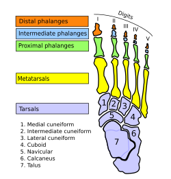
From above Ospied-en.svg
From above

To distribute the compressive forces exerted on the heel during gait, and especially the stance phase when the heel contacts the ground, the sole of the foot is covered by a layer of subcutaneous connective tissue up to 2 cm thick (under the heel). This tissue has a system of pressure chambers that both acts as a shock absorber and stabilises the sole. Each of these chambers contains fibrofatty tissue covered by a layer of tough connective tissue made of collagen fibers. These septa ("walls") are firmly attached both to the plantar aponeurosis above and the sole's skin below. The sole of the foot is one of the most highly vascularized regions of the body surface, and the dense system of blood vessels further stabilize the septa. [1]

The Achilles tendon is the muscle tendon of the triceps surae, a "three-headed" group of muscles—the soleus and the two heads of the gastrocnemius. The main function of the triceps surae is plantar flexion, i.e. to stretch the foot downward. It is accompanied by a "fourth head", the slight plantaris muscle, the long slender tendon of which is also attached to the heel bone but not visible. [2]

Function

The compressive forces applied to the foot are distributed along five rays, three medial (side of big toe) and two lateral (side of little toe). The lateral rays stretch over the cuboid bone to the heel bone and the medial rays over the three cuneiform bones and the navicular bone to the ankle bone. Because the ankle bone is placed over the heel bone, these rays are adjacent near the toes but overriding near the heel, and together they form the arches of the foot that are optimized to distributed compressive forces across an uneven terrain. In this context the heel thus forms the posterior point of support that together with the balls of the large and little toes bear the brunt of the loads. [3]

Cracked heels

Cracked heels is a common health problem and it may cause infections. It is caused by dryness of the foot skin, [4] and accumulation of dead skin. Over time, it may cause pain and irritations. Various moisturising creams and foot files are available to cure and prevent it.

Other animals

In the long-footed mammals, both the hoofed species (unguligrade) and the clawed forms which walk on the toes (digitigrade), the heel is well above the ground at the apex of the angular joint known as the hock. In plantigrade species, it rests on the ground. In birds, the heel is the backward-pointing joint which is often mistaken as the "knee" (the actual knee of birds is hidden under the plumage).

See also

Notes

  1. Thieme Atlas 2006, p 418
  2. Thieme Atlas 2006, p 434
  3. Thieme Atlas 2006, p 412
  4. "Dry, Cracked Feet: Treatment, Causes, and Home Remedies". www.footvitals.com.

Related Research Articles

<span class="mw-page-title-main">Foot</span> Anatomical structure found in vertebrates

The foot is an anatomical structure found in many vertebrates. It is the terminal portion of a limb which bears weight and allows locomotion. In many animals with feet, the foot is a separate organ at the terminal part of the leg made up of one or more segments or bones, generally including claws and/or nails.

<span class="mw-page-title-main">Human leg</span> Lower extremity or limb of the human body (foot, lower leg, thigh and hip)

The leg is the entire lower limb of the human body, including the foot, thigh or sometimes even the hip or buttock region. The major bones of the leg are the femur, tibia, and adjacent fibula. The thigh is between the hip and knee, while the calf (rear) and shin (front) are between the knee and foot.

<span class="mw-page-title-main">Fibularis longus</span> Superficial muscle in the lateral compartment of the leg

In human anatomy, the fibularis longus is a superficial muscle in the lateral compartment of the leg. It acts to tilt the sole of the foot away from the midline of the body (eversion) and to extend the foot downward away from the body at the ankle.

<span class="mw-page-title-main">Achilles tendon</span> Tendon at the back of the lower leg

The Achilles tendon or heel cord, also known as the calcaneal tendon, is a tendon at the back of the lower leg, and is the thickest in the human body. It serves to attach the plantaris, gastrocnemius (calf) and soleus muscles to the calcaneus (heel) bone. These muscles, acting via the tendon, cause plantar flexion of the foot at the ankle joint, and flexion at the knee.

<span class="mw-page-title-main">Calcaneus</span> Bone of the tarsus of the foot

In humans and many other primates, the calcaneus or heel bone is a bone of the tarsus of the foot which constitutes the heel. In some other animals, it is the point of the hock.

<span class="mw-page-title-main">Pes cavus</span> Medical condition

Pes cavus, also known as high arch, is a human foot type in which the sole of the foot is distinctly hollow when bearing weight. That is, there is a fixed plantar flexion of the foot. A high arch is the opposite of a flat foot and is somewhat less common.

<span class="mw-page-title-main">Plantar fascia</span> Aponeurosis of the sole of the foot

The plantar fascia or plantar aponeurosis is the thick connective tissue aponeurosis which supports the arch on the bottom of the foot. Recent studies suggest that the plantar fascia is actually an aponeurosis rather than true fascia. It runs from the tuberosity of the calcaneus forward to the heads of the metatarsal bones.

<span class="mw-page-title-main">Plantar fasciitis</span> Connective tissue disorder of the heel

Plantar fasciitis or plantar heel pain is a disorder of the plantar fascia, which is the connective tissue which supports the arch of the foot. It results in pain in the heel and bottom of the foot that is usually most severe with the first steps of the day or following a period of rest. Pain is also frequently brought on by bending the foot and toes up towards the shin. The pain typically comes on gradually, and it affects both feet in about one-third of cases.

<span class="mw-page-title-main">Diseases of the foot</span>

Diseases of the foot generally are not limited, that is they are related to or manifest elsewhere in the body. However, the foot is often the first place some of these diseases or a sign or symptom of others appear. This is because of the foot's distance from the central circulation, the heart and its constant exposure to pressures from the ground and the weight of the body.

<span class="mw-page-title-main">Tibialis posterior muscle</span> Muscle in the most central of all the leg muscles

The tibialis posterior muscle is the most central of all the leg muscles, and is located in the deep posterior compartment of the leg. It is the key stabilizing muscle of the lower leg.

<span class="mw-page-title-main">Soleus muscle</span> Powerful muscle in the back part of the lower leg

In humans and some other mammals, the soleus is a powerful muscle in the back part of the lower leg. It runs from just below the knee to the heel and is involved in standing and walking. It is closely connected to the gastrocnemius muscle, and some anatomists consider this combination to be a single muscle, the triceps surae. Its name is derived from the Latin word "solea", meaning "sandal".

<span class="mw-page-title-main">Triceps surae muscle</span> Pair of muscles: gastrocnemius and the soleus

The triceps surae consists of two muscles located at the calf – the two-headed gastrocnemius and the soleus. These muscles both insert into the calcaneus, the bone of the heel of the human foot, and form the major part of the muscle of the posterior leg, commonly known as the calf muscle.

<span class="mw-page-title-main">Gastrocnemius muscle</span> Calf muscle

The gastrocnemius muscle is a superficial two-headed muscle that is in the back part of the lower leg of humans. It is located superficial to the soleus in the posterior (back) compartment of the leg. It runs from its two heads just above the knee to the heel, extending across a total of three joints.

<span class="mw-page-title-main">Tibial nerve</span> Branch of the sciatic nerve

The tibial nerve is a branch of the sciatic nerve. The tibial nerve passes through the popliteal fossa to pass below the arch of soleus.

A leglock is a joint lock that is directed at joints of the leg such as the ankle, knee or hip joint. A leglock which is directed at joints in the foot is sometimes referred to as a foot lock and a lock at the hip as a hip lock. Leglocks are featured, with various levels of restrictions, in combat sports and martial arts such as Sambo, Brazilian Jiu-Jitsu, catch wrestling, mixed martial arts, Shootwrestling and submission wrestling, but are banned in some sports featuring joint locks such as judo. The technique has been seen across a wide range of different combat sports and is reportedly over 2,500 years old, having been seen in the lost art of Pankration in the original Olympic Games.

<span class="mw-page-title-main">Flexor hallucis longus muscle</span> One of the three deep muscles in the lower leg

The flexor hallucis longus muscle (FHL) attaches to the plantar surface of phalanx of the great toe and is responsible for flexing that toe. The FHL is one of the three deep muscles of the posterior compartment of the leg, the others being the flexor digitorum longus and the tibialis posterior. The tibialis posterior is the most powerful of these deep muscles. All three muscles are innervated by the tibial nerve which comprises half of the sciatic nerve.

<span class="mw-page-title-main">Flexor digitorum longus muscle</span> Muscle located on the tibial side of the leg

The flexor digitorum longus muscle is situated on the tibial side of the leg. At its origin it is thin and pointed, but it gradually increases in size as it descends. It serves to flex the second, third, fourth, and fifth toes.

<span class="mw-page-title-main">Sole (foot)</span> Bottom part of foot

In humans, the sole of the foot is anatomically referred to as the plantar aspect.

<span class="mw-page-title-main">Arches of the foot</span> Load-bearing curves in the tarsal and metatarsal bones of the feet

The arches of the foot, formed by the tarsal and metatarsal bones, strengthened by ligaments and tendons, allow the foot to support the weight of the body in the erect posture with the least weight.

<span class="mw-page-title-main">Fascial compartments of arm</span> Anatomical compartments

The fascial compartments of arm refers to the specific anatomical term of the compartments within the upper segment of the upper limb of the body. The upper limb is divided into two segments, the arm and the forearm. Each of these segments is further divided into two compartments which are formed by deep fascia – tough connective tissue septa (walls). Each compartment encloses specific muscles and nerves.

References