Extensor carpi radialis longus muscle

Last updated
Extensor carpi radialis longus
Extensor carpi radialis longus.png
Posterior view of the superficial muscles of the left forearm. Extensor carpi radialis longus visible in blue.
Gray422.png
Transverse section across the wrist and digits. (Ext. carp. rad. long. labeled at center left.)
Details
Origin Lateral supracondylar ridge
Insertion 2nd metacarpal
Artery Radial artery
Nerve Radial nerve
Actions Extensor at the wrist joint, abducts the hand at the wrist
Antagonist Flexor carpi ulnaris muscle
Identifiers
Latin musculus extensor carpi radialis longus
TA98 A04.6.02.040
TA2 2497
FMA 38494
Anatomical terms of muscle

The extensor carpi radialis longus is one of the five main muscles that control movements at the wrist. [1] This muscle is quite long, starting on the lateral side of the humerus, and attaching to the base of the second metacarpal bone (metacarpal of the index finger).

Contents

Structure

It originates from the lateral supracondylar ridge of the humerus, from the lateral intermuscular septum, and by a few fibers from the lateral epicondyle of the humerus. [2]

The fibers end at the upper third of the forearm in a flat tendon, which runs along the lateral border of the radius, beneath the abductor pollicis longus and extensor pollicis brevis; it then passes beneath the dorsal carpal ligament, where it lies in a groove on the back of the radius common to it and the extensor carpi radialis brevis, immediately behind the styloid process.

One of the three muscles of the radial forearm group, it initially lies beside the brachioradialis, but becomes mostly tendon early on. Passing between the brachioradialis and the extensor carpi radialis brevis, this tendon continues into the second tendon compartment together with the latter muscle. [2]

It is inserted into the dorsal surface of the base of the second metacarpal bone, on its radial side. [2]

Innervation

The extensor carpi radialis longus is a wrist extensor that is innervated by the radial nerve, [2] [3] from spinal roots C6 and C7. [4] All other major extensor muscles in the superficial layer of the posterior compartment (the extensor digitorum, extensor carpi radialis brevis, extensor carpi ulnaris, and extensor digiti minimi) are innervated by the posterior interosseous branch of the radial nerve.

Function

As the name suggests, this muscle is an extensor at the wrist joint and travels along the radial side of the arm, so it will also abduct (radial abduction) the hand at the wrist. [2] That is, it manipulates the wrist so as to move the hand towards the thumb (i.e. abduction—away from the mid-position of the hand) and away from the palmar side (i.e. extension—increased angle between the palm and the front of the forearm).

Exercises

The muscle, like all extensors of the forearm, can be strengthened by exercise that resist its extension.

Example exercises

Additional images

Notes

  1. Garten, Hans (2013-01-01), Garten, Hans (ed.), "M. extensor carpi radialis (longus and brevis)", The Muscle Test Handbook, Churchill Livingstone, pp. 58–59, doi:10.1016/b978-0-7020-3739-9.00029-8, ISBN   978-0-7020-3739-9 , retrieved 2020-10-22
  2. 1 2 3 4 5 Color atlas and textbook of human anatomy : in three volumes. Platzer, Werner, Kahle, Werner, Frotscher, Michael (5th rev. and enlarged [English] ed.). Stuttgart: Thieme. 2003. p. 164. ISBN   978-1-58890-159-0. OCLC   54767617.{{cite book}}: CS1 maint: date and year (link) CS1 maint: others (link)
  3. Genet, François; Autret, Katell; Schnitzler, Alexis; Lautridou, Christine; Bernuz, Benjamin; Denormandie, Philippe; Allieu, Yves; Parratte, Bernard (December 2012). "Motor Branch of Extensor Carpi Radialis Longus: Anatomic Localization". Archives of Physical Medicine and Rehabilitation. 93 (12): 2309–2312. doi:10.1016/j.apmr.2012.03.015. ISSN   0003-9993. PMID   22459176.
  4. Bradley Bowden, Illustrated Atlas of the Skeletal Muscles, 2005

Related Research Articles

<span class="mw-page-title-main">Carpal bones</span> Eight small bones that make up the wrist (or carpus) that connects the hand to the forearm

The carpal bones are the eight small bones that make up the wrist (carpus) that connects the hand to the forearm. The term "carpus" and "carpal" is derived from the Latin carpus and the Greek καρπός (karpós), meaning "wrist". In human anatomy, the main role of the carpal bones is to articulate with the radial and ulnar heads to form a highly mobile condyloid joint, to provide attachments for thenar and hypothenar muscles, and to form part of the rigid carpal tunnel which allows the median nerve and tendons of the anterior forearm muscles to be transmitted to the hand and fingers.

<span class="mw-page-title-main">Radial nerve</span> Nerve in the human body that supplies the posterior portion of the upper limb

The radial nerve is a nerve in the human body that supplies the posterior portion of the upper limb. It innervates the medial and lateral heads of the triceps brachii muscle of the arm, as well as all 12 muscles in the posterior osteofascial compartment of the forearm and the associated joints and overlying skin.

<span class="mw-page-title-main">Wrist</span> Part of the arm between the lower arm and the hand

In human anatomy, the wrist is variously defined as (1) the carpus or carpal bones, the complex of eight bones forming the proximal skeletal segment of the hand; (2) the wrist joint or radiocarpal joint, the joint between the radius and the carpus and; (3) the anatomical region surrounding the carpus including the distal parts of the bones of the forearm and the proximal parts of the metacarpus or five metacarpal bones and the series of joints between these bones, thus referred to as wrist joints. This region also includes the carpal tunnel, the anatomical snuff box, bracelet lines, the flexor retinaculum, and the extensor retinaculum.

In human anatomy, extensor carpi radialis brevis is a muscle in the forearm that acts to extend and abduct the wrist. It is shorter and thicker than its namesake extensor carpi radialis longus which can be found above the proximal end of the extensor carpi radialis brevis.

<span class="mw-page-title-main">Flexor carpi radialis muscle</span> Superficial muscle in the anterior side of the forearm

In anatomy, flexor carpi radialis is a muscle of the human forearm that acts to flex and (radially) abduct the hand. The Latin carpus means wrist; hence flexor carpi is a flexor of the wrist.

Intersection syndrome is a painful condition that affects the lateral side of the forearm when inflammation occurs at the intersection of the muscle bellies of the abductor pollicis longus and extensor pollicis brevis cross over the extensor carpi radialis longus and the extensor carpi radialis brevis. These 1st and 2nd dorsal muscle compartments intersect at this location, hence the name. The mechanism of injury is usually repetitive resisted extension, as with rowing, weight lifting, or pulling.

<span class="mw-page-title-main">Wrist drop</span> Medical condition

Wrist drop is a medical condition in which the wrist and the fingers cannot extend at the metacarpophalangeal joints. The wrist remains partially flexed due to an opposing action of flexor muscles of the forearm. As a result, the extensor muscles in the posterior compartment remain paralyzed.

<span class="mw-page-title-main">Upper limb</span> Consists of the arm, forearm, and hand

The upper limbs or upper extremities are the forelimbs of an upright-postured tetrapod vertebrate, extending from the scapulae and clavicles down to and including the digits, including all the musculatures and ligaments involved with the shoulder, elbow, wrist and knuckle joints. In humans, each upper limb is divided into the arm, forearm and hand, and is primarily used for climbing, lifting and manipulating objects.

In human anatomy, the extensor pollicis longus muscle (EPL) is a skeletal muscle located dorsally on the forearm. It is much larger than the extensor pollicis brevis, the origin of which it partly covers and acts to stretch the thumb together with this muscle.

<span class="mw-page-title-main">Supinator muscle</span> Muscle of the forearm in humans

In human anatomy, the supinator is a broad muscle in the posterior compartment of the forearm, curved around the upper third of the radius. Its function is to supinate the forearm.

In human anatomy, the abductor pollicis longus (APL) is one of the extrinsic muscles of the hand. Its major function is to abduct the thumb at the wrist. Its tendon forms the anterior border of the anatomical snuffbox.

<span class="mw-page-title-main">Adductor pollicis muscle</span> Muscle in the thenar compartment

In human anatomy, the adductor pollicis muscle is a muscle in the hand that functions to adduct the thumb. It has two heads: transverse and oblique.

<span class="mw-page-title-main">Extensor pollicis brevis muscle</span>

In human anatomy, the extensor pollicis brevis (EPB) is a skeletal muscle on the dorsal side of the forearm. It lies on the medial side of, and is closely connected with, the abductor pollicis longus. The extensor pollicis brevis belongs to the deep group of the posterior fascial compartment of the forearm. It is a part of the lateral border of the anatomical snuffbox.

<span class="mw-page-title-main">Posterior interosseous nerve</span> Nerve of the forearm

The posterior interosseous nerve is a nerve in the forearm. It is the continuation of the deep branch of the radial nerve, after this has crossed the supinator muscle. It is considerably diminished in size compared to the deep branch of the radial nerve. The nerve fibers originate from cervical segments C7 and C8 in the spinal column.

The posterior compartment of the forearm contains twelve muscles which primarily extend the wrist and digits. It is separated from the anterior compartment by the interosseous membrane between the radius and ulna.

<span class="mw-page-title-main">Mobile wad</span>

The mobile wad is a group of the following three muscles found in the lateral compartment of the forearm:

<span class="mw-page-title-main">Mucous sheaths on back of wrist</span>

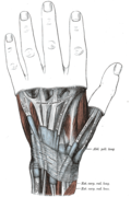
The mucous sheaths of the tendons on the back of the wrist are protective coverings for tendons in the wrist. Between the dorsal carpal ligament and the bones six compartments are formed for the passage of tendons, each compartment having a separate mucous sheath. One is found in each of the following positions:

  1. on the lateral side of the radial styloid process, for the tendons of the Abductor pollicis longus and Extensor pollicis brevis;
  2. behind the styloid process, for the tendons of the Extensores carpi radialis longus and brevis;
  3. about the middle of the dorsal surface of the radius, for the tendon of the Extensor pollicis longus;
  4. to the medial side of the latter, for the tendons of the Extensor digitorum communis and Extensor indicis proprius;
  5. opposite the interval between the radius and ulna, for the Extensor digiti quinti proprius;
  6. between the head and styloid process of the ulna, for the tendon of the Extensor carpi ulnaris.
<span class="mw-page-title-main">Extrinsic extensor muscles of the hand</span>

The extrinsic extensor muscles of the hand are located in the back of the forearm and have long tendons connecting them to bones in the hand, where they exert their action. Extrinsic denotes their location outside the hand. Extensor denotes their action which is to extend, or open flat, joints in the hand. They include the extensor carpi radialis longus (ECRL), extensor carpi radialis brevis (ECRB), extensor digitorum (ED), extensor digiti minimi (EDM), extensor carpi ulnaris (ECU), abductor pollicis longus (APL), extensor pollicis brevis (EPB), extensor pollicis longus (EPL), and extensor indicis (EI).

<span class="mw-page-title-main">Muscles of the thumb</span>

The muscles of the thumb are nine skeletal muscles located in the hand and forearm. The muscles allow for flexion, extension, adduction, abduction and opposition of the thumb. The muscles acting on the thumb can be divided into two groups: The extrinsic hand muscles, with their muscle bellies located in the forearm, and the intrinsic hand muscles, with their muscles bellies located in the hand proper.

References