Palmaris longus muscle

Last updated
Palmaris longus muscle
PL Tendon.png
Palmaris longus tendon as seen in surface anatomy
Details
Origin Medial epicondyle of humerus (common flexor tendon)
Insertion Palmar aponeurosis and flexor retinaculum of the hand
Artery Ulnar artery
Nerve Median nerve
Actions Wrist flexor
Antagonist Extensor carpi radialis brevis, extensor carpi radialis longus, extensor carpi ulnaris
Identifiers
Latin musculus palmaris longus
Greek Μακρύς παλαμικός μυς
TA98 A04.6.02.029
TA2 2482
FMA 38462
Anatomical terms of muscle

The palmaris longus is a muscle visible as a small tendon located between the flexor carpi radialis and the flexor carpi ulnaris, although it is not always present. It is absent in about 14 percent of the population; this number can vary in African, Asian, and Native American populations, however. [1] Absence of the palmaris longus does not have an effect on grip strength. [2] The lack of palmaris longus muscle does result in decreased pinch strength in fourth and fifth fingers. The absence of palmaris longus muscle is more prevalent in females than males. [3]

Contents

The palmaris longus muscle can be seen by touching the pads of the fourth finger and thumb and flexing the wrist. The tendon, if present, will be visible in the midline of the anterior wrist.

Structure

Palmaris longus is a slender, elongated, spindle shaped muscle, lying on the medial side of the flexor carpi radialis. It is widest in the middle, and narrowest at the proximal and distal attachments. [4]

It arises mainly from the medial epicondyle of the humerus via the common flexor tendon. It also takes origin from the adjacent intermuscular septa and from the antebrachial fascia. [4]

It ends in a slender, flattened tendon, which passes over the upper part of the flexor retinaculum and inserts onto the central part of the flexor retinaculum and lower part of the palmar aponeurosis. Frequently, it sends a tendinous slip to the short muscles of the thumb. [4]

Nerve supply

The palmaris longus is innervated by the median nerve. [5]

Variation

Palmaris Longus: misplaced or absent
The fleshy belly occasionally migrates distally and comes to lie close above the wrist, as here.
Palmaris Longus was absent in 98 of 716 dissected limbs (i.e., in 13.7% of 358 paired limbs, 26 times in both limbs, 26 in the right only, and 20 in the left only. (R. K. George). Grant 1962 97 D.png
Palmaris Longus: misplaced or absent
The fleshy belly occasionally migrates distally and comes to lie close above the wrist, as here.
Palmaris Longus was absent in 98 of 716 dissected limbs (i.e., in 13.7% of 358 paired limbs, 26 times in both limbs, 26 in the right only, and 20 in the left only. (R. K. George).

The palmaris longus muscle is a variable muscle. The most common variation is its absence. Several in vivo and in vitro studies have documented the prevalence or absence of the PL tendon in different ethnic groups. Between 5.5 and 24% of Caucasian populations (European and North American) and 4.6 to 26.6% of Asian populations (Chinese, Japanese, Indian, Turkish, Malaysian) have been reported to lack the PL tendon. [7]

There are also variations related to its form. It may be tendinous above and muscular below; or it may be muscular in the center with a tendon above and below; or it may present two muscular bundles with a central tendon; or finally it may consist solely of a tendinous band. The muscle may be double, or missing entirely. Slips of origin from the coronoid process or from the radius have been seen. Partial or complete insertion into the fascia of the forearm, into the tendon of the flexor carpi ulnaris and pisiform bone, into the scaphoid, and into the muscles of the little finger have been observed.

Clinical significance

Use in tendon grafts

Cross-section through the middle of the forearm Gray417 color.PNG
Cross-section through the middle of the forearm

The palmaris longus muscle is the most popular for use in tendon grafts for the wrist due to the length and diameter of the palmaris longus tendon, and the fact that it can be used without producing any functional deformities. When a tendon becomes ruptured in the wrist, the palmaris longus tendon may be removed from the flexor retinaculum and grafted to take the place of the ruptured tendon. The tendons most commonly replaced or supplemented by the palmaris longus tendon when ruptured are the long flexors of the fingers and the flexor pollicis longus tendon. [8]

The palmaris longus muscle itself is a weak flexor, and provides no substantial flexing force that would inhibit movement in the wrist if its tendon were cut and moved elsewhere. The palmaris longus may contribute and assist in thumb abduction movements; an action necessary to open the hand. [9] [10] If the palmaris longus muscle is not available for harvesting in an individual, the anatomically homologous plantaris muscle in the leg may be taken instead. [11] Using the patient’s own tendon is advantageous, as it does not introduce foreign material into the body.

Carpal tunnel syndrome and palmaris longus variants

Of the known anatomical variants of the palmaris longus, the reverse belly of the palmaris longus may be localized within the carpal tunnel producing symptoms of carpal tunnel syndrome. Knowledge of this variation is important to prevent unnecessary carpal tunnel release surgery, in which the median nerve compression may remain unresolved due to the presence of this palmaris longus variant. [12]

Other animals

Evolution

The evolutionary interpretation of the muscle's absence is that humans inherited the muscle through common descent, and numerous animals that humans share a common ancestor with (such as the orangutan) still actively employ the muscle. [13] Close primate relatives (such as the chimpanzee and gorilla) also do not actively employ the muscle, and hence they also demonstrate the same variability. [14] The common descent principle suggests that at some stage our ancestors employed the muscle actively. The thumb apparatus (and particularly the thenar muscle group) then started developing in the hominin branch, and consequently the Palmaris longus became vestigial. As there is no apparent evolutionary pressure (positive or negative) concerning the muscle, it has remained largely unaffected by evolutionary processes. [15]

See also

Related Research Articles

<span class="mw-page-title-main">Carpal bones</span> Eight small bones that make up the wrist (or carpus) that connects the hand to the forearm

The carpal bones are the eight small bones that make up the wrist (carpus) that connects the hand to the forearm. The term "carpus" and "carpal" is derived from the Latin carpus and the Greek καρπός (karpós), meaning "wrist". In human anatomy, the main role of the carpal bones is to articulate with the radial and ulnar heads to form a highly mobile condyloid joint, to provide attachments for thenar and hypothenar muscles, and to form part of the rigid carpal tunnel which allows the median nerve and tendons of the anterior forearm muscles to be transmitted to the hand and fingers.

<span class="mw-page-title-main">Median nerve</span> Nerve of the upper limb

The median nerve is a nerve in humans and other animals in the upper limb. It is one of the five main nerves originating from the brachial plexus.

<span class="mw-page-title-main">Wrist</span> Part of the arm between the lower arm and the hand

In human anatomy, the wrist is variously defined as (1) the carpus or carpal bones, the complex of eight bones forming the proximal skeletal segment of the hand; (2) the wrist joint or radiocarpal joint, the joint between the radius and the carpus and; (3) the anatomical region surrounding the carpus including the distal parts of the bones of the forearm and the proximal parts of the metacarpus or five metacarpal bones and the series of joints between these bones, thus referred to as wrist joints. This region also includes the carpal tunnel, the anatomical snuff box, bracelet lines, the flexor retinaculum, and the extensor retinaculum.

<span class="mw-page-title-main">Thenar eminence</span> Mound at the base of the thumb formed by the intrinsic group of muscles

The thenar eminence is the mound formed at the base of the thumb on the palm of the hand by the intrinsic group of muscles of the thumb. The skin overlying this region is the area stimulated when trying to elicit a palmomental reflex. The word thenar comes from Ancient Greek θέναρ (thenar) 'palm of the hand'.

In human anatomy, extensor carpi radialis brevis is a muscle in the forearm that acts to extend and abduct the wrist. It is shorter and thicker than its namesake extensor carpi radialis longus which can be found above the proximal end of the extensor carpi radialis brevis.

<span class="mw-page-title-main">Wrist drop</span> Medical condition

Wrist drop is a medical condition in which the wrist and the fingers cannot extend at the metacarpophalangeal joints. The wrist remains partially flexed due to an opposing action of flexor muscles of the forearm. As a result, the extensor muscles in the posterior compartment remain paralyzed.

<span class="mw-page-title-main">Upper limb</span> Consists of the arm, forearm, and hand

The upper limbs or upper extremities are the forelimbs of an upright-postured tetrapod vertebrate, extending from the scapulae and clavicles down to and including the digits, including all the musculatures and ligaments involved with the shoulder, elbow, wrist and knuckle joints. In humans, each upper limb is divided into the arm, forearm and hand, and is primarily used for climbing, lifting and manipulating objects.

<span class="mw-page-title-main">Flexor carpi ulnaris muscle</span> Muscle of the forearm

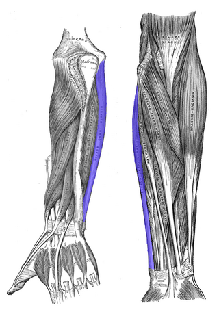
The flexor carpi ulnaris (FCU) is a muscle of the forearm that flexes and adducts at the wrist joint.

In human anatomy, the extensor pollicis longus muscle (EPL) is a skeletal muscle located dorsally on the forearm. It is much larger than the extensor pollicis brevis, the origin of which it partly covers and acts to stretch the thumb together with this muscle.

<span class="mw-page-title-main">Flexor pollicis brevis muscle</span> Muscle in the thenar compartment

The flexor pollicis brevis is a muscle in the hand that flexes the thumb. It is one of three thenar muscles. It has both a superficial part and a deep part.

The flexor pollicis longus is a muscle in the forearm and hand that flexes the thumb. It lies in the same plane as the flexor digitorum profundus. This muscle is unique to humans, being either rudimentary or absent in other primates. A meta-analysis indicated accessory flexor pollicis longus is present in around 48% of the population.

<span class="mw-page-title-main">Abductor digiti minimi muscle of hand</span> Muscle in the hypothenar compartment

In human anatomy, the abductor digiti minimi is a skeletal muscle situated on the ulnar border of the palm of the hand. It forms the ulnar border of the palm and its spindle-like shape defines the hypothenar eminence of the palm together with the skin, connective tissue, and fat surrounding it. Its main function is to pull the little finger away from the other fingers.

<span class="mw-page-title-main">Flexor retinaculum of the hand</span> Thickened fascia over the carpal tunnel

The flexor retinaculum is a fibrous band on the palmar side of the hand near the wrist. It arches over the carpal bones of the hands, covering them and forming the carpal tunnel.

<span class="mw-page-title-main">Extensor retinaculum of the hand</span> A thickened fascia holding the tendons of the hand extensor muscles in place

The extensor retinaculum is a thickened portion of the antebrachial fascia that holds the tendons of the extensor muscles in place. It is located on the back of the forearm, just proximal to the hand. It is continuous with the palmar carpal ligament.

<span class="mw-page-title-main">Carpal tunnel</span> Structure of human wrist

In the human body, the carpal tunnel or carpal canal is a flattened body cavity on the flexor (palmar/volar) side of the wrist, bounded by the carpal bones and flexor retinaculum. It forms the passageway that transmits the median nerve and the tendons of the extrinsic flexor muscles of the hand from the forearm to the hand. There are described cases of the anatomical variant median artery occurrence.

<span class="mw-page-title-main">Median nerve palsy</span> Medical condition

Injuries to the arm, forearm or wrist area can lead to various nerve disorders. One such disorder is median nerve palsy. The median nerve controls the majority of the muscles in the forearm. It controls abduction of the thumb, flexion of hand at wrist, flexion of digital phalanx of the fingers, is the sensory nerve for the first three fingers, etc. Because of this major role of the median nerve, it is also called the eye of the hand. If the median nerve is damaged, the ability to abduct and oppose the thumb may be lost due to paralysis of the thenar muscles. Various other symptoms can occur which may be repaired through surgery and tendon transfers. Tendon transfers have been very successful in restoring motor function and improving functional outcomes in patients with median nerve palsy.

<span class="mw-page-title-main">Extrinsic extensor muscles of the hand</span>

The extrinsic extensor muscles of the hand are located in the back of the forearm and have long tendons connecting them to bones in the hand, where they exert their action. Extrinsic denotes their location outside the hand. Extensor denotes their action which is to extend, or open flat, joints in the hand. They include the extensor carpi radialis longus (ECRL), extensor carpi radialis brevis (ECRB), extensor digitorum (ED), extensor digiti minimi (EDM), extensor carpi ulnaris (ECU), abductor pollicis longus (APL), extensor pollicis brevis (EPB), extensor pollicis longus (EPL), and extensor indicis (EI).

<span class="mw-page-title-main">Muscles of the thumb</span>

The muscles of the thumb are nine skeletal muscles located in the hand and forearm. The muscles allow for flexion, extension, adduction, abduction and opposition of the thumb. The muscles acting on the thumb can be divided into two groups: The extrinsic hand muscles, with their muscle bellies located in the forearm, and the intrinsic hand muscles, with their muscles bellies located in the hand proper.

<span class="mw-page-title-main">Extensor pollicis et indicis communis muscle</span>

In human anatomy, the extensor pollicis et indicis communis is an aberrant muscle in the posterior compartment of forearm. It was first described in 1863. The muscle has a prevalence from 0.5% to 4%.

Linburg–Comstock variation is an occasional tendinous connection between the flexor pollicis longus and the flexor digitorum profundus of the index, the middle finger or both. It is found in around 21% of the population. It is an anatomical variation in human, which may be viewed as a pathology if causes symptoms. It was recognised as early as the 1800s, but was first described by Linburg and Comstock in 1979.

References

PD-icon.svgThis article incorporates text in the public domain from page 446 of the 20th edition of Gray's Anatomy (1918)

  1. Sebastin SJ, Puhaindran ME, Lim AY, Lim IJ, Bee WH (October 2005). "The prevalence of absence of the palmaris longus--a study in a Chinese population and a review of the literature". Journal of Hand Surgery. 30 (5): 525–7. doi:10.1016/j.jhsb.2005.05.003. PMID   16006020. S2CID   12394636.
  2. Sebastin SJ, Lim AY, Bee WH, Wong TC, Methil BV (August 2005). "Does the absence of the palmaris longus affect grip and pinch strength?". Journal of Hand Surgery. 30 (4): 406–8. doi:10.1016/j.jhsb.2005.03.011. PMID   15935531. S2CID   35394120.
  3. Cetin, A.; Genc, M.; Sevil, S.; Coban, Y. K. (2013). "Prevalence of the palmaris longus muscle and its relationship with grip and pinch strength". Hand (New York, N.Y.). 8 (2): 215–220. doi:10.1007/s11552-013-9509-6. PMC   3652998 . PMID   24426922.
  4. 1 2 3 Gray's Anatomy (1918), see infobox
  5. Drake, Richard L.; Vogl, Wayne; Mitchell, Adam W. M. (2005). Gray's anatomy for students. Philadelphia: Elsevier/Churchill Livingstone. p. 693. ISBN   978-0-8089-2306-0.
  6. Grant's Atlas of Anatomy. 1962.
  7. Kose O, Adanir O, Cirpar M, Kurklu M, Komurcu M (May 2009). "The prevalence of absence of the palmaris longus: a study in Turkish population". Archives of Orthopaedic and Trauma Surgery. 129 (5): 609–11. doi:10.1007/s00402-008-0631-9. PMID   18418616. S2CID   26235241.
  8. Thejodhar P, Potu BK, Vasavi RG (January 2008). "Unusual palmaris longus muscle". Indian Journal of Plastic Surgery. 41 (1): 95–6. doi: 10.4103/0970-0358.41125 . PMC   2739544 . PMID   19753215.
  9. Moore, Colin W.; Fanous, Jacob; Rice, Charles L. (27 December 2017). "Revisiting the functional anatomy of the palmaris longus as a thenar synergist". Clinical Anatomy. 31 (6): 760–770. doi:10.1002/ca.23023. PMID   29178622. S2CID   21747288.
  10. Moore, Colin W.; Fanous, Jacob; Rice, Charles L. (1 January 2021). "Fiber type composition of contiguous palmaris longus and abductor pollicis brevis muscles: Morphological evidence of a functional synergy". Journal of Anatomy. 238 (1): 53–62. doi:10.1111/joa.13289. PMC   7754940 . PMID   32790091.
  11. Wheeless, Clifford R (December 2009). "Palmaris Longus Tendon Graft Harvest". Wheeless' Textbook of Orthopaedics. Retrieved February 25, 2012.
  12. DEPUYDT, K. H.; SCHUURMAN, A. H.; KON, M. (2016-08-29). "Reversed Palmaris Longus Muscle Causing Effort-Related Median Nerve Compression". Journal of Hand Surgery. 23 (1): 117–119. doi:10.1016/S0266-7681(98)80241-6. PMID   9571503. S2CID   26397516.
  13. "Proof of evolution that you can find on your own body". 17 March 2016.
  14. Thejodhar et al, Indian J Plast Surg Vol 41.1 (2008)
  15. Maughan et al: The Roles of Mutation Accumulation and Selection in Loss of Sporulation in Experimental Populations of Bacillus subtilis Genetics. 2007 Oct;177(2):937-48