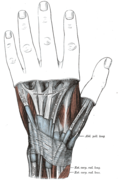
Extensor indicis muscle

Last updated
Extensor indicis proprius
Gray421.png
Transverse section across distal ends of radius and ulna. (Label "Ext. indic. prop." visible at bottom center.)
Extensor indicis muscle.png
Posterior surface of the forearm. Deep muscles. Extensor indicis muscle is labeled in purple.
Details
Origin Posterior distal third of ulna and interosseous membrane
Insertion Index finger (extensor hood)
Artery Posterior interosseous artery
Nerve Posterior interosseous nerve
Actions Extends index finger, wrist
Identifiers
Latin musculus extensor indicis
TA98 A04.6.02.052
TA2 2515
FMA 38524
Anatomical terms of muscle

In human anatomy, the extensor indicis (proprius) is a narrow, elongated skeletal muscle in the deep layer of the dorsal forearm, placed medial to, and parallel with, the extensor pollicis longus. Its tendon goes to the index finger, which it extends.

Contents

Structure

It arises from the distal third of the dorsal part of the body of ulna and from the interosseous membrane. It runs through the fourth tendon compartment together with the extensor digitorum, from where it projects into the dorsal aponeurosis of the index finger. [1]

Opposite the head of the second metacarpal bone, it joins the ulnar side of the tendon of the extensor digitorum which belongs to the index finger.

Like the extensor digiti minimi (i.e. the extensor of the little finger), the tendon of the extensor indicis runs and inserts on the ulnar side of the tendon of the common extensor digitorum. [2] The extensor indicis lacks the juncturae tendinum interlinking the tendons of the extensor digitorum on the dorsal side of the hand. [3]

Variation

The extensor indicis proprius does not show much variation. It exists as a single tendon most of the time. [4] Double tendons of the extensor indicis proprius was also reported. [2] [5] [6]

It is known that the extensor indicis proprius inserts to the index finger on the ulnar side of the extensor digitorum. [7] However, the insertion on the radial side of the common extensor digitorum infrequently seen, namely the extensor indicis radialis. [2] Split tendons of the muscle inserting on both ulnar and the radial side of the common extensor digitorum was also reported. [2]

Anomalous hand extensors including the extensor medii proprius and the extensor indicis et medii communis are often seen as variations of the extensor indicis [2] due to the shared characteristics and embryonic origin. [8]

Function

The extensor indicis extends the index finger, and by its continued action assists in extending (dorsiflexion) the wrist and the midcarpal joints. [1]

Because the index finger and little finger have separate extensors, these fingers can be moved more independently than the other fingers. [3]

Additional images

See also

Notes

  1. 1 2 Platzer 2004 , p. 168
  2. 1 2 3 4 5 Komiyama, M.; Nwe, T. M.; Toyota, N.; Shimada, Y. (1999). "Variations of the Extensor Indicis Muscle and Tendon" (PDF). Journal of Hand Surgery, British Volume. 24 (5): 575–578. doi:10.1054/jhsb.1999.0239. PMID   10597935. S2CID   23240783 . Retrieved 2015-06-09.
  3. 1 2 Ross & Lamperti 2006 , p. 300
  4. Dass, Prameela; Prabhu, Latha V.; Pai, Mangala M.; Nayak, Varsha; Kumar, Ganesh; Janardhanan, Jiji P. (Nov–Dec 2011). "A comprehensive study of the extensor tendons to the medial four digits of the hand". Chang Gung Medical Journal. 34 (6): 612–619. ISSN   2309-835X. PMID   22196064.
  5. "Double tendon of the Human Extensor Indicis Muscle provides "insight' into individual development -- Kumka 22 (1): 983.2 -- The FASEB Journal". www.fasebj.org. Retrieved 2015-06-09.
  6. Aziz, M. Ashraf; Dunlap, Samuel Strong (1986-07-01). "The human extensor digitorum profundus muscle with comments on the evolution of the primate hand". Primates. 27 (3): 293–319. doi:10.1007/BF02382073. ISSN   0032-8332. S2CID   39525970.
  7. "Anatomy of the human body". archive.org. Retrieved 2015-06-09.
  8. Straus, W.I. (1941). "Phylogeny of human forearm extensors". Ann Hum Biol (13): 203–238.

Related Research Articles

<span class="mw-page-title-main">Carpal bones</span> Eight small bones that make up the wrist (or carpus) that connects the hand to the forearm

The carpal bones are the eight small bones that make up the wrist (carpus) that connects the hand to the forearm. The term "carpus" and "carpal" is derived from the Latin carpus and the Greek καρπός (karpós), meaning "wrist". In human anatomy, the main role of the carpal bones is to articulate with the radial and ulnar heads to form a highly mobile condyloid joint, to provide attachments for thenar and hypothenar muscles, and to form part of the rigid carpal tunnel which allows the median nerve and tendons of the anterior forearm muscles to be transmitted to the hand and fingers.

<span class="mw-page-title-main">Wrist</span> Part of the arm between the lower arm and the hand

In human anatomy, the wrist is variously defined as (1) the carpus or carpal bones, the complex of eight bones forming the proximal skeletal segment of the hand; (2) the wrist joint or radiocarpal joint, the joint between the radius and the carpus and; (3) the anatomical region surrounding the carpus including the distal parts of the bones of the forearm and the proximal parts of the metacarpus or five metacarpal bones and the series of joints between these bones, thus referred to as wrist joints. This region also includes the carpal tunnel, the anatomical snuff box, bracelet lines, the flexor retinaculum, and the extensor retinaculum.

The flexor digitorum profundus is a muscle in the forearm of humans that flexes the fingers. It is considered an extrinsic hand muscle because it acts on the hand while its muscle belly is located in the forearm.

<span class="mw-page-title-main">Extensor carpi radialis longus muscle</span>

The extensor carpi radialis longus is one of the five main muscles that control movements at the wrist. This muscle is quite long, starting on the lateral side of the humerus, and attaching to the base of the second metacarpal bone.

In human anatomy, extensor carpi radialis brevis is a muscle in the forearm that acts to extend and abduct the wrist. It is shorter and thicker than its namesake extensor carpi radialis longus which can be found above the proximal end of the extensor carpi radialis brevis.

<span class="mw-page-title-main">Radius (bone)</span> One of the two long bones of the forearm

The radius or radial bone is one of the two large bones of the forearm, the other being the ulna. It extends from the lateral side of the elbow to the thumb side of the wrist and runs parallel to the ulna. The ulna is longer than the radius, but the radius is thicker. The radius is a long bone, prism-shaped and slightly curved longitudinally.

<span class="mw-page-title-main">Upper limb</span> Consists of the arm, forearm, and hand

The upper limbs or upper extremities are the forelimbs of an upright-postured tetrapod vertebrate, extending from the scapulae and clavicles down to and including the digits, including all the musculatures and ligaments involved with the shoulder, elbow, wrist and knuckle joints. In humans, each upper limb is divided into the arm, forearm and hand, and is primarily used for climbing, lifting and manipulating objects.

<span class="mw-page-title-main">Extensor digiti minimi muscle</span>

The extensor digiti minimi is a slender muscle of the forearm, placed on the ulnar side of the extensor digitorum communis, with which it is generally connected.

<span class="mw-page-title-main">Extensor digitorum muscle</span> Muscle of the posterior forearm

The extensor digitorum muscle is a muscle of the posterior forearm present in humans and other animals. It extends the medial four digits of the hand. Extensor digitorum is innervated by the posterior interosseous nerve, which is a branch of the radial nerve.

In human anatomy, the extensor pollicis longus muscle (EPL) is a skeletal muscle located dorsally on the forearm. It is much larger than the extensor pollicis brevis, the origin of which it partly covers and acts to stretch the thumb together with this muscle.

The flexor pollicis longus is a muscle in the forearm and hand that flexes the thumb. It lies in the same plane as the flexor digitorum profundus. This muscle is unique to humans, being either rudimentary or absent in other primates. A meta-analysis indicated accessory flexor pollicis longus is present in around 48% of the population.

<span class="mw-page-title-main">Extensor pollicis brevis muscle</span>

In human anatomy, the extensor pollicis brevis (EPB) is a skeletal muscle on the dorsal side of the forearm. It lies on the medial side of, and is closely connected with, the abductor pollicis longus. The extensor pollicis brevis belongs to the deep group of the posterior fascial compartment of the forearm. It is a part of the lateral border of the anatomical snuffbox.

<span class="mw-page-title-main">Abductor digiti minimi muscle of hand</span> Muscle in the hypothenar compartment

In human anatomy, the abductor digiti minimi is a skeletal muscle situated on the ulnar border of the palm of the hand. It forms the ulnar border of the palm and its spindle-like shape defines the hypothenar eminence of the palm together with the skin, connective tissue, and fat surrounding it. Its main function is to pull the little finger away from the other fingers.

The posterior compartment of the forearm contains twelve muscles which primarily extend the wrist and digits. It is separated from the anterior compartment by the interosseous membrane between the radius and ulna.

Extensor digitorum brevis manus is an extra or accessory muscle on the backside (dorsum) of the hand. It was first described by Albinus in 1758. The muscles lies in the fourth extensor compartment of the wrist, and is relatively rare. It has a prevalence of 4% in the general population according to a meta-analysis. This muscle is commonly misdiagnosed as a ganglion cyst, synovial nodule or cyst.

<span class="mw-page-title-main">Hand</span> Extremity at the end of an arm or forelimb

A hand is a prehensile, multi-fingered appendage located at the end of the forearm or forelimb of primates such as humans, chimpanzees, monkeys, and lemurs. A few other vertebrates such as the koala are often described as having "hands" instead of paws on their front limbs. The raccoon is usually described as having "hands" though opposable thumbs are lacking.

<span class="mw-page-title-main">Extrinsic extensor muscles of the hand</span>

The extrinsic extensor muscles of the hand are located in the back of the forearm and have long tendons connecting them to bones in the hand, where they exert their action. Extrinsic denotes their location outside the hand. Extensor denotes their action which is to extend, or open flat, joints in the hand. They include the extensor carpi radialis longus (ECRL), extensor carpi radialis brevis (ECRB), extensor digitorum (ED), extensor digiti minimi (EDM), extensor carpi ulnaris (ECU), abductor pollicis longus (APL), extensor pollicis brevis (EPB), extensor pollicis longus (EPL), and extensor indicis (EI).

<span class="mw-page-title-main">Extensor medii proprius muscle</span>

The extensor medii proprius is a rare anatomical variant in the extensor compartment of the forearm. The aberrant muscle is analogous to the extensor indicis with the insertion being the middle finger instead of the index finger.

<span class="mw-page-title-main">Extensor indicis et medii communis muscle</span>

The extensor indicis et medii communis is a rare anatomical variant in the extensor compartment of forearm. This additional muscle lies in the deep extensor layer next to the extensor indicis proprius and the extensor pollicis longus. The characteristics of this anomalous muscle resemble those of the extensor indicis proprius, with split tendons to the index and the middle finger. This muscle can also be considered as a variation of the aberrant extensor medii proprius.

<span class="mw-page-title-main">Extensor pollicis et indicis communis muscle</span>

In human anatomy, the extensor pollicis et indicis communis is an aberrant muscle in the posterior compartment of forearm. It was first described in 1863. The muscle has a prevalence from 0.5% to 4%.

References

PD-icon.svgThis article incorporates text in the public domain from page 456 of the 20th edition of Gray's Anatomy (1918)