Related Research Articles

The latissimus dorsi is a large, flat muscle on the back that stretches to the sides, behind the arm, and is partly covered by the trapezius on the back near the midline.

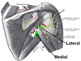
The axillary nerve or the circumflex nerve is a nerve of the human body, that originates from the brachial plexus at the level of the axilla (armpit) and carries nerve fibers from C5 and C6. The axillary nerve travels through the quadrangular space with the posterior circumflex humeral artery and vein to innervate the deltoid and teres minor.

The deltoid muscle is the muscle forming the rounded contour of the human shoulder. It is also known as the 'common shoulder muscle', particularly in other animals such as the domestic cat. Anatomically, the deltoid muscle is made up of three distinct sets of muscle fibers, namely the
- anterior or clavicular part
- posterior or scapular part
- intermediate or acromial part

Pectoralis minor muscle is a thin, triangular muscle, situated at the upper part of the chest, beneath the pectoralis major in the human body. It arises from ribs III-V; it inserts onto the coracoid process of the scapula. It is innervated by the medial pectoral nerve. Its function is to stabilise the scapula by holding it fast in position against the chest wall.

The axilla is the area on the human body directly under the shoulder joint. It includes the axillary space, an anatomical space within the shoulder girdle between the arm and the thoracic cage, bounded superiorly by the imaginary plane between the superior borders of the first rib, clavicle and scapula, medially by the serratus anterior muscle and thoracolumbar fascia, anteriorly by the pectoral muscles and posteriorly by the subscapularis, teres major and latissimus dorsi muscle.
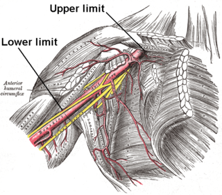
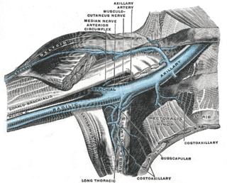
In human anatomy, the axillary artery is a large blood vessel that conveys oxygenated blood to the lateral aspect of the thorax, the axilla (armpit) and the upper limb. Its origin is at the lateral margin of the first rib, before which it is called the subclavian artery.
In human anatomy, the axillary vein is a large blood vessel that conveys blood from the lateral aspect of the thorax, axilla (armpit) and upper limb toward the heart. There is one axillary vein on each side of the body.
Axillary means "related to the axilla (armpit)" or "related to the leaf axils".

The subclavius is a small triangular muscle, placed between the clavicle and the first rib. Along with the pectoralis major and pectoralis minor muscles, the subclavius muscle makes up the anterior axioappendicular muscles, also known as anterior wall of the axilla.

The intercostobrachial nerve is the name applied to the lateral cutaneous branch of the second intercostal nerve. It arises anterior to the long thoracic nerve. It provides sensory innervation to the skin of the axilla, and a variable region of the medial side of the upper arm.

The axillary sheath is a fibrous sheath that encloses the axillary artery and the three cords of the brachial plexus to form the neurovascular bundle. It is surrounded by the axillary fat. It is an extension of the prevertebral fascia of the deep cervical fascia and is continuous with the carotid sheath at the venous angle.
The clavipectoral fascia is a strong fascia situated under cover of the clavicular portion of the pectoralis major.
The tail of Spence has historically been described as an extension of the tissue of the upper outer quadrant of the breast traveling into the axilla. The "axillary tail" has been reported to pass into the axilla through an opening in the deep fascia called foramen of Langer. The "tail of Spence" was named after the Scottish surgeon James Spence, who served as a President of the Royal College of Surgeons in Edinburgh in the latter half of the 19th Century.

The pectoral fascia is a thin lamina, covering the surface of the pectoralis major, and sending numerous prolongations between its fasciculi: it is attached, in the middle line, to the front of the sternum; above, to the clavicle; laterally and below it is continuous with the fascia of the shoulder, axilla, and thorax.

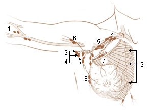
The axillary lymph nodes or armpit lymph nodes are lymph nodes in the human armpit. Between 20 and 49 in number, they drain lymph vessels from the lateral quadrants of the breast, the superficial lymph vessels from thin walls of the chest and the abdomen above the level of the navel, and the vessels from the upper limb. They are divided in several groups according to their location in the armpit. These lymph nodes are clinically significant in breast cancer, and metastases from the breast to the axillary lymph nodes are considered in the staging of the disease.

The prevertebral fascia is the layer of deep cervical fascia that surrounds the vertebral column. It is the deepest layer of deep cervical fascia.
The quadrangular space, also known as the quadrilateral space (of Velpeau) and the foramen humerotricipitale, is one of the three spaces in the axillary space. The other two spaces are: triangular space and triangular interval.
The axillary spaces are anatomic spaces. through which axillary contents leave the axilla. They consist of the quadrangular space, triangular space, and triangular interval. It is bounded by teres major, teres minor, medial border of the humerus, and long head of triceps brachii.
The suspensory ligament of axilla, or Gerdy's ligament, is a suspensory ligament that connects the clavipectoral fascia to the axillary fascia. This union shapes the axilla (underarm).

The axillary arch is a variant of the latissimus dorsi muscle in humans. It is found as a slip of muscle or fascia extending between the latissimus dorsi muscle and the pectoralis major. There is considerable variation in the exact position of its origin and insertions as well as its blood and nerve supply. The arch may occur on one or both sides of the body. A meta-analysis revealed that the axillary arch had an overall prevalence of 5.3% of limbs.
References
![]() This article incorporates text in the public domain from page 436 of the 20th edition of Gray's Anatomy (1918)
This article incorporates text in the public domain from page 436 of the 20th edition of Gray's Anatomy (1918)
| | This anatomy article is a stub. You can help Wikipedia by expanding it. |