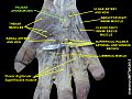
Palmar aponeurosis

Last updated
Palmar aponeurosis.
Gray425.png
The palmar aponeurosis
Details
Identifiers
Latin aponeurosis palmaris
TA98 A04.6.03.012
TA2 2549
FMA 42435
Anatomical terminology

The palmar aponeurosis (palmar fascia) invests the muscles of the palm, and consists of central, lateral, and medial portions.

Contents

Structure

The central portion occupies the middle of the palm, is triangular in shape, and of great strength

Its apex is continuous with the lower margin of the transverse carpal ligament, and receives the expanded tendon of the palmaris longus.

Its base divides below into four slips, one for each finger. Each slip gives off superficial fibers to the skin of the palm and finger, those to the palm joining the skin at the furrow corresponding to the metacarpophalangeal articulations, and those to the fingers passing into the skin at the transverse fold at the bases of the fingers.

The deeper part of each slip subdivides into two processes, which are inserted into the fibrous sheaths of the flexor tendons. From the sides of these processes offsets are attached to the transverse metacarpal ligament.

By this arrangement short channels are formed on the front of the heads of the metacarpal bones; through these the flexor tendons pass. The intervals between the four slips transmit the digital vessels and nerves, and the tendons of the lumbricales.

At the points of division into the slips mentioned, numerous strong, transverse fasciculi bind the separate processes together.

The central part of the palmar aponeurosis is intimately bound to the integument by dense fibroareolar tissue forming the superficial palmar fascia, and gives origin by its medial margin to the palmaris brevis.

It covers the superficial volar arch, the tendons of the flexor muscles, and the branches of the median and ulnar nerves; and on either side it gives off a septum, which is continuous with the interosseous aponeurosis, and separates the intermediate from the collateral groups of muscles.

Lateral and medial portions

The lateral and medial portions of the palmar aponeurosis are thin, fibrous layers, which cover, on the radial side, the muscles of the ball of the thumb, and, on the ulnar side, the muscles of the little finger; they are continuous with the central portion and with the fascia on the dorsum of the hand.

Additional images

Related Research Articles

<span class="mw-page-title-main">Median nerve</span> Nerve of the upper limb

The median nerve is a nerve in humans and other animals in the upper limb. It is one of the five main nerves originating from the brachial plexus.

<span class="mw-page-title-main">Ulnar nerve</span> Nerve which runs near the ulna bone

In human anatomy, the ulnar nerve is a nerve that runs near the ulna bone. The ulnar collateral ligament of elbow joint is in relation with the ulnar nerve. The nerve is the largest in the human body unprotected by muscle or bone, so injury is common. This nerve is directly connected to the little finger, and the adjacent half of the ring finger, innervating the palmar aspect of these fingers, including both front and back of the tips, perhaps as far back as the fingernail beds.

<span class="mw-page-title-main">Upper limb</span> Consists of the arm, forearm, and hand

The upper limbs or upper extremities are the forelimbs of an upright-postured tetrapod vertebrate, extending from the scapulae and clavicles down to and including the digits, including all the musculatures and ligaments involved with the shoulder, elbow, wrist and knuckle joints. In humans, each upper limb is divided into the arm, forearm and hand, and is primarily used for climbing, lifting and manipulating objects.

<span class="mw-page-title-main">Aponeurosis</span> Tissue which connects muscles to other organs

An aponeurosis is a flattened tendon by which muscle attaches to bone or fascia. Aponeuroses exhibit an ordered arrangement of collagen fibres, thus attaining high tensile strength in a particular direction while being vulnerable to tensional or shear forces in other directions. They have a shiny, whitish-silvery color, are histologically similar to tendons, and are very sparingly supplied with blood vessels and nerves. When dissected, aponeuroses are papery and peel off by sections. The primary regions with thick aponeuroses are in the ventral abdominal region, the dorsal lumbar region, the ventriculus in birds, and the palmar (palms) and plantar (soles) regions.

<span class="mw-page-title-main">Cubital fossa</span> The human elbow pit

The cubital fossa, chelidon or inside of elbow is the area on the anterior side of the upper part between the arm and forearm of a human or other hormid animals. It lies anteriorly to the elbow when in standard anatomical position.

<span class="mw-page-title-main">Ulnar artery</span> Artery of the forearm

The ulnar artery is the main blood vessel, with oxygenated blood, of the medial aspects of the forearm. It arises from the brachial artery and terminates in the superficial palmar arch, which joins with the superficial branch of the radial artery. It is palpable on the anterior and medial aspect of the wrist.

<span class="mw-page-title-main">Flexor carpi ulnaris muscle</span> Muscle of the forearm

The flexor carpi ulnaris (FCU) is a muscle of the forearm that flexes and adducts at the wrist joint.

<span class="mw-page-title-main">Extensor digitorum muscle</span> Muscle of the posterior forearm

The extensor digitorum muscle is a muscle of the posterior forearm present in humans and other animals. It extends the medial four digits of the hand. Extensor digitorum is innervated by the posterior interosseous nerve, which is a branch of the radial nerve.

<span class="mw-page-title-main">Adductor pollicis muscle</span> Muscle in the thenar compartment

In human anatomy, the adductor pollicis muscle is a muscle in the hand that functions to adduct the thumb. It has two heads: transverse and oblique.

<span class="mw-page-title-main">Palmar interossei muscles</span> Muscles between the metacarpals

In human anatomy, the palmar or volar interossei are four muscles, one on the thumb that is occasionally missing, and three small, unipennate, central muscles in the hand that lie between the metacarpal bones and are attached to the index, ring, and little fingers. They are smaller than the dorsal interossei of the hand.

<span class="mw-page-title-main">Palmaris brevis muscle</span>

Palmaris brevis muscle is a thin, quadrilateral muscle, placed beneath the integument of the ulnar side of the hand. It acts to fold the skin of the hypothenar eminence transversally.

<span class="mw-page-title-main">Abductor digiti minimi muscle of hand</span> Muscle in the hypothenar compartment

In human anatomy, the abductor digiti minimi is a skeletal muscle situated on the ulnar border of the palm of the hand. It forms the ulnar border of the palm and its spindle-like shape defines the hypothenar eminence of the palm together with the skin, connective tissue, and fat surrounding it. Its main function is to pull the little finger away from the other fingers.

The palmar carpal ligament is a thickened portion of antebrachial fascia on anterior/palmar side of the wrist which - together with the flexor retinaculum of the hand - retains the tendons of most of the flexor muscles of the hand.

<span class="mw-page-title-main">Flexor retinaculum of the hand</span> Thickened fascia over the carpal tunnel

The flexor retinaculum is a fibrous band on the palmar side of the hand near the wrist. It arches over the carpal bones of the hands, covering them and forming the carpal tunnel.

<span class="mw-page-title-main">Superficial palmar arch</span>

The superficial palmar arch is formed predominantly by the ulnar artery, with a contribution from the superficial palmar branch of the radial artery. However, in some individuals the contribution from the radial artery might be absent, and instead anastomoses with either the princeps pollicis artery, the radialis indicis artery, or the median artery, the former two of which are branches from the radial artery.

The antebrachial fascia continuous above with the brachial fascia, is a dense, membranous investment, which forms a general sheath for the muscles in this region; it is attached, behind, to the olecranon and dorsal border of the ulna, and gives off from its deep surface numerous intermuscular septa, which enclose each muscle separately.

<span class="mw-page-title-main">Proper palmar digital nerves of median nerve</span> Nerve

In the palm of the hand the median nerve is covered by the skin and the palmar aponeurosis, and rests on the tendons of the flexor muscles. Immediately after emerging from under the transverse carpal ligament the median nerve becomes enlarged and flattened and splits into a smaller, lateral, and a larger, medial portion.

<span class="mw-page-title-main">Common palmar digital nerves of median nerve</span>

In the palm of the hand, the median nerve is covered by the skin and the palmar aponeurosis, and rests on the tendons of the Flexor muscles. Immediately after emerging from under the transverse carpal ligament the median nerve becomes enlarged and flattened and splits into a smaller, lateral, and a larger, medial portion.

<span class="mw-page-title-main">Outline of human anatomy</span> Overview of and topical guide to human anatomy

The following outline is provided as an overview of and topical guide to human anatomy:

<span class="mw-page-title-main">Muscles of the thumb</span>

The muscles of the thumb are nine skeletal muscles located in the hand and forearm. The muscles allow for flexion, extension, adduction, abduction and opposition of the thumb. The muscles acting on the thumb can be divided into two groups: The extrinsic hand muscles, with their muscle bellies located in the forearm, and the intrinsic hand muscles, with their muscles bellies located in the hand proper.

References

PD-icon.svgThis article incorporates text in the public domain from page 460 of the 20th edition of Gray's Anatomy (1918)