Palmar interossei muscles

Last updated
Palmar interossei muscles
1121 Intrinsic Muscles of the Hand PIL.png
Three of the palmar interossei of the left hand, inferior view
Details
Origin Sides of metacarpals facing midline
Insertion Bases of proximal phalanges, extensor expansions
Artery Palmar metacarpal artery of deep palmar arch
Nerve Deep branch of ulnar nerve
Actions Adduction, flexion and extension
Antagonist Dorsal interossei
Identifiers
Latin musculi interossei palmares
TA98 A04.6.02.067
TA2 2534
FMA 37419
Anatomical terms of muscle

In human anatomy, the palmar or volar interossei (interossei volares in older literature) are four muscles, one on the thumb that is occasionally missing, and three small, unipennate, central muscles in the hand that lie between the metacarpal bones and are attached to the index, ring, and little fingers. [1] They are smaller than the dorsal interossei of the hand.

Contents

Structure

All palmar interossei originate along the shaft of the metacarpal bone of the digit on which they act. They are inserted into the base of the proximal phalanx and the extensor expansion of the extensor digitorum of the same digit. [1]

Pollical palmar interosseous

The first palmar interosseous is located at the thumb's medial side. Passing between the first dorsal interosseous and the oblique head of adductor pollicis, it is inserted on the base of the thumb's proximal phalanx together with adductor pollicis. [1]

The "pollical" palmar interosseous muscle (PPIM), is present in more than 80% of individuals and was first described by Henle 1858. Its presence has been verified by numerous anatomists since, but others have either failed to mention it or considered it part of either adductor pollicis or flexor pollicis brevis. [2] However, the deep head of the flexor pollicis brevis originates on the thumb's ulnar sesamoid bone and the oblique portion of the adductor pollicis on several carpal bones, as well as the bases of the second and third metacarpal bones and not on the first metacarpal. [3]

Central palmar interossei

The other three palmar interossei originate on the side of the metacarpal facing the hand's midline (ray of long finger); [4] the second is attached to the medial side of the index finger; the third to the lateral side of the ring finger; and the fourth to the lateral side of the little finger. The tendons of these three muscles pass posterior to the deep transverse ligament before being inserted onto the extensor expansion. [1]

Innervation

All of the interosseous muscles of the hand are innervated by the deep branch of the ulnar nerve. [4] [5] [6]

Blood supply

The palmar interossei are supplied by the palmar metacarpal artery of the deep palmar arch. [4]

Function

The palmar interosseous muscles adduct the fingers towards the middle finger. This is in contrast to the dorsal interossei, which abduct the fingers away from the middle finger. In addition (like dorsal interossei) they flex the finger at the metacarpo-phalangeal joint and extend the finger at the interphalangeal joint and thus assist the lumbricals. [7]

The palmar interossei, together with the dorsal interossei and the lumbricals, are active components of the finger's extensor mechanism. Fibers from some of the interossei contribute directly to the extensor hoods that wrap around the proximal phalanges, while other fibers may contribute to the central tendon and lateral bands of the mechanism. All three intrinsic groups of muscles pass palmar to the axis of the metacarpophalangeal joints, and therefore contribute to flexion there. Extension at the interphalangeal joints cannot be produced by the extensor digitorum alone, but active contraction of one of the three aforementioned intrinsic groups will because of their direct contribution to the extensor mechanism. [8]

Other animals

The pollical palmar interosseous muscle (PPIM) is absent in non-human primates, and is probably an autapomorphic muscle unique to the human thumb (together with flexor pollicis longus) which probably evolved from the oblique portion of adductor pollicis. In African apes, adductor pollicis is notably well-developed, with an origin on the carpus and its ligaments, and an insertion that has migrated distally, in some cases as far as the distal phalanx. The insertion of the PPIM into the extensor mechanism is likely to have evolved with tool usage in early hominids. [3] As comparative anatomy studies of the human PPIM strongly suggest that the muscle is evolutionarily derived from the adductor pollicis, it has been proposed that PPIM should be designated by the name musculus adductor pollicis accessorius, which indicates that the muscle is most likely a de novo structure derived from the adductor pollicis. [9]

Additional images

See also

Notes

  1. 1 2 3 4 Palastanga & Soames 2012 , p. 92
  2. Susman, Nyati & Jassal 1999 , Abstract and Table 1
  3. 1 2 Susman, Nyati & Jassal 1999 , Discussion
  4. 1 2 3 "Brachium to Hand Musculature". PTCentral. Archived from the original on 2012-02-04. Retrieved 8 December 2012.
  5. Neal, Sara (2015-01-01), Tubbs, R. Shane; Rizk, Elias; Shoja, Mohammadali M.; Loukas, Marios (eds.), "Chapter 33 - Peripheral Nerve Injury of the Upper Extremity", Nerves and Nerve Injuries, San Diego: Academic Press, pp. 505–524, doi:10.1016/b978-0-12-802653-3.00082-8, ISBN   978-0-12-802653-3 , retrieved 2020-10-25
  6. Campbell, W. W. (2014-01-01), "Ulnar Neuropathy", in Aminoff, Michael J.; Daroff, Robert B. (eds.), Encyclopedia of the Neurological Sciences (Second Edition), Oxford: Academic Press, pp. 564–569, doi:10.1016/b978-0-12-385157-4.00703-x, ISBN   978-0-12-385158-1 , retrieved 2020-10-25
  7. "Actions of interossei muscles of hand". lifehugger. 2008-11-01. Archived from the original on October 17, 2009. Retrieved 2009-04-26.
  8. Austin 2005 , pp. 329–31
  9. Bello-Hellegouarch, Gaelle; Aziz, M. Ashraf; Ferrero, Eva M.; Kern, Michael; Francis, Nadia; Diogo, Rui (2013-03-01). ""Pollical palmar interosseous muscle" (musculus adductor pollicis accessorius): Attachments, innervation, variations, phylogeny, and implications for human evolution and medicine". Journal of Morphology. 274 (3): 275–293. doi:10.1002/jmor.20090. ISSN   1097-4687. PMID   23109102. S2CID   206091796.

Related Research Articles

<span class="mw-page-title-main">Finger</span> Organ of manipulation and sensation found in the hands of humans and other primates

A finger is a prominent digit on the forelimbs of most tetrapod vertebrate animals, especially those with prehensile extremities such as humans and other primates. Most tetrapods have five digits (pentadactyly), and short digits are typically referred to as toes, while those that are notably elongated are called fingers. In humans, the fingers are flexibly articulated and opposable, serving as an important organ of tactile sensation and fine movements, which are crucial to the dexterity of the hands and the ability to grasp and manipulate objects.

<span class="mw-page-title-main">Carpal bones</span> Eight small bones that make up the wrist (or carpus) that connects the hand to the forearm

The carpal bones are the eight small bones that make up the wrist (carpus) that connects the hand to the forearm. The term "carpus" and "carpal" is derived from the Latin carpus and the Greek καρπός (karpós), meaning "wrist". In human anatomy, the main role of the carpal bones is to articulate with the radial and ulnar heads to form a highly mobile condyloid joint, to provide attachments for thenar and hypothenar muscles, and to form part of the rigid carpal tunnel which allows the median nerve and tendons of the anterior forearm muscles to be transmitted to the hand and fingers.

<span class="mw-page-title-main">Thumb</span> First digit of the hand

The thumb is the first digit of the hand, next to the index finger. When a person is standing in the medical anatomical position, the thumb is the outermost digit. The Medical Latin English noun for thumb is pollex, and the corresponding adjective for thumb is pollical.

<span class="mw-page-title-main">Median nerve</span> Nerve of the upper limb

The median nerve is a nerve in humans and other animals in the upper limb. It is one of the five main nerves originating from the brachial plexus.

<span class="mw-page-title-main">Wrist</span> Part of the arm between the lower arm and the hand

In human anatomy, the wrist is variously defined as (1) the carpus or carpal bones, the complex of eight bones forming the proximal skeletal segment of the hand; (2) the wrist joint or radiocarpal joint, the joint between the radius and the carpus and; (3) the anatomical region surrounding the carpus including the distal parts of the bones of the forearm and the proximal parts of the metacarpus or five metacarpal bones and the series of joints between these bones, thus referred to as wrist joints. This region also includes the carpal tunnel, the anatomical snuff box, bracelet lines, the flexor retinaculum, and the extensor retinaculum.

<span class="mw-page-title-main">Ulnar nerve</span> Nerve which runs near the ulna bone

In human anatomy, the ulnar nerve is a nerve that runs near the ulna bone. The ulnar collateral ligament of elbow joint is in relation with the ulnar nerve. The nerve is the largest in the human body unprotected by muscle or bone, so injury is common. This nerve is directly connected to the little finger, and the adjacent half of the ring finger, innervating the palmar aspect of these fingers, including both front and back of the tips, perhaps as far back as the fingernail beds.

<span class="mw-page-title-main">Upper limb</span> Consists of the arm, forearm, and hand

The upper limbs or upper extremities are the forelimbs of an upright-postured tetrapod vertebrate, extending from the scapulae and clavicles down to and including the digits, including all the musculatures and ligaments involved with the shoulder, elbow, wrist and knuckle joints. In humans, each upper limb is divided into the arm, forearm and hand, and is primarily used for climbing, lifting and manipulating objects.

In human anatomy, the extensor pollicis longus muscle (EPL) is a skeletal muscle located dorsally on the forearm. It is much larger than the extensor pollicis brevis, the origin of which it partly covers and acts to stretch the thumb together with this muscle.

<span class="mw-page-title-main">Adductor pollicis muscle</span> Muscle in the thenar compartment

In human anatomy, the adductor pollicis muscle is a muscle in the hand that functions to adduct the thumb. It has two heads: transverse and oblique.

<span class="mw-page-title-main">Carpometacarpal joint</span>

The carpometacarpal (CMC) joints are five joints in the wrist that articulate the distal row of carpal bones and the proximal bases of the five metacarpal bones.

<span class="mw-page-title-main">Dorsal interossei of the hand</span> Muscles between the metacarpals

In human anatomy, the dorsal interossei (DI) are four muscles in the back of the hand that act to abduct (spread) the index, middle, and ring fingers away from hand's midline and assist in flexion at the metacarpophalangeal joints and extension at the interphalangeal joints of the index, middle and ring fingers.

<span class="mw-page-title-main">Deep palmar arch</span>

The deep palmar arch is an arterial network found in the palm. It is usually primarily formed from the terminal part of the radial artery. The ulnar artery also contributes through an anastomosis. This is in contrast to the superficial palmar arch, which is formed predominantly by the ulnar artery.

<span class="mw-page-title-main">Princeps pollicis artery</span> Artery of the human thumb

The princeps pollicis artery, or principal artery of the thumb, arises from the radial artery just as it turns medially towards the deep part of the hand; it descends between the first dorsal interosseous muscle and the oblique head of the adductor pollicis, along the medial side of the first metacarpal bone to the base of the proximal phalanx, where it lies beneath the tendon of the flexor pollicis longus muscle and divides into two branches.

<span class="mw-page-title-main">Cervical spinal nerve 8</span> Spinal nerve of the cervical segment

The cervical spinal nerve 8 (C8) is a spinal nerve of the cervical segment.

<span class="mw-page-title-main">Muscles of the hand</span> Muscles of the human hand

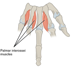
The muscles of the hand are the skeletal muscles responsible for the movement of the hand and fingers. The muscles of the hand can be subdivided into two groups: the extrinsic and intrinsic muscle groups. The extrinsic muscle groups are the long flexors and extensors. They are called extrinsic because the muscle belly is located on the forearm. The intrinsic group are the smaller muscles located within the hand itself. The muscles of the hand are innervated by the radial, median, and ulnar nerves from the brachial plexus.

<span class="mw-page-title-main">Ulnar claw</span> Deformity of the hand that develops due to ulnar nerve damage

An ulnar claw, also known as claw hand or ‘Spinster’s Claw’, is a deformity or an abnormal attitude of the hand that develops due to ulnar nerve damage causing paralysis of the lumbricals. A claw hand presents with a hyperextension at the metacarpophalangeal joints and flexion at the proximal and distal interphalangeal joints of the 4th and 5th fingers. The patients with this condition can make a full fist but when they extend their fingers, the hand posture is referred to as claw hand. The ring- and little finger can usually not fully extend at the proximal interphalangeal joint (PIP).

<span class="mw-page-title-main">Hand</span> Extremity at the end of an arm or forelimb

A hand is a prehensile, multi-fingered appendage located at the end of the forearm or forelimb of primates such as humans, chimpanzees, monkeys, and lemurs. A few other vertebrates such as the koala are often described as having "hands" instead of paws on their front limbs. The raccoon is usually described as having "hands" though opposable thumbs are lacking.

<span class="mw-page-title-main">Extrinsic extensor muscles of the hand</span>

The extrinsic extensor muscles of the hand are located in the back of the forearm and have long tendons connecting them to bones in the hand, where they exert their action. Extrinsic denotes their location outside the hand. Extensor denotes their action which is to extend, or open flat, joints in the hand. They include the extensor carpi radialis longus (ECRL), extensor carpi radialis brevis (ECRB), extensor digitorum (ED), extensor digiti minimi (EDM), extensor carpi ulnaris (ECU), abductor pollicis longus (APL), extensor pollicis brevis (EPB), extensor pollicis longus (EPL), and extensor indicis (EI).

<span class="mw-page-title-main">Muscles of the thumb</span>

The muscles of the thumb are nine skeletal muscles located in the hand and forearm. The muscles allow for flexion, extension, adduction, abduction and opposition of the thumb. The muscles acting on the thumb can be divided into two groups: The extrinsic hand muscles, with their muscle bellies located in the forearm, and the intrinsic hand muscles, with their muscles bellies located in the hand proper.

References