Rhomboid minor muscle

Last updated

Rhomboid minor
Rhomboideus minor.png
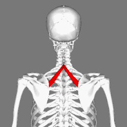
Muscles connecting the upper extremity to the vertebral column. (Rhomboid minor in red)
Details
Origin Nuchal ligaments and spinous processes of C7-T1
Insertion Medial border of scapula, superior to the insertion of rhomboid major muscle
Artery Deep branch of transverse cervical artery
Nerve Dorsal scapular nerve (C45)
Actions Retracts and rotates scapula, fixes scapula to thoracic wall
Antagonist Serratus anterior
Identifiers
Latin musculus rhomboideus minor
TA98 A04.3.01.008
TA2 2233
FMA 13380
Anatomical terms of muscle

In human anatomy, the rhomboid minor is a small skeletal muscle of the back that connects the scapula to the vertebrae of the spinal column.[ citation needed ] It arises from the nuchal ligament, and the 7th cervical and 1st thoracic vertebrae and intervening supraspinous ligaments; it inserts onto the medial border of the scapula. It is innervated by the dorsal scapular nerve.

Contents

It acts together with the rhomboid major to keep the scapula pressed against the thoracic wall. [1]

Anatomy

Origin

The rhomboid minor arises from the inferior border of the nuchal ligament, from the spinous processes of the vertebrae C7–T1, and from the intervening supraspinous ligaments. [2]

Insertion

It inserts onto a small area of the medial border of the scapula at the level of the scapular spine. [3]

Innervation

It is innervated by the dorsal scapular nerve (a branch of the brachial plexus), with most of its fibers derived from the C5 nerve root and only minor contribution from C4 or C6. [4]

Blood supply

The rhomboid minor receives arterial blood supply from the dorsal scapular artery.[ citation needed ]

Relations

It is located inferior to levator scapulae, and superior to rhomboid major.[ citation needed ]

It lies deep to trapezius, and superficial to the long spinal muscles. [2]

Variation

It is usually separated from the rhomboid major by a slight interval, but the adjacent margins of the two muscles are occasionally united. [5]

Actions/movements

Together with the rhomboid major, the rhomboid minor retracts the scapula when trapezius is contracted. Acting as a synergist to the trapezius, the rhomboid major and minor elevate the medial border of the scapula medially and upward, working in tandem with the levator scapulae muscle to rotate the scapulae downward. While other shoulder muscles are active, the rhomboid major and minor stabilize the scapula. [6]

Additional images

Related Research Articles

<span class="mw-page-title-main">Trapezius</span> Muscle between the lower spine and the shoulder blade

The trapezius is a large paired trapezoid-shaped surface muscle that extends longitudinally from the occipital bone to the lower thoracic vertebrae of the spine and laterally to the spine of the scapula. It moves the scapula and supports the arm.

<span class="mw-page-title-main">Scapula</span> Bone that connects the humerus (upper arm bone) with the clavicle (collar bone)

The scapula, also known as the shoulder blade, is the bone that connects the humerus with the clavicle. Like their connected bones, the scapulae are paired, with each scapula on either side of the body being roughly a mirror image of the other. The name derives from the Classical Latin word for trowel or small shovel, which it was thought to resemble.

Articles related to anatomy include:

<span class="mw-page-title-main">Spinal nerve</span> Nerve that carries signals between the spinal cord and the body

A spinal nerve is a mixed nerve, which carries motor, sensory, and autonomic signals between the spinal cord and the body. In the human body there are 31 pairs of spinal nerves, one on each side of the vertebral column. These are grouped into the corresponding cervical, thoracic, lumbar, sacral and coccygeal regions of the spine. There are eight pairs of cervical nerves, twelve pairs of thoracic nerves, five pairs of lumbar nerves, five pairs of sacral nerves, and one pair of coccygeal nerves. The spinal nerves are part of the peripheral nervous system.

<span class="mw-page-title-main">Dorsal scapular nerve</span> Branch of the brachial plexus that supplies rhomboid muscles and levator scapulae

The dorsal scapular nerve is a branch of the brachial plexus, usually derived from the ventral ramus of cervical nerve C5. It provides motor innervation to the rhomboid major muscle, rhomboid minor muscle, and levator scapulae muscle.

<span class="mw-page-title-main">Upper limb</span> Consists of the arm, forearm, and hand

The upper limbs or upper extremities are the forelimbs of an upright-postured tetrapod vertebrate, extending from the scapulae and clavicles down to and including the digits, including all the musculatures and ligaments involved with the shoulder, elbow, wrist and knuckle joints. In humans, each upper limb is divided into the arm, forearm and hand, and is primarily used for climbing, lifting and manipulating objects.

<span class="mw-page-title-main">Levator scapulae muscle</span> Slender skeletal muscle at the back and side of the neck

The levator scapulae is a slender skeletal muscle situated at the back and side of the neck. It originates from the transverse processes of the four uppermost cervical vertebrae; it inserts onto the upper portion of the medial border of the scapula. It is innervated by the cervical nerves C3-C4, and frequently also by the dorsal scapular nerve. As the Latin name suggests, its main function is to lift the scapula.

<span class="mw-page-title-main">Rhomboid major muscle</span> Skeletal muscle in the human back

The rhomboid major is a skeletal muscle of the back that connects the scapula with the vertebrae of the spinal column. It originates from the spinous processes of the thoracic vertebrae T2–T5 and supraspinous ligament; it inserts onto the lower portion of the medial border of the scapula. It acts together with the rhomboid minor to keep the scapula pressed against thoracic wall and to retract the scapula toward the vertebral column.

<span class="mw-page-title-main">Serratus anterior muscle</span> Muscle on the surface of the ribs

The serratus anterior is a muscle of the chest. It originates at the side of the chest from the upper 8 or 9 ribs; it inserts along the entire length of the anterior aspect of the medial border of the scapula. It is innervated by the long thoracic nerve from the brachial plexus. The serratus anterior acts to pull the scapula forward around the thorax.

<span class="mw-page-title-main">Rhomboid muscles</span> Upper back muscles

The rhomboid muscles, often simply called the rhomboids, are rhombus-shaped muscles associated with the scapula. There are two rhomboid muscles on each side of the upper back:

<span class="mw-page-title-main">Human back</span> Body part

The human back, also called the dorsum, is the large posterior area of the human body, rising from the top of the buttocks to the back of the neck. It is the surface of the body opposite from the chest and the abdomen. The vertebral column runs the length of the back and creates a central area of recession. The breadth of the back is created by the shoulders at the top and the pelvis at the bottom.

<span class="mw-page-title-main">Posterior triangle of the neck</span>

The posterior triangle is a region of the neck.

<span class="mw-page-title-main">Transverse cervical artery</span> Artery of the neck

The transverse cervical artery is an artery in the neck and a branch of the thyrocervical trunk, running at a higher level than the suprascapular artery.

<span class="mw-page-title-main">Suprascapular artery</span> Artery of the neck

The suprascapular artery is a branch of the thyrocervical trunk on the neck.

<span class="mw-page-title-main">Supraspinous fossa</span> Part of the shoulderblade

The supraspinous fossa of the posterior aspect of the scapula is smaller than the infraspinous fossa, concave, smooth, and broader at its vertebral than at its humeral end. Its medial two-thirds give origin to the Supraspinatus.

<span class="mw-page-title-main">Scapular anastomosis</span> Circulatory system around the shoulder blade

The scapular anastomosis is a system connecting certain subclavian artery and their corresponding axillary artery, forming a circulatory anastomosis around the scapula. It allows blood to flow past the joint in case of occlusion, damage, or pinching of the following scapular arteries:

<span class="mw-page-title-main">Winged scapula</span> Skeletal muscle condition around the shoulder blade

A winged scapula is a skeletal medical condition in which the shoulder blade protrudes from a person's back in an abnormal position.

<span class="mw-page-title-main">Outline of human anatomy</span> Overview of and topical guide to human anatomy

The following outline is provided as an overview of and topical guide to human anatomy:

The Eden–Lange procedure is an orthopedic procedure to alleviate the symptoms of trapezius palsy when more conservative measures, such as spontaneous resolution and surgical nerve repair are not promising. The rhomboid major, rhomboid minor, and levator scapulae muscles are transferred laterally along the scapula to replace the functions of the lower, middle, and upper fibers of the trapezius, respectively. The transferred muscles hold the scapula in a more medial and upwardly rotated position, without winging.

References

PD-icon.svgThis article incorporates text in the public domain from page 434 of the 20th edition of Gray's Anatomy (1918)

  1. Platzer, W (2004). Color Atlas of Human Anatomy, Vol. 1: Locomotor System (5th ed.). Thieme. p. 144. ISBN   1-58890-159-9.
  2. 1 2 "rhomboid minor (anatomy)" . GPnotebook.
  3. Origin, insertion and nerve supply of the muscle at Loyola University Chicago Stritch School of Medicine
  4. Martin, R. M.; Fish, D. E. (2007). "Scapular winging: anatomical review, diagnosis, and treatments". Current Reviews in Musculoskeletal Medicine. 1 (1): 1–11. doi:10.1007/s12178-007-9000-5. PMC   2684151 . PMID   19468892., p. 4
  5. Gray's Anatomy (1918), see infobox
  6. "Function (of rhomboid muscles)". GP Notebook. Retrieved January 28, 2011.