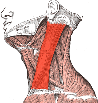
The sternocleidomastoid muscle is one of the largest and most superficial cervical muscles. The primary actions of the muscle are rotation of the head to the opposite side and flexion of the neck. The sternocleidomastoid is innervated by the accessory nerve.

A dermatome is an area of skin that is mainly supplied by afferent nerve fibres from the dorsal root of any given spinal nerve. There are 8 cervical nerves , 12 thoracic nerves, 5 lumbar nerves and 5 sacral nerves. Each of these nerves relays sensation from a particular region of skin to the brain.

The popliteal fossa is a shallow depression located at the back of the knee joint. The bones of the popliteal fossa are the femur and the tibia. Like other flexion surfaces of large joints, it is an area where blood vessels and nerves pass relatively superficially, and with an increased number of lymph nodes.

In human anatomy, the pterygopalatine fossa is a fossa in the skull. A human skull contains two pterygopalatine fossae—one on the left side, and another on the right side. Each fossa is a cone-shaped paired depression deep to the infratemporal fossa and posterior to the maxilla on each side of the skull, located between the pterygoid process and the maxillary tuberosity close to the apex of the orbit. It is the indented area medial to the pterygomaxillary fissure leading into the sphenopalatine foramen. It communicates with the nasal and oral cavities, infratemporal fossa, orbit, pharynx, and middle cranial fossa through eight foramina.
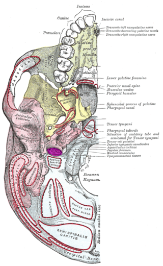
The jugular fossa is a deep depression in the inferior part of the temporal bone at the base of the skull. It lodges the bulb of the internal jugular vein.

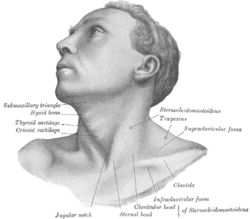
The posterior triangle is a region of the neck.

The pterygoid processes of the sphenoid, one on either side, descend perpendicularly from the regions where the body and the greater wings of the sphenoid bone unite.

The supraclavicular nerve is a cutaneous (sensory) nerve of the cervical plexus that arises from the third and fourth cervical (spinal) nerves. It emerges from beneath the posterior border of the sternocleidomastoid muscle, then split into multiple branches. Together, these innervate the skin over the shoulder.
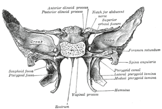
The pterygoid fossa is an anatomical term for the fossa formed by the divergence of the lateral pterygoid plate and the medial pterygoid plate of the sphenoid bone.

The infraspinousfossa of the scapula is much larger than the supraspinatous fossa; toward its vertebral margin a shallow concavity is seen at its upper part; its center presents a prominent convexity, while near the axillary border is a deep groove which runs from the upper toward the lower part.

The supraspinous fossa of the posterior aspect of the scapula is smaller than the infraspinous fossa, concave, smooth, and broader at its vertebral than at its humeral end. Its medial two-thirds give origin to the Supraspinatus.

The subclavian triangle, the smaller division of the posterior triangle, is bounded, above, by the inferior belly of the omohyoideus; below, by the clavicle; its base is formed by the posterior border of the sternocleidomastoideus.

The Infraclavicular fossa is an indentation, or fossa, immediately below the clavicle, above the third rib and between the deltoid muscle laterally and medioclavicular line medially.

The navicular fossa is a short dilated portion of the male urethra within the glans penis just proximal to the external urethral meatus. The roof of the fossa is especially dilated, forming a lacuna; medical instruments being inserted into the male urethra should initially be directed towards the floor of the fossa so as not to get snagged at the fossa. It is one of three dilations of the male urethra.

Supraclavicular lymph nodes are lymph nodes found above the clavicle, that can be felt in the supraclavicular fossa. The supraclavicular lymph nodes on the left side are called Virchow's nodes. It leads to an appreciable mass that can be recognized clinically, called Troisier sign.

The following outline is provided as an overview of and topical guide to human anatomy:

The superficial lateral cervical lymph nodes are found along the course of the external jugular vein, between the inferior aspect of the parotid gland and the supraclavicular nodes. The nodes are intercalated along the course of the vessels draining the parotid nodes and the infraauricular nodes. These nodes drain into the supraclavicular nodes, and on to the jugular trunk, followed by the thoracic duct on the left or the right lymphatic duct.
In anatomy, a fossa is a depression or hollow usually in a bone, such as the hypophyseal fossa. Some examples include:
Ambesh maneuver is a technique that involves the simple external compression of internal jugular vein in supraclavicular fossa to prevent and diagnose misplacement of the subclavian vein catheter into the internal jugular vein (IJV). The subclavian vein is a big vessel that drains the blood from the hand, forearm and the upper arm into the right side of the heart through superior vena cava. The subclavian veins lie just behind the clavicle on each side and therefore known as subclavian vein.

Brachial plexus block is a regional anesthesia technique that is sometimes employed as an alternative or as an adjunct to general anesthesia for surgery of the upper extremity. This technique involves the injection of local anesthetic agents in close proximity to the brachial plexus, temporarily blocking the sensation and ability to move the upper extremity. The subject can remain awake during the ensuing surgical procedure, or they can be sedated or even fully anesthetized if necessary.