Omohyoid muscle

Last updated
Omohyoid muscle
Musculi colli omohyoideus.svg
The omohyoid muscle, highlighted.
Musculus omohyoideus.png
Muscles of the neck. Anterior view. Omohyoid is labeled on both sides.
Details
Origin Upper border of the scapula (inferior belly), intermediate tendon (superior belly)
Insertion Intermediate tendon (inferior belly), hyoid bone (superior belly)
Artery Branches from the inferior thyroid artery (ITA)
Nerve Ansa cervicalis (C1-C3)
Actions Depresses the larynx and hyoid bone. Also carries hyoid bone backward and to the side.
Identifiers
Latin musculus omohyoideus
TA98 A04.2.04.003
TA2 2169
FMA 13342
Anatomical terms of muscle

The omohyoid muscle is a muscle in the neck. It is one of the infrahyoid muscles. It consists of two bellies separated by an intermediate tendon. Its inferior belly is attached to the scapula; its superior belly is attached to the hyoid bone. Its intermediate tendon is anchored to the clavicle and first rib by a fascial sling. The omohyoid is innervated by the ansa cervicalis of the cervical plexus. It acts to depress the hyoid bone.

Contents

Anatomy

Structure

The omohyoid muscle consists of muscle bellies that meet at an angle at the muscle's intermediate tendon. [1]

Inferior belly

The inferior belly is narrow and flat band. [1]

It arises from the superior border of scapula (near the scapular notch). It sometimes also arises from the superior transverse scapular ligament. [1]

It is directed anteriorly and somewhat superiorly from its origin, extending across the inferior portion of the neck. It passes posterior to the sternocleidomastoid muscle to insert at the intermediate tendon. [1]

Superior belly

The superior belly arises at the intermediate tendon. It extends nearly vertically superior-ward close to the lateral border of sternocleidomastoid muscle to insert at the inferior border of the body of hyoid bone (its site of insertion is lateral to that of the sternohyoid muscle). [1]

Intermediate tendon

The intermediate tendon is variable in length and form, and contains a variable amount of muscle tissue. It is anchored inferiorly to the clavicle and first rib by a band of deep cervical fascia that surrounds the intermediate tendon; this fascial fixation maintains the angle of the muscle. [1]

It is typically situated at the level of the arch of cricoid cartilage, adjacent to the internal jugular vein. [1]

Innervation

The omohyoid is innervated by the ansa cervicalis of the cervical plexus. [2] The inferior belly is innervated by the ansa cervicalis itself (which is derived from cervical spinal nerves C1-C3), and the superior belly is innervated by the superior root of ansa cervicalis (which is derived from the first cervical spinal nerve (C1)). [1]

Relations

The inferior belly of the omohyoid divides the posterior triangle of the neck into a occipital triangle (above) and a subclavian triangle (below). [3] [ verification needed ]

Its superior belly divides the anterior triangle into an carotid triangle (above) and a muscular triangle (below). [3] [4] [ verification needed ] Its superior belly serves as the most lateral member of the infrahyoid muscles, located lateral to both the sternothyroid muscles and the thyrohyoid muscles. [5] [ verification needed ]

The tendon is related to the internal jugular vein and can be used as a landmark for this vein during surgery.[ citation needed ]

Variation

The omohyoid muscle may be doubled or completely absent in some people.[ citation needed ] Either belly may be doubled or absent. [6]

The inferior belly may be attached to the clavicle directly (rather than by fascia). [7]

The superior belly may be fused with the sternohyoid muscle. [8]

Actions/movements

The muscle depresses the hyoid bone when the bone is in an elevated position. [1]

A putative action of the muscle is tension of the inferior portion of deep cervical fascia to prevent soft tissues from collapsing inward into the upper airway during deep inspiration. [1]

Etymology

The name "omohyoid" derives from the Greek "omos" meaning shoulder, giving one of its attachments, and "hyoid", giving the other attachment – the hyoid bone.[ citation needed ]

Related Research Articles

<span class="mw-page-title-main">Infrahyoid muscles</span> Group of muscles

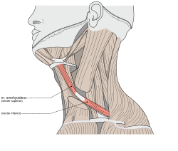
The infrahyoid muscles, or strap muscles, are a group of four pairs of muscles in the anterior (frontal) part of the neck. The four infrahyoid muscles are the sternohyoid, sternothyroid, thyrohyoid and omohyoid muscles.

<span class="mw-page-title-main">Levator scapulae muscle</span> Slender skeletal muscle at the back and side of the neck

The levator scapulae is a slender skeletal muscle situated at the back and side of the neck. It originates from the transverse processes of the four uppermost cervical vertebrae; it inserts onto the upper portion of the medial border of the scapula. It is innervated by the cervical nerves C3-C4, and frequently also by the dorsal scapular nerve. As the Latin name suggests, its main function is to lift the scapula.

<span class="mw-page-title-main">Cervical plexus</span> Network of nerves in the neck

The cervical plexus is a nerve plexus of the anterior rami of the first four cervical spinal nerves C1-C4. The cervical plexus provides motor innervation to some muscles of the neck, and the diaphragm; it provides sensory innervation to parts of the head, neck, and chest.

<span class="mw-page-title-main">Digastric muscle</span> Small muscle located under the jaw in mammals

The digastric muscle is a bilaterally paired suprahyoid muscle located under the jaw. Its posterior belly is attached to the mastoid notch of temporal bone, and its anterior belly is attached to the digastric fossa of mandible; the two bellies are united by an intermediate tendon which is held in a loop that attaches to the hyoid bone. The anterior belly is innervated via the mandibular nerve, and the posterior belly is innervated via the facial nerve. It may act to depress the mandible or elevate the hyoid bone.

<span class="mw-page-title-main">Obliquus capitis inferior muscle</span>

The obliquus capitis inferior muscle is a muscle in the upper back of the neck. It is one of the suboccipital muscles. Its inferior attachment is at the spinous process of the axis; its superior attachment is at the transverse process of the atlas. It is innervated by the suboccipital nerve. The muscle rotates the head to its side.

<span class="mw-page-title-main">Obliquus capitis superior muscle</span>

The obliquus capitis superior muscle is a small muscle in the upper back part of the neck. It is one of the suboccipital muscles. It attaches inferiorly at the transverse process of the atlas ; it attaches superiorly at the external surface of the occipital bone. The muscle is innervated by the suboccipital nerve.

<span class="mw-page-title-main">Stylohyoid muscle</span> Muscle

The stylohyoid muscle is one of the suprahyoid muscles. Its originates from the styloid process of the temporal bone; it inserts onto hyoid bone. It is innervated by a branch of the facial nerve. It acts draw the hyoid bone upwards and backwards.

<span class="mw-page-title-main">Ansa cervicalis</span> From cervical plexus

The ansa cervicalis is a loop formed by muscular branches of the cervical plexus formed by branches of cervical spinal nerves C1-C3. The ansa cervicalis has two roots - a superior root and an inferior root - that unite distally, forming a loop. It is situated within the carotid sheath.

<span class="mw-page-title-main">Sternohyoid muscle</span>

The sternohyoid muscle is a bilaterally paired, long, thin, narrow strap muscle of the anterior neck. It is one of the infrahyoid muscles. It is innervated by the ansa cervicalis. It acts to depress the hyoid bone.

<span class="mw-page-title-main">Sternothyroid muscle</span>

The sternothyroid muscle is an infrahyoid muscle of the neck. It acts to depress the hyoid bone.

<span class="mw-page-title-main">Thyrohyoid muscle</span> Neck muscle that depresses the hyoid bone and elevates the larynx

The thyrohyoid muscle is a small skeletal muscle of the neck. Above, it attaches onto the greater cornu of the hyoid bone; below, it attaches onto the oblique line of the thyroid cartilage. It is innervated by fibres derived from the cervical spinal nerve 1 that run with the hypoglossal nerve to reach this muscle. The thyrohyoid muscle depresses the hyoid bone and elevates the larynx during swallowing. By controlling the position and shape of the larynx, it aids in making sound.

<span class="mw-page-title-main">Rectus capitis posterior major muscle</span> Tendon

The rectus capitis posterior major is a muscle in the upper back part of the neck. It is one of the suboccipital muscles. Its inferior attachment is at the spinous process of the axis ; its superior attachment is onto the outer surface of the occipital bone on and around the side part of the inferior nuchal line. The muscle is innervated by the suboccipital nerve. The muscle acts to extend the head and rorate the head to its side.

<span class="mw-page-title-main">Rectus capitis posterior minor muscle</span> Tendon

The rectus capitis posterior minor is a muscle in the upper back part of the neck. It is one of the suboccipital muscles. Its inferior attachment is at the posterior arch of atlas; its superior attachment is onto the occipital bone at and below the inferior nuchal line. The muscle is innervated by the suboccipital nerve. The muscle acts as a weak extensor of the head.

<span class="mw-page-title-main">Carotid sheath</span>

The carotid sheath is a condensation of the deep cervical fascia enveloping multiple vital neurovascular structures of the neck, including the common and internal carotid arteries, the internal jugular vein, the vagus nerve, and ansa cervicalis. The carotid sheath helps protects the structures contained therein.

<span class="mw-page-title-main">Inferior thyroid artery</span> Artery of the neck

The inferior thyroid artery is an artery in the neck. It arises from the thyrocervical trunk and passes upward, in front of the vertebral artery and longus colli muscle. It then turns medially behind the carotid sheath and its contents, and also behind the sympathetic trunk, the middle cervical ganglion resting upon the vessel.

<span class="mw-page-title-main">Anterior triangle of the neck</span>

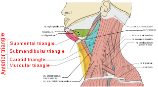
The anterior triangle is a region of the neck.

<span class="mw-page-title-main">Carotid triangle</span>

The carotid triangle is a portion of the anterior triangle of the neck.

<span class="mw-page-title-main">Prevertebral fascia</span> Layer of deep cervical fascia that surrounds the vertebral column

The prevertebral fascia is the layer of deep cervical fascia that surrounds the vertebral column. It is the deepest layer of deep cervical fascia.

<span class="mw-page-title-main">Posterior atlantooccipital membrane</span> Membrane at the base of the skull

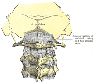
The posterior atlantooccipital membrane is a broad but thin membrane extending between the to the posterior margin of the foramen magnum above, and posterior arch of atlas below. It forms the floor of the suboccipital triangle.

<span class="mw-page-title-main">Thyrohyoid branch</span> Nervous system structure

The thyrohyoid branch (also: thyrohyoid branch of ansa cervicalis, or nerve to thyrohyoid (muscle)) is a motor branch derived from the cervical plexus formed by fibres of (the anterior ramus of) the cervical spinal nerve 1 (C1) (and - according to some sources - cervical spinal nerve 2 (C2) as well) that join and travel with the hypoglossal nerve (cranial nerve XII) to reach the suprahyoid region, branching away from CN XII distal to the superior root of ansa cervicalis (which is a branching other fibres of C1-C2 that had traveled with the CN XII), near the posterior border of the hyoglossus muscle. The thyrohyoid branch of ansa cervicalis innervates the thyrohyoid muscle.

References

PD-icon.svgThis article incorporates text in the public domain from page 392 of the 20th edition of Gray's Anatomy (1918)

  1. 1 2 3 4 5 6 7 8 9 10 Standring, Susan (2020). Gray's Anatomy: The Anatomical Basis of Clinical Practice (42nd ed.). New York. pp. 581–582. ISBN   978-0-7020-7707-4. OCLC   1201341621.{{cite book}}: CS1 maint: location missing publisher (link)
  2. McHanwell, Steve; Watson, Charles (2009-01-01), Watson, Charles; Paxinos, George; Kayalioglu, Gulgun (eds.), "Chapter 7 - Localization of Motoneurons in the Spinal Cord", The Spinal Cord, San Diego: Academic Press, pp. 94–114, doi:10.1016/b978-0-12-374247-6.50011-0, ISBN   978-0-12-374247-6 , retrieved 2020-11-20
  3. 1 2 Luna, Mario A.; Pfaltz, Madeleine (2009-01-01), Gnepp, Douglas R. (ed.), "Chapter 11 - Cysts of the Neck, Unknown Primary Tumor, and Neck Dissection", Diagnostic Surgical Pathology of the Head and Neck (Second Edition), Philadelphia: W.B. Saunders, pp. 839–881, doi:10.1016/b978-1-4160-2589-4.00011-5, ISBN   978-1-4160-2589-4 , retrieved 2020-11-20
  4. Human anatomy, Jacobs, Elsevier, 2008, page 189
  5. Illustrated Anatomy of the Head and Neck, Fehrenbach and Herring, Elsevier, 2012, page 102
  6. Standring, Susan (2020). Gray's Anatomy: The Anatomical Basis of Clinical Practice (42nd ed.). New York. p. 581. ISBN   978-0-7020-7707-4. OCLC   1201341621.{{cite book}}: CS1 maint: location missing publisher (link)
  7. Standring, Susan (2020). Gray's Anatomy: The Anatomical Basis of Clinical Practice (42nd ed.). New York. p. 581. ISBN   978-0-7020-7707-4. OCLC   1201341621.{{cite book}}: CS1 maint: location missing publisher (link)
  8. Standring, Susan (2020). Gray's Anatomy: The Anatomical Basis of Clinical Practice (42nd ed.). New York. p. 581. ISBN   978-0-7020-7707-4. OCLC   1201341621.{{cite book}}: CS1 maint: location missing publisher (link)