The two mylohyoid muscles arise from the mandible at the mylohyoid line, which extends from the mandibular symphysis in front to the last molar tooth behind. The posterior fibers pass inferomedially and insert at anterior surface of the hyoid bone. The medial fibres of the two mylohyoid muscles unite in a midline raphe (where the two muscles intermesh).[3]
The mylohyoid muscle separates the sublingual space from the submandibular space, which communicate via a lateral gap between the mylohyoid and hyoglossus muscles at the posterior free margin of mylohyoid muscle.[4] The submandibular gland wraps around the edges of the mylohyoid, and is divided into superficial and deep lobes above and below the muscle.[5]
Nerve supply
The mylohyoid muscle is supplied by a branch of the mandibular nerve, the inferior alveolar nerve. The mylohyoid nerve is a branch of the inferior alveolar nerve. The mylohyoid nerve emerges to give motor supply to the mylohyoid muscle.[1]
The mylohyoid muscle may be united to or replaced by the anterior belly of the digastric muscle; accessory slips to other hyoid muscles are frequent. This median raphe is sometimes absent; the fibers of the two muscles are then continuous.[citation needed] Variations in the mylohyoid muscle itself are not common.[6] Accessory mylohyoid muscles have been seen in some people, which have the same attachments, nerve supply, and function.[6] The mylohyoid muscle may also be split into an anterior portion and a posterior portion, with the sublingual gland occupying the space between these portions.[7]
An area of herniation of the sublingual gland, blood vessels, or fat, may be present, with studies reporting this in 10-50% of people.[4]
Function
The mylohyoid muscle elevates the hyoid bone and the tongue. This is particularly important during swallowing and speaking. Alternatively, if other muscles are used to keep the position of the hyoid bone fixed, then the mylohyoid muscle depresses the mandible.[1] It also functions as reinforcing the floor of mouth.[1]
Clinical significance
The mylohyoid muscle may be imaged by CT or MRI.[4] The mylohyoid separates the submandibular space below from the sublingual space above. Around the posterior border of the mylohoid muscle, these spaces communicate. Infections, especially odontogenic infections can spread from one space to the other via this communication, or alternatively penetrate the mylohyoid muscle, which is a poor barrier to the spread of infection. Because the attachment of the mylohyoid muscle (the mylohoid line of the mandible) becomes more superior towards the posterior of the mandible, posterior infected teeth are more likely to drain into the submandibular space, and infected anterior teeth are more likely to drain into the sublingual space, since the apices of the teeth are more likely to be below and above the mylohoid line respectively (see diagram).
History
The mylohyoid muscle may also be known as the diaphragma oris muscle.[8][9] It is named after its two attachments near the molar teeth ("mylo" comes from the Greek word for "molar").[10]
Additional images
Anterior view
Medial view
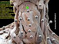
The origin of the mylohyoid muscle, inferior view.
The insertion of the mylohyoid muscle on the hyoid bone.
Drake, Richard L.; Vogl, Wayne; Tibbitts, Adam W.M. Mitchell (2005). Gray's anatomy for students. Philadelphia: Elsevier/Churchill Livingstone. ISBN978-0-443-06612-2.
Herring, Margaret J.; Fehrenbach, Susan W. (2013). Illustrated anatomy of the head and neck (4thed.). St. Louis, MO: Elsevier/Saunders. ISBN978-1-4377-2419-6.
This page is based on this Wikipedia article Text is available under the CC BY-SA 4.0 license; additional terms may apply. Images, videos and audio are available under their respective licenses.