Thyrohyoid muscle

Last updated
Thyrohyoid muscle
Thyrohyoid muscle.PNG
Muscles of the neck. Lateral view. (Thyrohyoideus labeled center-left.)
Thyrohyoideus.png
Muscles of the neck. Anterior view. (Thyrohyoideus visible center-left.)
Details
Origin Thyroid cartilage of the larynx
Insertion Hyoid bone
Artery Superior thyroid artery
Nerve First cervical nerve (C1) via hypoglossal nerve
Actions Elevates thyroid and depresses the hyoid bone
Identifiers
Latin musculus thyrohyoideus
TA98 A04.2.04.007
TA2 2174
FMA 13344
Anatomical terms of muscle

The thyrohyoid muscle is a small skeletal muscle of the neck. Above, it attaches onto the greater cornu of the hyoid bone; below, it attaches onto the oblique line of the thyroid cartilage. It is innervated by fibres derived from the cervical spinal nerve 1 that run with the hypoglossal nerve (CN XII) to reach this muscle. The thyrohyoid muscle depresses the hyoid bone and elevates the larynx during swallowing. By controlling the position and shape of the larynx, it aids in making sound.

Contents

Structure

The thyrohyoid muscle is a small, [1] broad and short muscle. [2] It is quadrilateral in shape. [1] It may be considered a superior-ward continuation of sternothyroid muscle. [1]

It belongs to the infrahyoid muscles group and the outer laryngeal muscle group. [3] :567–568

Attachments

Its superior attachment [note 1] [1] is the inferior border [1] of the greater cornu of the hyoid bone [2] [1] [3] :538 and adjacent portions of the body of hyoid bone. [1]

Its inferior attachment [note 2] [1] is the oblique line of the thyroid cartilage (alongside the sternothyroid muscle [1] ). [2] [1] [3] :538

Innervation

The thyrohyoid muscle is innervated (along with the geniohyoid muscle [1] [3] :457, 709) by a branch of the cervical plexus [4] [3] :538 - the nerve to thyrohyoid muscle (thyrohyoid branch of ansa cervicalis) [4] - which is formed by fibres of the cervical spinal nerve 1 (C1) [1] [2] [3] :538 (and - according to some sources - cervical spinal nerve 2 as well [4] [3] :457) that join and travel with the hypoglossal nerve (CN XII) [2] before splitting away from it [1] [2] distal to the superior root of ansa cervicalis. [1] [3] :709 The thyrohyoid muscle is the only infrahyoid muscle that is not innervated via the ansa cervicalis. [1]

Blood supply

The muscle is provided with arterial blood by branches of the superior thyroid artery, and of the lingual artery. [1]

Relations

The thyrohyoid muscle forms the inferior boundary of the carotid triangle. [5] It is situated deep to (beneath) the (depending upon the source) superior portion of [2] /superior belly of [3] :538 the sternohyoid muscle, [2] [3] :538 and the superior portion of [2] the omohyoid muscle. [2] [3] :538

Function

The thyrohyoid muscle depresses and fixates the hyoid bone. It elevates the larynx during swallowing. [3] :538 By controlling the position and shape of the larynx, it aids in making sound. [6]

Additional images

See also

Notes

  1. Described as either the origin or as the insertion.
  2. Described as either the origin or as the insertion.

Related Research Articles

Articles related to anatomy include:

<span class="mw-page-title-main">Infrahyoid muscles</span> Group of muscles

The infrahyoid muscles, or strap muscles, are a group of four pairs of muscles in the anterior (frontal) part of the neck. The four infrahyoid muscles are the sternohyoid, sternothyroid, thyrohyoid and omohyoid muscles.

<span class="mw-page-title-main">Omohyoid muscle</span> Human neck muscle

The omohyoid muscle is a muscle in the neck. It is one of the infrahyoid muscles. It consists of two bellies separated by an intermediate tendon. Its inferior belly is attached to the scapula; its superior belly is attached to the hyoid bone. Its intermediate tendon is anchored to the clavicle and first rib by a fascial sling. The omohyoid is innervated by the ansa cervicalis of the cervical plexus. It acts to depress the hyoid bone.

<span class="mw-page-title-main">Cervical plexus</span> Network of nerves in the neck

The cervical plexus is a nerve plexus of the anterior rami of the first four cervical spinal nerves C1-C4. The cervical plexus provides motor innervation to some muscles of the neck, and the diaphragm; it provides sensory innervation to parts of the head, neck, and chest.

<span class="mw-page-title-main">Digastric muscle</span> Small muscle located under the jaw in mammals

The digastric muscle is a bilaterally paired suprahyoid muscle located under the jaw. Its posterior belly is attached to the mastoid notch of temporal bone, and its anterior belly is attached to the digastric fossa of mandible; the two bellies are united by an intermediate tendon which is held in a loop that attaches to the hyoid bone. The anterior belly is innervated via the mandibular nerve, and the posterior belly is innervated via the facial nerve. It may act to depress the mandible or elevate the hyoid bone.

<span class="mw-page-title-main">Geniohyoid muscle</span> Muscle of the head

The geniohyoid muscle is a narrow paired muscle situated superior to the medial border of the mylohyoid muscle. It is named for its passage from the chin to the hyoid bone.

<span class="mw-page-title-main">Ansa cervicalis</span> From cervical plexus

The ansa cervicalis is a loop formed by muscular branches of the cervical plexus formed by branches of cervical spinal nerves C1-C3. The ansa cervicalis has two roots - a superior root and an inferior root - that unite distally, forming a loop. It is situated within the carotid sheath.

<span class="mw-page-title-main">Sternohyoid muscle</span>

The sternohyoid muscle is a bilaterally paired, long, thin, narrow strap muscle of the anterior neck. It is one of the infrahyoid muscles. It is innervated by the ansa cervicalis. It acts to depress the hyoid bone.

<span class="mw-page-title-main">Sternothyroid muscle</span>

The sternothyroid muscle is an infrahyoid muscle of the neck. It acts to depress the hyoid bone.

<span class="mw-page-title-main">Hyoglossus</span> Muscle

The hyoglossus is a thin and quadrilateral extrinsic muscle of the tongue. It originates from the hyoid bone; it inserts onto the side of the tongue. It is innervated by the hypoglossal nerve. It acts to depress and retract the tongue.

<span class="mw-page-title-main">Middle pharyngeal constrictor muscle</span> Muscle in the neck

The middle pharyngeal constrictor is a fan-shaped muscle located in the neck. It is one of three pharyngeal constrictor muscles. It is smaller than the inferior pharyngeal constrictor muscle.
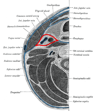
<span class="mw-page-title-main">Carotid sheath</span>

The carotid sheath is a condensation of the deep cervical fascia enveloping multiple vital neurovascular structures of the neck, including the common and internal carotid arteries, the internal jugular vein, the vagus nerve, and ansa cervicalis. The carotid sheath helps protects the structures contained therein.

<span class="mw-page-title-main">Thyrohyoid membrane</span> Elastic membrane in the larynx

The thyrohyoid membrane is a broad, fibro-elastic sheet of the larynx. It connects the upper border of the thyroid cartilage to the hyoid bone.
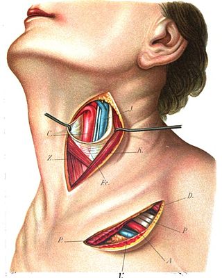
<span class="mw-page-title-main">Superior thyroid artery</span>

The superior thyroid artery arises from the external carotid artery just below the level of the greater cornu of the hyoid bone and ends in the thyroid gland.

<span class="mw-page-title-main">Superior laryngeal nerve</span> Branch of the vagus nerve

The superior laryngeal nerve is a branch of the vagus nerve. It arises from the middle of the inferior ganglion of vagus nerve and additionally also receives a sympathetic branch from the superior cervical ganglion.
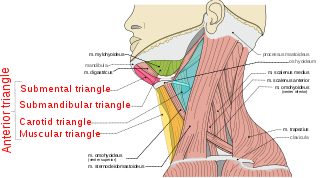
<span class="mw-page-title-main">Anterior triangle of the neck</span>

The anterior triangle is a region of the neck.

<span class="mw-page-title-main">Carotid triangle</span>

The carotid triangle is a portion of the anterior triangle of the neck.

<span class="mw-page-title-main">Muscular triangle</span>

The inferior carotid triangle, is bounded, in front, by the median line of the neck from the hyoid bone to the sternum; behind, by the anterior margin of the sternocleidomastoid; above, by the superior belly of the omohyoid.

<span class="mw-page-title-main">Cervical spinal nerve 1</span> Spinal nerve of the cervical segment

The cervical spinal nerve 1 (C1) is a spinal nerve of the cervical segment. C1 carries predominantly motor fibres, but also a small meningeal branch that supplies sensation to parts of the dura around the foramen magnum.

<span class="mw-page-title-main">Thyrohyoid branch</span> Nervous system structure

The thyrohyoid branch (also: thyrohyoid branch of ansa cervicalis, or nerve to thyrohyoid (muscle)) is a motor branch derived from the cervical plexus formed by fibres of (the anterior ramus of) the cervical spinal nerve 1 (C1) (and - according to some sources - cervical spinal nerve 2 (C2) as well) that join and travel with the hypoglossal nerve (cranial nerve XII) to reach the suprahyoid region, branching away from CN XII distal to the superior root of ansa cervicalis (which is a branching other fibres of C1-C2 that had traveled with the CN XII), near the posterior border of the hyoglossus muscle. The thyrohyoid branch of ansa cervicalis innervates the thyrohyoid muscle.

References

PD-icon.svgThis article incorporates text in the public domain from page 394 of the 20th edition of Gray's Anatomy (1918)

  1. 1 2 3 4 5 6 7 8 9 10 11 12 13 14 15 16 Standring, Susan (2020). Gray's Anatomy: The Anatomical Basis of Clinical Practice (42th ed.). New York. p. 582. ISBN   978-0-7020-7707-4. OCLC   1201341621.{{cite book}}: CS1 maint: location missing publisher (link)
  2. 1 2 3 4 5 6 7 8 9 10 Sinnatamby, Chummy S. (2011). Last's Anatomy (12th ed.). Elsevier Australia. p. 337. ISBN   978-0-7295-3752-0.
  3. 1 2 3 4 5 6 7 8 9 10 11 12 Waschke, Jens; Böckers, Tobias M.; Paulsen, Friedrich; Arnold, Wolfgang; Bechmann, Ingo, eds. (2018). Sobotta Anatomy Textbook: English Edition with Latin Nomenclature (1st ed.). München: Elsevier. ISBN   978-0-7020-6760-0.
  4. 1 2 3 "ramus thyrohyoideus ansae cervicalis". TheFreeDictionary.com. Retrieved 2023-06-30.
  5. Luna, Mario A.; Pfaltz, Madeleine (2009). "11 - Cysts of the Neck, Unknown Primary Tumor, and Neck Dissection". Diagnostic Surgical Pathology of the Head and Neck (2nd ed.). Philadelphia: Saunders. pp. 839–881. doi:10.1016/B978-1-4160-2589-4.00011-5. ISBN   978-1-4377-1951-2. OCLC   460904310.
  6. Hage, Steffen R. (2010). "8.3 - Neuronal networks involved in the generation of vocalization". Handbook of Behavioral Neuroscience. Vol. 19. Stefan Brudzynski. London: Academic Press. pp. 339–349. doi:10.1016/B978-0-12-374593-4.00032-2. ISBN   978-0-12-374593-4. ISSN   1569-7339. OCLC   528610774.