Inferior pharyngeal constrictor muscle

Last updated
Inferior pharyngeal constrictor muscle
Musculusconstrictorpharyngisinferior.png
Muscles of the pharynx and cheek. (Constrictor pharyngis inferior visible at bottom left.)
Gray1031.png
Muscles of the pharynx, viewed from behind, together with the associated vessels and nerves. (Inf. const. labeled at bottom center.)
Details
Origin Cricoid cartilage and thyroid cartilage
Insertion Pharyngeal raphe
Nerve Pharyngeal plexus of vagus nerve, recurrent laryngeal nerve and superior laryngeal nerve
Actions Swallowing
Identifiers
Latin musculus constrictor pharyngis inferior
TA98 A05.3.01.111
TA2 2187
FMA 46623
Anatomical terms of muscle

The inferior pharyngeal constrictor muscle is a skeletal muscle of the neck. It is the thickest of the three outer pharyngeal muscles. It arises from the sides of the cricoid cartilage and the thyroid cartilage. It is supplied by the vagus nerve (CN X). It is active during swallowing, and partially during breathing and speech. It may be affected by Zenker's diverticulum.

Contents

Structure

The inferior pharyngeal constrictor muscle is composed of two parts. The first part (and more superior) arises from the thyroid cartilage (thyropharyngeal part), and the second part arises from the cricoid cartilage (cricopharyngeal part). [1]

From these origins, the fibers spread backward and medially to insert with the muscle of the opposite side into the fibrous pharyngeal raphe in the posterior median line of the pharynx. The thyropharyngeal part mainly uses type 2 fibres (anaerobic), while the cricopharyngeal part mainly uses type 1 fibres (aerobic). [2] Fibre type may change after birth. [3]

The inferior fibers are horizontal and continuous with the circular fibers of the esophagus; the rest ascend, increasing in obliquity, and overlap the middle pharyngeal constrictor muscle. The cricopharyngeal part is synonymous with the upper esophageal sphincter (UES), which controls the opening of the cervical esophagus. [4] It is sometimes referred to as the cricopharyngeal inlet.

Nerve supply

The inferior pharyngeal constrictor muscle can be supplied by branches from the pharyngeal plexus, [4] the recurrent laryngeal nerve, the external branch of the superior laryngeal nerve, or a combination of these (the recurrent laryngeal nerve being the most common innervation of the cricopharyngeal part). [5] All these branches and nerves come from the vagus nerve (CN X). [4]

Variation

The inferior pharyngeal constrictor muscle can merge with superior pharyngeal constrictor, or posterior part of pharyngobasilar fascia. [6]

Function

The inferior pharyngeal constrictor muscle has a broad role in moving the lower part of the pharynx. [7]

Swallowing

As soon as a bolus of food is received in the pharynx, elevator muscles relax, and the pharynx descends. The inferior pharyngeal constrictor muscle, along with the other constrictors, contract upon the bolus, and convey it downward into the esophagus. [4] [7] During swallowing, they contract and cause peristalsis in the pharynx. [4]

Breathing

The inferior pharyngeal constrictor muscle is partially used during breathing and speech. [4] It helps to keep the pharynx open, particularly during sleep. [8]

Clinical significance

Zenker's diverticulum

Uncoordinated muscle contraction, cricopharyngeal spasm, or impaired relaxation of the inferior pharyngeal constrictor muscle are currently considered the main factors in development of a Zenker's diverticulum. Zenker's diverticulum develops between the two muscular bellies (the thyropharyngeal part and the cricopharyngeal part) in a small gap called Killian's dehiscence. A diverticulum can form where a balloon of mucosa becomes trapped outside the pharyngeal boundaries. Food or other materials may reside here, which may lead to infection. Motor incoordination of the cricopharyngeal part can cause difficulty swallowing.

In extreme cases, this can be related to retrograde cricopharyngeal dysfunction (R-CPD) which causes the inability to burp; this is in part due to the muscle not being able to relax. Botox or a cricopharyngeal myotomy are used to treat the condition. [9] [10]

Radiological damage

The inferior pharyngeal constrictor muscle may be damaged by chemotherapy-intensity modulated radiotherapy. [11] This may lead to dysphagia, causing continued use of a feeding tube rather than independent swallowing. [11]

Additional images

See also

Related Research Articles

<span class="mw-page-title-main">Larynx</span> Voice box, an organ in the neck of amphibians, reptiles, and mammals

The larynx, commonly called the voice box, is an organ in the top of the neck involved in breathing, producing sound and protecting the trachea against food aspiration. The opening of larynx into pharynx known as the laryngeal inlet is about 4–5 centimeters in diameter. The larynx houses the vocal cords, and manipulates pitch and volume, which is essential for phonation. It is situated just below where the tract of the pharynx splits into the trachea and the esophagus. The word 'larynx' comes from the Ancient Greek word lárunx ʻlarynx, gullet, throat.ʼ

<span class="mw-page-title-main">Esophagus</span> Vertebrate organ through which food passes to the stomach

The esophagus or oesophagus, colloquially known also as the food pipe, food tube, or gullet, is an organ in vertebrates through which food passes, aided by peristaltic contractions, from the pharynx to the stomach. The esophagus is a fibromuscular tube, about 25 cm (10 in) long in adults, that travels behind the trachea and heart, passes through the diaphragm, and empties into the uppermost region of the stomach. During swallowing, the epiglottis tilts backwards to prevent food from going down the larynx and lungs. The word oesophagus is from Ancient Greek οἰσοφάγος (oisophágos), from οἴσω (oísō), future form of φέρω + ἔφαγον.

Swallowing, also called deglutition or inglutition in scientific contexts, is the process in the body of a human or other animal that allows for a substance to pass from the mouth, to the pharynx, and into the esophagus, while shutting the epiglottis. Swallowing is an important part of eating and drinking. If the process fails and the material goes through the trachea, then choking or pulmonary aspiration can occur. In the human body the automatic temporary closing of the epiglottis is controlled by the swallowing reflex.

A Zenker's diverticulum, also pharyngeal pouch, is a diverticulum of the mucosa of the human pharynx, just above the cricopharyngeal muscle. It is a pseudo diverticulum or false diverticulum, also known as a pulsion diverticulum.

<span class="mw-page-title-main">Recurrent laryngeal nerve</span> Nerve in the human body

The recurrent laryngeal nerve (RLN) is a branch of the vagus nerve that supplies all the intrinsic muscles of the larynx, with the exception of the cricothyroid muscles. There are two recurrent laryngeal nerves, right and left. The right and left nerves are not symmetrical, with the left nerve looping under the aortic arch, and the right nerve looping under the right subclavian artery then traveling upwards. They both travel alongside the trachea. Additionally, the nerves are among the few nerves that follow a recurrent course, moving in the opposite direction to the nerve they branch from, a fact from which they gain their name.

<span class="mw-page-title-main">Posterior cricoarytenoid muscle</span> Muscle of the larynx

The posterior cricoarytenoid muscle is a intrinsic muscle of the larynx. It arises from the cricoid cartilage; it inserts onto the arytenoid cartilage of the same side. It is innervated by the recurrent laryngeal nerve. Each acts to open the vocal folds by pulling the vocal fold of the same side laterally. It participates in the production of sounds.

<span class="mw-page-title-main">Cricoid cartilage</span> Complete ring of cartilage around the trachea

The cricoid cartilage, or simply cricoid or cricoid ring, is the only complete ring of cartilage around the trachea. It forms the back part of the voice box and functions as an attachment site for muscles, cartilages, and ligaments involved in opening and closing the airway and in producing speech.

<span class="mw-page-title-main">Common carotid artery</span> One of the two arteries that supply the head and neck with blood

In anatomy, the left and right common carotid arteries (carotids) are arteries that supply the head and neck with oxygenated blood; they divide in the neck to form the external and internal carotid arteries.

<span class="mw-page-title-main">Levator veli palatini</span> Muscle of the soft palate

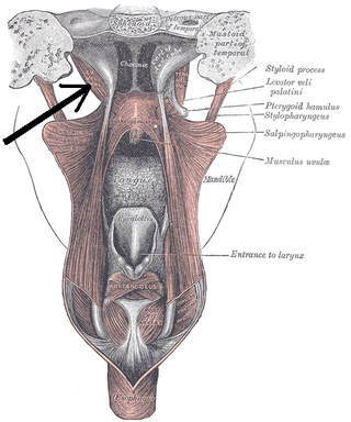
The levator veli palatini is a muscle of the soft palate and pharynx. It is innervated by the vagus nerve via its pharyngeal plexus. During swallowing, it contracts, elevating the soft palate to help prevent food from entering the nasopharynx.

<span class="mw-page-title-main">Salpingopharyngeus muscle</span> Muscle of the pharynx

The salpingopharyngeus muscle is a muscle of the pharynx. It arises from the lower part of the cartilage of the Eustachian tube, and inserts into the palatopharyngeus muscle by blending with its posterior fasciculus. It is innervated by vagus nerve via the pharyngeal plexus. It raises the pharynx and larynx during deglutition (swallowing) and laterally draws the pharyngeal walls up. It opens the pharyngeal orifice of the Eustachian tube during swallowing to allow for the equalization of pressure between it and the pharynx.

<span class="mw-page-title-main">Middle pharyngeal constrictor muscle</span> Muscle in the neck

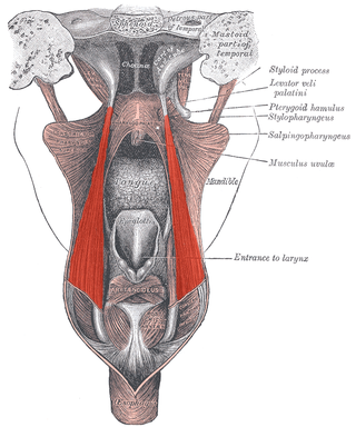
The middle pharyngeal constrictor is a fan-shaped muscle located in the neck. It is one of three pharyngeal constrictor muscles. It is smaller than the inferior pharyngeal constrictor muscle.

<span class="mw-page-title-main">Superior pharyngeal constrictor muscle</span> Muscle

The superior pharyngeal constrictor muscle is a quadrilateral muscle of the pharynx. It is the uppermost and thinnest of the three pharyngeal constrictors.

<span class="mw-page-title-main">Stylopharyngeus muscle</span> Muscle

The stylopharyngeus muscle is a muscle in the head. It originates from the temporal styloid process. Some of its fibres insert onto the thyroid cartilage, while others end by intermingling with proximal structures. It is innervated by the glossopharyngeal nerve. It acts to elevate the larynx and pharynx, and dilate the pharynx, thus facilitating swallowing.

<span class="mw-page-title-main">Inferior thyroid artery</span> Artery of the neck

The inferior thyroid artery is an artery in the neck. It arises from the thyrocervical trunk and passes upward, in front of the vertebral artery and longus colli muscle. It then turns medially behind the carotid sheath and its contents, and also behind the sympathetic trunk, the middle cervical ganglion resting upon the vessel.

<span class="mw-page-title-main">Superior laryngeal nerve</span> Branch of the vagus nerve

The superior laryngeal nerve is a branch of the vagus nerve. It arises from the middle of the inferior ganglion of vagus nerve and additionally also receives a sympathetic branch from the superior cervical ganglion.

<span class="mw-page-title-main">Pharyngeal plexus of vagus nerve</span> Nerve fibers innervating most of the palate and pharynx

The pharyngeal plexus is a nerve plexus located upon the outer surface of the pharynx. It contains a motor component, a sensory component, and sympathetic component.

<span class="mw-page-title-main">Killian's dehiscence</span>

Killian's dehiscence is a triangular area in the wall of the pharynx between the cricopharyngeus and thyropharyngeus which are the two parts of the inferior constrictors(also see Pharyngeal pouch). It can be seen as a locus minoris resistentiae. Similar triangular area between circular fibres of cricopharyngeus and longitudinal fibres of esophagus is Lamier'striangle or Lamier-hackermann's area.

<span class="mw-page-title-main">Pharynx</span> Part of the throat that is behind the mouth and nasal cavity

The pharynx is the part of the throat behind the mouth and nasal cavity, and above the esophagus and trachea. It is found in vertebrates and invertebrates, though its structure varies across species. The pharynx carries food to the esophagus and air to the larynx. The flap of cartilage called the epiglottis stops food from entering the larynx.

Cricopharyngeal spasms occur in the cricopharyngeus muscle of the pharynx. Cricopharyngeal spasm is an uncomfortable but harmless and temporary disorder.

The face and neck development of the human embryo refers to the development of the structures from the third to eighth week that give rise to the future head and neck. They consist of three layers, the ectoderm, mesoderm and endoderm, which form the mesenchyme, neural crest and neural placodes. The paraxial mesoderm forms structures named somites and somitomeres that contribute to the development of the floor of the brain and voluntary muscles of the craniofacial region. The lateral plate mesoderm consists of the laryngeal cartilages. The three tissue layers give rise to the pharyngeal apparatus, formed by six pairs of pharyngeal arches, a set of pharyngeal pouches and pharyngeal grooves, which are the most typical feature in development of the head and neck. The formation of each region of the face and neck is due to the migration of the neural crest cells which come from the ectoderm. These cells determine the future structure to develop in each pharyngeal arch. Eventually, they also form the neurectoderm, which forms the forebrain, midbrain and hindbrain, cartilage, bone, dentin, tendon, dermis, pia mater and arachnoid mater, sensory neurons, and glandular stroma.

References

PD-icon.svgThis article incorporates text in the public domain from page 1142 of the 20th edition of Gray's Anatomy (1918)

  1. Origin, insertion and nerve supply of the muscle at Loyola University Chicago Stritch School of Medicine
  2. Hyodo, Masamitsu; Aibara, Ryuichi; Kawakita, Seiji; Yumoto, Eiji (1998-01-01). "Histochemical Study of the Canine Inferior Pharyngeal Constrictor Muscle: Implications for its Function". Acta Oto-Laryngologica. 118 (2): 272–279. doi:10.1080/00016489850155017. ISSN   0001-6489. PMID   9583798.
  3. Masamitsu Hyodo, Eiji Yumoto, Seiji Kawakita, Takahiko Yamagata (1999-01-01). "Postnatal Changes in the Types of Muscle Fibre in the Canine Inferior Pharyngeal Constrictor". Acta Oto-Laryngologica. 119 (7): 843–846. doi:10.1080/00016489950180522. ISSN   0001-6489. PMID   10687945.{{cite journal}}: CS1 maint: multiple names: authors list (link)
  4. 1 2 3 4 5 6 Mu, Liancai; Sanders, Ira (2001). "Neuromuscular compartments and fiber-type regionalization in the human inferior pharyngeal constrictor muscle". The Anatomical Record . 264 (4): 367–377. doi: 10.1002/ar.10020 . ISSN   1097-0185. PMID   11745092. S2CID   22153114.
  5. Uludag, Mehmet; Aygun, Nurcihan (2016). "Innervation of the human cricopharyngeal muscle by the recurrent laryngeal nerve and external branch of the superior laryngeal nerve". Langenbecks Arch Surg . 402: 683–690. doi:10.1007/s00423-016-1376-5. PMID   26843022.
  6. Bui, Toai; M Das, Joe (2022), "Anatomy, Head and Neck, Pharyngeal Muscles", StatPearls, Treasure Island (FL): StatPearls Publishing, PMID   31869118 , retrieved 2022-07-22
  7. 1 2 Yamaoka, Minoru; Furusawa, Kiyofumi (May 1997). "Function of the Inferior Pharyngeal Constrictor Muscle". Dysphagia. 12 (3): 171. doi: 10.1007/BF03500907 . ISSN   0179-051X. PMID   9190104. S2CID   44633854.
  8. Sherrey, John H.; Pollard, Marcus J.; Megirian, David (1 April 1986). "Respiratory functions of the inferior pharyngeal constrictor and sternohyoid muscles during sleep". Experimental Neurology . 92 (1): 267–277. doi:10.1016/0014-4886(86)90140-8. ISSN   0014-4886. PMID   3956654. S2CID   7751627.
  9. Bastian, Robert W.; Hoesli, Rebecca C. (2020). "Partial Cricopharyngeal Myotomy for Treatment of Retrograde Cricopharyngeal Dysfunction". Oto Open. 4 (2): 2473974X2091764. doi:10.1177/2473974X20917644. PMC   7163242 . PMID   32328538.
  10. Bastian, R. W.; Smithson, M. L. (2019). "Inability to Belch and Associated Symptoms Due to Retrograde Cricopharyngeus Dysfunction: Diagnosis and Treatment". Oto Open. 3 (1): 2473974X19834553. doi:10.1177/2473974X19834553. PMC   6572913 . PMID   31236539.
  11. 1 2 Vlacich, Gregory; Spratt, Daniel E.; Diaz, Roberto; Phillips, John G.; Crass, Jostin; Li, Chung-I; Shyr, Yu; Cmelak, Anthony J. (1 March 2014). "Dose to the inferior pharyngeal constrictor predicts prolonged gastrostomy tube dependence with concurrent intensity-modulated radiation therapy and chemotherapy for locally-advanced head and neck cancer". Radiotherapy and Oncology. 110 (3): 435–440. doi:10.1016/j.radonc.2013.12.007. ISSN   0167-8140. PMID   24440043.