Rectus capitis posterior minor muscle

Last updated
Rectus capitis posterior minor muscle
Rectus capitis posterior minor muscle back.png
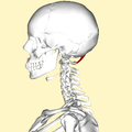
Human skull seen from back (rectus capitis posterior minor shown in red.)
Rectus capitis posterior minor.png
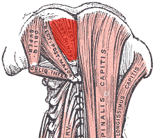
Deep muscles of the back. (rectus capitis posterior minor labeled at top center.)
Details
Origin Tubercle on the posterior arch of the atlas
Insertion Medial part of the inferior nuchal line of the occipital bone and the surface between it and the foramen magnum
Nerve Branch of the dorsal primary division of the suboccipital nerve
Actions Extends the head at the neck, but is now considered to be more of a sensory organ than a muscle
Identifiers
Latin musculus rectus capitis posterior minor
TA98 A04.2.02.005
TA2 2250
FMA 32526
Anatomical terms of muscle

The rectus capitis posterior minor (or rectus capitis posticus minor[ citation needed ]) is a muscle in the upper back part of the neck. It is one of the suboccipital muscles. Its inferior attachment is at the posterior arch of atlas; its superior attachment is onto the occipital bone at and below the inferior nuchal line. The muscle is innervated by the suboccipital nerve (the posterior ramus of first cervical spinal nerve). The muscle acts as a weak extensor of the head.

Contents

Anatomy

The rectus capitis posterior major muscle is one of the suboccipital muscles. [1]

The muscle extends vertically superior-ward from its inferiro attachment to its superior attachment. [2] The muscle becomes broader superiorly. [1]

Attachments

The inferior attachment is (by a narrow tendon) onto the posterior tubercle [1] of the posterior arch of atlas. [2]

Its superior attachment is onto the medial portion of the inferior nuchal line [1] and the external surface of the occipital bone inferior to it [2] [1] (between this line superiorly and the foramen magnum inferiorly [1] ).

The muscle usually also additionally attaches onto the posterior atlantooccipital membrane (which is in turn attached onto adjacent dura mater of the spinal canal). [1]

Innervation

The muscle receives motor innervation from the suboccipital nerve (the posterior ramus of cervical spinal nerve C1). [2] [1]

Variation

The muscle of either side may be doubled (along its length). [1]

Actions/movements

The muscle is a weak [2] extensor of the head. [2] [1]

The synergists are the rectus capitis posterior major and the obliquus capitis superior.[ citation needed ]

Research

Role in headache

Connective tissue bridges were noted at the atlanto-occipital joint between the rectus capitis posterior minor (RCPm) muscle and the dorsal spinal dura. [3] Similar connective tissue connections of the rectus capitis posterior major have been reported recently as well. [4] The perpendicular arrangement of these fibers appears to restrict dural movement toward the spinal cord. The ligamentum nuchae was found to be continuous with the posterior cervical spinal dura and the lateral portion of the occipital bone. Anatomic structures innervated by cervical nerves C1-C3 have the potential to cause headache pain. Included are the joint complexes of the upper three cervical segments, the dura mater, and spinal cord.[ citation needed ]

The dura-muscular (myodural) and dura-ligamentous connections in the upper cervical spine and occipital areas may provide anatomic and physiologic answers to the cause of the cervicogenic headache. [5] The level of strain at which RCPm muscle fibers began to tear as a result of overstretching has been estimated to be 30%. [6] This would be expected to put them at risk of injury during whiplash-type distortions when the occipitoatlantal (OA) joint is flexed upon impact. [7] Tearing of the muscle fibers would result in fatty infiltration (FI) that would be expected to impact the functional relationship between the RCPm muscles and the pain sensitive spinal dura. [8] While FI and/or a reduction in the cross sectional area (CSA) of active muscle would not be expected to be the direct cause of chronic headache, it is known that muscle pathology will result in functional deficits. Pathologies in RCPm muscles in conjunction with the myodural bridge can compromise the normal functional relationship between the RCPm and the pain sensitive dura mater [9] and result in referred head and neck pain. This could help to explain manipulation's efficacy in the treatment of cervicogenic headache.[ citation needed ]

Additional images

See also

Related Research Articles

<span class="mw-page-title-main">Atlas (anatomy)</span> First cervical vertebra of the spine which supports the skull

In anatomy, the atlas (C1) is the most superior (first) cervical vertebra of the spine and is located in the neck.

Articles related to anatomy include:

<span class="mw-page-title-main">Suboccipital nerve</span>

The suboccipital nerve is the dorsal primary ramus of the first cervical nerve (C1). It exits the spinal cord between the skull and the first cervical vertebra, the atlas.

<span class="mw-page-title-main">Greater occipital nerve</span> Nerve of the head

The greater occipital nerve is a nerve of the head. It is a spinal nerve, specifically the medial branch of the dorsal primary ramus of cervical spinal nerve 2. It arises from between the first and second cervical vertebrae, ascends, and then passes through the semispinalis muscle. It ascends further to supply the skin along the posterior part of the scalp to the vertex. It supplies sensation to the scalp at the top of the head, over the ear and over the parotid glands.

<span class="mw-page-title-main">Dura mater</span> Outermost layer of the protective tissues around the central nervous system (meninges)

In neuroanatomy, dura mater is a thick membrane made of dense irregular connective tissue that surrounds the brain and spinal cord. It is the outermost of the three layers of membrane called the meninges that protect the central nervous system. The other two meningeal layers are the arachnoid mater and the pia mater. It envelops the arachnoid mater, which is responsible for keeping in the cerebrospinal fluid. It is derived primarily from the neural crest cell population, with postnatal contributions of the paraxial mesoderm.

<span class="mw-page-title-main">Levator scapulae muscle</span> Slender skeletal muscle at the back and side of the neck

The levator scapulae is a slender skeletal muscle situated at the back and side of the neck. It originates from the transverse processes of the four uppermost cervical vertebrae; it inserts onto the upper portion of the medial border of the scapula. It is innervated by the cervical nerves C3-C4, and frequently also by the dorsal scapular nerve. As the Latin name suggests, its main function is to lift the scapula.

<span class="mw-page-title-main">Obliquus capitis inferior muscle</span>

The obliquus capitis inferior muscle is a muscle in the upper back of the neck. It is one of the suboccipital muscles. Its inferior attachment is at the spinous process of the axis; its superior attachment is at the transverse process of the atlas. It is innervated by the suboccipital nerve. The muscle rotates the head to its side.

<span class="mw-page-title-main">Obliquus capitis superior muscle</span>

The obliquus capitis superior muscle is a small muscle in the upper back part of the neck. It is one of the suboccipital muscles. It attaches inferiorly at the transverse process of the atlas ; it attaches superiorly at the external surface of the occipital bone. The muscle is innervated by the suboccipital nerve.

<span class="mw-page-title-main">Vertebral artery</span> Major arteries of the neck

The vertebral arteries are major arteries of the neck. Typically, the vertebral arteries originate from the subclavian arteries. Each vessel courses superiorly along each side of the neck, merging within the skull to form the single, midline basilar artery. As the supplying component of the vertebrobasilar vascular system, the vertebral arteries supply blood to the upper spinal cord, brainstem, cerebellum, and posterior part of brain.

<span class="mw-page-title-main">Rectus capitis posterior major muscle</span> Tendon

The rectus capitis posterior major is a muscle in the upper back part of the neck. It is one of the suboccipital muscles. Its inferior attachment is at the spinous process of the axis ; its superior attachment is onto the outer surface of the occipital bone on and around the side part of the inferior nuchal line. The muscle is innervated by the suboccipital nerve. The muscle acts to extend the head and rorate the head to its side.

<span class="mw-page-title-main">Occipital artery</span>

The occipital artery is a branch of the external carotid artery that provides arterial supply to the back of the scalp, sternocleidomastoid muscles, and deep muscles of the back and neck.

<span class="mw-page-title-main">Suboccipital triangle</span>

The suboccipital triangle is a region of the neck bounded by the following three muscles of the suboccipital group of muscles:

<span class="mw-page-title-main">Anterior atlantooccipital membrane</span>

The anterior atlantooccipital membrane is a broad, dense membrane extending between the anterior margin of the foramen magnum (superiorly), and the anterior arch of atlas (inferiorly).

<span class="mw-page-title-main">Posterior atlantooccipital membrane</span> Membrane at the base of the skull

The posterior atlantooccipital membrane is a broad but thin membrane extending between the to the posterior margin of the foramen magnum above, and posterior arch of atlas below. It forms the floor of the suboccipital triangle.

<span class="mw-page-title-main">Posterior branches of cervical nerves</span>

The posterior branches of cervical nerves branch from the dorsal rami of the cervical nerves.

<span class="mw-page-title-main">Outline of human anatomy</span> Overview of and topical guide to human anatomy

The following outline is provided as an overview of and topical guide to human anatomy:

<span class="mw-page-title-main">Cervical spinal nerve 2</span> Spinal nerve of the cervical segment

The cervical spinal nerve 2 (C2) is a spinal nerve of the cervical segment. It is a part of the ansa cervicalis along with the C1 and C3 nerves sometimes forming part of superior root of the ansa cervicalis. it also connects into the inferior root of the ansa cervicalis with the C3.

<span class="mw-page-title-main">Suboccipital muscles</span>

The suboccipital muscles are a group of muscles defined by their location to the occiput. Suboccipital muscles are located below the occipital bone. These are four paired muscles on the underside of the occipital bone; the two straight muscles (rectus) and the two oblique muscles (obliquus).

<span class="mw-page-title-main">Vertebra</span> Bone in the vertebral column

Each vertebra is an irregular bone with a complex structure composed of bone and some hyaline cartilage, that make up the vertebral column or spine, of vertebrates. The proportions of the vertebrae differ according to their spinal segment and the particular species.

The myodural bridge or miodural ligament is a bridge of connective tissue that extends between the suboccipital muscles and the cervical spinal dura mater, the outer membrane that envelops the spinal cord. It provides a physical connection between the musculoskeletal and nervous systems, and the circulation of cerebrospinal fluid. Its importance has been highlighted by various authors.

References

PD-icon.svgThis article incorporates text in the public domain from page 401 of the 20th edition of Gray's Anatomy (1918)

  1. 1 2 3 4 5 6 7 8 9 10 Standring, Susan (2020). Gray's Anatomy: The Anatomical Basis of Clinical Practice (42th ed.). New York. pp. 848–849. ISBN   978-0-7020-7707-4. OCLC   1201341621.{{cite book}}: CS1 maint: location missing publisher (link)
  2. 1 2 3 4 5 6 Sinnatamby, Chummy S. (2011). Last's Anatomy (12th ed.). Elsevier Australia. p. 430. ISBN   978-0-7295-3752-0.
  3. Hack, Gary D.; Koritzer, Richard T.; Robinson, Walker L.; Hallgren, Richard C.; Greenman, Philip E. "Anatomic Relation between the Rectus Capitis Posterior Minor Muscle and the Dura Mater". Spine. 20 (23): 2484. ISSN   0362-2436.
  4. Scali, Frank; Marsili, Eric S.; Pontell, Matthew E. (2011-12-01). "Anatomical Connection Between the Rectus Capitis Posterior Major and the Dura Mater". Spine. 36 (25): E1612. doi:10.1097/BRS.0b013e31821129df. ISSN   0362-2436.
  5. Gary D. Hack; Peter Ratiu; John P. Kerr; Gwendolyn F. Dunn; Mi Young Toh. "Visualization of the Muscle-Dural Bridge in the Visible Human Female Data Set". The Visible Human Project, National Library of Medicine.
  6. Hallgren, Richard C. (2017-02-01). "Injury Threshold of Rectus Capitis Muscles at the Atlanto-occipital Joint". Journal of Manipulative and Physiological Therapeutics. 40 (2): 71–76. doi: 10.1016/j.jmpt.2016.11.001 . ISSN   0161-4754.
  7. Grauer, Jonathan N.; Panjabi, Manohar M.; Cholewicki, Jacek; Nibu, Kimio; Dvorak, Jiri (1997-11-01). "Whiplash Produces an S-Shaped Curvature of the Neck With Hyperextension at Lower Levels". Spine. 22 (21): 2489. ISSN   0362-2436.
  8. Kong, Andrew; Van der Vliet, Andrew; Zadow, Steven (2007-07-01). "MRI and US of gluteal tendinopathy in greater trochanteric pain syndrome". European Radiology. 17 (7): 1772–1783. doi:10.1007/s00330-006-0485-x. ISSN   1432-1084.
  9. Enix, Dennis E.; Scali, Frank; Pontell, Matthew E. (June 2014). "The cervical myodural bridge, a review of literature and clinical implications". The Journal of the Canadian Chiropractic Association. 58 (2): 184–192. ISSN   0008-3194. PMC   4025088 . PMID   24932022.