Middle pharyngeal constrictor muscle

Last updated
Middle pharyngeal constrictor muscle
Musculusconstrictorpharyngismedius.png
Muscles of the pharynx and cheek (middle pharyngeal constrictor muscle labeled as constrictor pharyngis medius at center left)
Gray1031.png
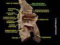
Muscles of the pharynx, viewed from behind, together with the associated vessels and nerves (middle pharyngeal constrictor muscle labeled as Mid. constr. at center)
Details
Origin Hyoid bone
Insertion Pharyngeal raphe
Artery Ascending pharyngeal artery
Nerve Pharyngeal plexus of vagus nerve
Actions Swallowing
Identifiers
Latin musculus constrictor pharyngis medius
TA98 A05.3.01.108
TA2 2184
FMA 46622
Anatomical terms of muscle

The middle pharyngeal constrictor is a fan-shaped muscle located in the neck. It is one of three pharyngeal constrictor muscles. It is smaller than the inferior pharyngeal constrictor muscle.

Contents

The middle pharyngeal constrictor originates from the greater cornu and lesser cornu of the hyoid bone, and the stylohyoid ligament. It inserts onto the pharyngeal raphe. It is innervated by a branch of the vagus nerve through the pharyngeal plexus. It acts to propel a bolus downwards along the pharynx towards the esophagus, facilitating swallowing.

Structure

The middle pharyngeal constrictor is a sheet-like, fan-shaped muscle. [1]

The muscle's fibers diverge from their origin: the more inferior fibres descend deep to the inferior pharyngeal constrictor muscle; the middle portion of fibres pass transversely; the more superior fibers ascend and overlap the superior pharyngeal constrictor muscle. [1]

Origin

Two parts of the middle pharyngeal constrictor muscle are distinguished according to its sites of origin:

Insertion

The muscle inserts (posteriorly) into the pharyngeal raphe, [1] blending with its contralateral partner at the midline.[ citation needed ]

Innervation

Similarly to the superior and inferior pharyngeal constrictor muscles, it is innervated by a branch of the vagus nerve through the pharyngeal plexus.[ citation needed ]

Actions/movements

The contraction of the muscle constricts the middle portion of the pharynx. [1]

Function

The muscle contracts during swallowing: [1] as soon as the bolus of food is received in the pharynx, the elevator muscles relax, the pharynx descends, and the constrictors contract upon the bolus, and convey it downward towards the esophagus. [2] [3]

They also have respiratory mechanical effects. [4]

Additional images

Related Research Articles

Swallowing, also called deglutition or inglutition in scientific contexts, is the process in the body of a human or other animal that allows for a substance to pass from the mouth, to the pharynx, and into the esophagus, while shutting the epiglottis. Swallowing is an important part of eating and drinking. If the process fails and the material goes through the trachea, then choking or pulmonary aspiration can occur. In the human body the automatic temporary closing of the epiglottis is controlled by the swallowing reflex.

<span class="mw-page-title-main">Facial nerve</span> Cranial nerve VII, for the face and tasting

The facial nerve, also known as the seventh cranial nerve, cranial nerve VII, or simply CN VII, is a cranial nerve that emerges from the pons of the brainstem, controls the muscles of facial expression, and functions in the conveyance of taste sensations from the anterior two-thirds of the tongue. The nerve typically travels from the pons through the facial canal in the temporal bone and exits the skull at the stylomastoid foramen. It arises from the brainstem from an area posterior to the cranial nerve VI and anterior to cranial nerve VIII.

<span class="mw-page-title-main">Hyoid bone</span> Bone situated in the neck between the chin and the thyroid cartilage

The hyoid bone is a horseshoe-shaped bone situated in the anterior midline of the neck between the chin and the thyroid cartilage. At rest, it lies between the base of the mandible and the third cervical vertebra.

<span class="mw-page-title-main">Sternothyroid muscle</span>

The sternothyroid muscle is an infrahyoid muscle of the neck. It acts to depress the hyoid bone.

<span class="mw-page-title-main">Thyrohyoid muscle</span> Neck muscle that depresses the hyoid bone and elevates the larynx

The thyrohyoid muscle is a small skeletal muscle of the neck. Above, it attaches onto the greater cornu of the hyoid bone; below, it attaches onto the oblique line of the thyroid cartilage. It is innervated by fibres derived from the cervical spinal nerve 1 that run with the hypoglossal nerve to reach this muscle. The thyrohyoid muscle depresses the hyoid bone and elevates the larynx during swallowing. By controlling the position and shape of the larynx, it aids in making sound.

<span class="mw-page-title-main">Hyoglossus</span> Muscle

The hyoglossus is a thin and quadrilateral extrinsic muscle of the tongue. It originates from the hyoid bone; it inserts onto the side of the tongue. It is innervated by the hypoglossal nerve. It acts to depress and retract the tongue.

<span class="mw-page-title-main">Styloglossus</span>

The styloglossus muscle is a bilaterally paired muscle of the tongue. It originates at the styloid process of the temporal bone. It inserts onto the side of the tongue. It acts to elevate and retract the tongue. It is innervated by the hypoglossal nerve.

<span class="mw-page-title-main">Levator veli palatini</span> Muscle of the soft palate

The levator veli palatini is a muscle of the soft palate and pharynx. It is innervated by the vagus nerve via its pharyngeal plexus. During swallowing, it contracts, elevating the soft palate to help prevent food from entering the nasopharynx.

<span class="mw-page-title-main">Salpingopharyngeus muscle</span> Muscle of the pharynx

The salpingopharyngeus muscle is a muscle of the pharynx. It arises from the lower part of the cartilage of the Eustachian tube, and inserts into the palatopharyngeus muscle by blending with its posterior fasciculus. It is innervated by vagus nerve via the pharyngeal plexus. It raises the pharynx and larynx during deglutition (swallowing) and laterally draws the pharyngeal walls up. It opens the pharyngeal orifice of the Eustachian tube during swallowing to allow for the equalization of pressure between it and the pharynx.

<span class="mw-page-title-main">Inferior pharyngeal constrictor muscle</span> Skeletal muscle of the pharynx

The inferior pharyngeal constrictor muscle is a skeletal muscle of the neck. It is the thickest of the three outer pharyngeal muscles. It arises from the sides of the cricoid cartilage and the thyroid cartilage. It is supplied by the vagus nerve. It is active during swallowing, and partially during breathing and speech. It may be affected by Zenker's diverticulum.

<span class="mw-page-title-main">Superior pharyngeal constrictor muscle</span> Muscle

The superior pharyngeal constrictor muscle is a quadrilateral muscle of the pharynx. It is the uppermost and thinnest of the three pharyngeal constrictors.

<span class="mw-page-title-main">Stylopharyngeus muscle</span> Muscle

The stylopharyngeus muscle is a muscle in the head. It originates from the temporal styloid process. Some of its fibres insert onto the thyroid cartilage, while others end by intermingling with proximal structures. It is innervated by the glossopharyngeal nerve. It acts to elevate the larynx and pharynx, and dilate the pharynx, thus facilitating swallowing.

<span class="mw-page-title-main">Pharyngeal arch</span> Embryonic precursor structures in vertebrates

The pharyngeal arches, also known as visceral arches, are structures seen in the embryonic development of vertebrates that are recognisable precursors for many structures. In fish, the arches are known as the branchial arches, or gill arches.

<span class="mw-page-title-main">Superior thyroid artery</span>

The superior thyroid artery arises from the external carotid artery just below the level of the greater cornu of the hyoid bone and ends in the thyroid gland.

<span class="mw-page-title-main">Superior laryngeal nerve</span> Branch of the vagus nerve

The superior laryngeal nerve is a branch of the vagus nerve. It arises from the middle of the inferior ganglion of vagus nerve and additionally also receives a sympathetic branch from the superior cervical ganglion.

<span class="mw-page-title-main">Pharyngeal muscles</span>

The pharyngeal muscles are a group of muscles that form the pharynx, which is posterior to the oral cavity, determining the shape of its lumen, and affecting its sound properties as the primary resonating cavity.

<span class="mw-page-title-main">Carotid triangle</span>

The carotid triangle is a portion of the anterior triangle of the neck.

<span class="mw-page-title-main">Pharyngeal plexus of vagus nerve</span> Nerve fibers innervating most of the palate and pharynx

The pharyngeal plexus is a nerve plexus located upon the outer surface of the pharynx. It contains a motor component, a sensory component, and sympathetic component.

The pharyngobasilar fascia is a fascia of the pharynx. It is situated between the mucous and muscular layers of the pharynx. It is formed as a thickening of the pharyngeal mucosa superior to the superior pharyngeal constrictor muscle. It attaches to the basilar part of occipital bone, the petrous part of the temporal bone, the medial pterygoid plate, and the pterygomandibular raphe. It diminishes in thickness inferiorly. Posteriorly, it is reinforced by the pharyngeal raphe. It reinforces the pharyngeal wall where muscle is deficient.

<span class="mw-page-title-main">Pharynx</span> Part of the throat that is behind the mouth and nasal cavity

The pharynx is the part of the throat behind the mouth and nasal cavity, and above the esophagus and trachea. It is found in vertebrates and invertebrates, though its structure varies across species. The pharynx carries food to the esophagus and air to the larynx. The flap of cartilage called the epiglottis stops food from entering the larynx.

References

PD-icon.svgThis article incorporates text in the public domain from page 1143 of the 20th edition of Gray's Anatomy (1918)

  1. 1 2 3 4 5 6 7 Standring, Susan (2020). Gray's Anatomy: The Anatomical Basis of Clinical Practice (42th ed.). New York. pp. 712–713. ISBN   978-0-7020-7707-4. OCLC   1201341621.{{cite book}}: CS1 maint: location missing publisher (link)
  2. Rowe LD, Miller AJ, Chierici G, Clendenning D (August 1984). "Adaptation in the function of pharyngeal constrictor muscles". Otolaryngology–Head and Neck Surgery . 92 (4): 392–401. doi:10.1177/019459988409200404. PMID   6435057. S2CID   32361287.
  3. Donner, Martin W.; Bosnia, James F.; Robertson, Diane L. (1985). "Anatomy and physiology of the pharynx". Gastrointestinal Radiology. 10 (1): 197–212. doi:10.1007/BF01893103. ISSN   0364-2356. PMID   4029536. S2CID   37515662.
  4. Kuna, Samuel T (2000). "Respiratory-related activation and mechanical effects of the pharyngeal constrictor muscles". Respiration Physiology. 119 (2–3): 155–161. doi:10.1016/S0034-5687(99)00110-3. ISSN   0034-5687. PMID   10722858.

Further reading