Sublingual space

Last updated
Sublingual space
Gray507.png
Sublingual space situated deep to mylohyoid muscle
Submandibular and sublingual space.jpg
Sublingual and submandibular space, coronal plane illustration and MRI
Anatomical terminology

The sublingual space is a fascial space of the head and neck (sometimes also termed fascial spaces or tissue spaces). It is a potential space [1] located below the mouth and above the mylohyoid muscle, and is part of the suprahyoid group of fascial spaces.

Contents

Location and structure

Anatomic boundaries

The sublingual space is V-shaped, with the apex pointing to the anterior. Its boundaries are: [2]

Communications

The sublingual space communicates posteriorly around the posterior free border of the mylohyoid muscle with the submandibular space. [2] Infections of the sublingual space may also erode through the mylohyoid, or spread via the lymphatics to the submandibular and submental spaces.
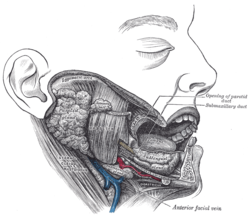
Contents

The sublingual space contains: [2]

Clinical relevance

Diagram of medial surface of the mandible, showing the slanting attachment of mylohyoid (the mylohyoid line). This arrangement means that the apices of posterior teeth are more likely to be below the level of mylohyoid, so a periapical abscess associated with posterior teeth is more likely to spread into the submandibular space. Gray177.png
Diagram of medial surface of the mandible, showing the slanting attachment of mylohyoid (the mylohyoid line). This arrangement means that the apices of posterior teeth are more likely to be below the level of mylohyoid, so a periapical abscess associated with posterior teeth is more likely to spread into the submandibular space.
Diagram showing sublingual gland in sublingual space. Gray1024.png
Diagram showing sublingual gland in sublingual space.

This space may be created by pathology, such as the spread of pus in an infection, e.g. odontogenic infections. A periapical abscess may spread into the sublingual space if the apex of the tooth is above the level of attachment of mylohyoid, and the infection erodes through the lingual cortical plate of the mandible.

Signs and symptoms of a sublingual space infection might include a firm, painful swelling in the anterior part of the floor of the mouth. A sublingual abscess may elevate the tongue and cause drooling or dysphagia (difficulty swallowing). There is usually little swelling visible on the face outside the mouth.

If the space contains pus, the usual treatment is by incision and drainage. The site of the incision is intra-oral, made lateral to sublingual plica. Incision of the plica itself can result in a ranula, or an incision placed medial to the plica can damage Wharton's duct, the sublingual artery and veins and the lingual nerve.

Pathology arising from the sublingual gland is rare, however, sublingual gland neoplasms are predominantly malignant and thus important to recognize.

Ludwig's angina is a serious infection involving the submandibular, sublingual and submental spaces bilaterally. [3] Ludwig's angina may extend into the pharyngeal and cervical spaces, and the swelling can compress the airway and cause dyspnoea (difficulty breathing). [3] Collectively, the submandibular, sublingual and submental spaces are sometimes termed the perimandibular spaces, or the submaxillary space. [4]

Related Research Articles

Submandibular gland

The paired submandibular glands are major salivary glands located beneath the floor of the mouth. They each weigh about 15 grams and contribute some 60–67% of unstimulated saliva secretion; on stimulation their contribution decreases in proportion as the parotid secretion rises to 50%.

Sublingual gland major salivary glands in the mouth

The paired sublingual glands are major salivary glands in the mouth. They are the smallest, most diffuse, and the only unencapsulated major salivary glands. They provide only 3-5% of the total salivary volume. There are also two other types of salivary glands; they are submandibular and Parotid glands.

Ludwigs angina human disease

Ludwig's angina is a type of severe cellulitis involving the floor of the mouth. Early on the floor of the mouth is raised and there is difficulty swallowing saliva, which may run from the person's mouth. As the condition worsens, the airway may be compromised with hardening of the spaces on both sides of the tongue. This condition has a rapid onset over hours.

Mylohyoid muscle paired muscle

The mylohyoid muscle is a paired muscle running from the mandible to the hyoid bone, forming the floor of the oral cavity of the mouth. It is named after its two attachments near the molar teeth. These muscles are mesodermal in embryologic origin. The mylohyoid muscle is derived from the first pharyngeal arch.

Hyoglossus

The hyoglossus, thin and quadrilateral, arises from the side of the body and from the whole length of the greater cornu of the hyoid bone, and passes almost vertically upward to enter the side of the tongue, between the styloglossus and the inferior longitudinal muscle of the tongue. It forms a part of the floor of submandibular triangle.

Ranula human disease

A ranula is a mucus extravasation cyst involving a sublingual gland and is a type of mucocele found on the floor of the mouth. Ranulae present as a swelling of connective tissue consisting of collected mucin from a ruptured salivary gland caused by local trauma. If small and asymptomatic further treatment may not be needed, otherwise minor oral surgery may be indicated.

Lingual artery

The lingual artery arises from the external carotid between the superior thyroid artery and facial artery. It can be located easily in the tongue.

Submandibular duct

The submandibular duct or Wharton duct or submaxillary duct, is one of the salivary excretory ducts. It is about 5 cm. long, and its wall is much thinner than that of the parotid duct. It drains saliva from each bilateral submandibular gland and sublingual gland to the sublingual caruncle at the base of the tongue.

Submandibular ganglion

The submandibular ganglion is part of the human autonomic nervous system. It is one of four parasympathetic ganglia of the head and neck..

Sialolithiasis condition where a calcified mass or sialolith forms within a salivary gland, usually in the duct of the submandibular gland

Sialolithiasis, is a condition where a calcified mass or sialolith forms within a salivary gland, usually in the duct of the submandibular gland. Less commonly the parotid gland or rarely the sublingual gland or a minor salivary gland may develop salivary stones.

Buccal space fascial space of the head and neck

The buccal space is a fascial space of the head and neck. It is a potential space in the cheek, and is paired on each side. The buccal space is superficial to the buccinator muscle and deep to the platysma muscle and the skin. The buccal space is part of the subcutaneous space, which is continuous from head to toe.

Mouth infections, also known as oral infections, are a group of infections that occur around the oral cavity. They include dental infection, dental abscess, and Ludwig's angina. Mouth infections typically originate from dental caries at the root of molars and premolars that spread to adjacent structures. In otherwise healthy patients, removing the offending tooth to allow drainage will usually resolve the infection. In cases that spread to adjacent structures or in immunocompromised patients, surgical drainage and systemic antibiotics may be required in addition to tooth extraction. Since bacteria that normally reside in the oral cavity cause mouth infections, proper dental hygiene can prevent most cases of infection. As such, mouth infections are more common in populations with poor access to dental care or populations with health-related behaviors that damage one's teeth and oral mucosa. This is a common problem, representing nearly 36% of all encounters within the emergency department related to dental conditions.

Submandibular space fascial space of the head and neck

The submandibular space is a fascial space of the head and neck. It is a potential space, and is paired on either side, located on the superficial surface of the mylohyoid muscle between the anterior and posterior bellies of the digastric muscle. The space corresponds to the anatomic region termed the submandibular triangle, part of the anterior triangle of the neck.

Submental space fascial space of the head and neck

The submental space is a fascial space of the head and neck. It is a potential space located between the mylohyoid muscle superiorly, the platysma muscle inferiorly, under the chin in the midline. The space coincides with the anatomic region termed the submental triangle, part of the anterior triangle of the neck.

Fascial spaces are potential spaces that exist between the fasciae and underlying organs and other tissues. In health, these spaces do not exist; they are only created by pathology, e.g. the spread of pus or cellulitis in an infection. The fascial spaces can also be opened during the dissection of a cadaver. The fascial spaces are different from the fasciae themselves, which are bands of connective tissue that surround structures, e.g. muscles. The opening of fascial spaces may be facilitated by pathogenic bacterial release of enzymes which cause tissue lysis. The spaces filled with loose areolar connective tissue may also be termed clefts. Other contents such as salivary glands, blood vessels, nerves and lymph nodes are dependent upon the location of the space. Those containing neurovascular tissue may also be termed compartments.

Submasseteric space fascial space of the head and neck

The submasseterric space is a fascial space of the head and neck. It is a potential space in the face over the angle of the jaw, and is paired on each side. It is located between the lateral aspect of the mandible and the medial aspect of the masseter muscle and its investing fascia. The term is derived from sub- meaning "under" in Latin and masseteric which refers to the masseter muscle. The submasseteric space is one of the four compartments of the masticator space. Sometimes the submasseteric space is described as a series of spaces, created because the masseter muscle has multiple insertions that cover most of the lateral surface of the ramus of the mandible.

Pterygomandibular space fascial space of the head and neck

The pterygomandibular space is a fascial space of the head and neck. It is a potential space in the head and is paired on each side. It is located between the medial pterygoid muscle and the medial surface of the ramus of the mandible. The pterygomandibular space is one of the four compartments of the masticator space.

Canine space

The canine space, is a fascial space of the head and neck. It is a thin potential space on the face, and is paired on either side. It is located between the levator anguli oris muscle inferiorly and the levator labii superioris muscle superiorly. The term is derived from the fact that the space is in the region of the canine fossa, and that infections originating from the maxillary canine tooth may spread to involve the space. Infra-orbital is derived from infra- meaning below and orbit which refers to the eye socket.

Infratemporal space

The Infratemporal space is a fascial space of the head and neck. It is a potential space in the side of the head, and is paired on either side. It is located posterior to the maxilla, between the lateral pterygoid plate of the sphenoid bone medially and by the base of skull superiorly. The term is derived from infra- meaning below and temporal which refers to the temporalis muscle.

Salivary gland disease Human disease

Salivary gland diseases (SGD) are multiple and varied in cause.

References

  1. "Sublingual space". Medcyclopaedia. GE.
  2. 1 2 3 Neil Norton; et al. (2007). Netter's head and neck anatomy for dentistry. Philadelphia, Pa.: Saunders Elsevier. p. 466. ISBN   9781929007882.
  3. 1 2 Kenneth M. Hargreaves; Stephen Cohen, eds. (2010). Cohen's pathways of the pulp (10th ed.). St. Louis, Mo.: Mosby Elsevier. pp. 590, 592. ISBN   978-0323064897.
  4. Hupp JR, Ellis E, Tucker MR (2008). Contemporary oral and maxillofacial surgery (5th ed.). St. Louis, Mo.: Mosby Elsevier. pp. 317–333. ISBN   9780323049030.