The greater occipital nerve is a spinal nerve, specifically the medial branch of the dorsal primary ramus of cervical spinal nerve 2. This nerve arises from between the first and second cervical vertebrae, along with the lesser occipital nerve. It ascends after emerging from below the suboccipital triangle beneath the obliquus capitis inferior muscle. It then passes through the semispinalis muscle before ascending to innervate the skin along the posterior part of the scalp to the vertex. It innervates the scalp at the top of the head, over the ear and over the parotid glands.

The scalp is the anatomical area bordered by the human face at the front, and by the neck at the sides and back.

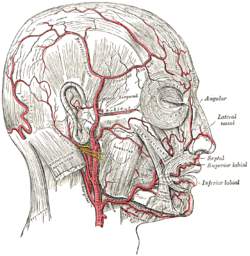
The middle meningeal artery is typically the third branch of the first portion of the maxillary artery, one of the two terminal branches of the external carotid artery. After branching off the maxillary artery in the infratemporal fossa, it runs through the foramen spinosum to supply the dura mater the outer meningeal layer, and the calvaria. The middle meningeal artery is the largest of the three (paired) arteries that supply the meninges, the others being the anterior meningeal artery and the posterior meningeal artery.

The occipitalis muscle is a muscle which covers parts of the skull. Some sources consider the occipital muscle to be a distinct muscle. However, Terminologia Anatomica currently classifies it as part of the occipitofrontalis muscle along with the frontalis muscle.

The epicranial aponeurosis is an aponeurosis which covers the upper part of the cranium in humans and various other animals. In humans, it is attached in the interval between its union with the occipitofrontalis muscle, to the external occipital protuberance and highest nuchal lines of the occipital bone; in front, it forms a short and narrow prolongation between its union with the frontalis muscle or frontal part of the occipitofrontalis muscle.

In human anatomy, the superficial temporal artery is a major artery of the head. It arises from the external carotid artery when it splits into the superficial temporal artery and maxillary artery.

The occipitofrontalis muscle is a muscle which covers parts of the skull. It consists of two parts or bellies: The occipital belly, near the occipital bone, and the frontal belly, near the frontal bone. In humans, the occipitofrontalis only serves for facial expressions.

The occipital vein begins as a plexus at the posterior aspect of the scalp from the external occipital protuberance and superior nuchal line to the back part of the vertex of the skull.

The mastoid foramen is a hole in the posterior border of the temporal bone. It transmits a Mastoid emissary vein to the sigmoid sinus and a small branch of the occipital artery, the posterior meningeal artery to the dura mater.
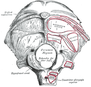
The condylar canal is a canal in the condyloid fossa of the lateral parts of occipital bone behind the occipital condyle. Resection of the rectus capitis posterior major and minor muscles reveals the bony recess leading to the condylar canal, which is situated posterior and lateral to the occipital condyle. It is immediately superior to the extradural vertebral artery, which makes a loop above the posterior C1 ring to enter the foramen magnum. The anteriomedial wall of the condylar canal thickens to join the foramen magnum rim and connect to the occipital condyle.

The posterior auricular nerve arises from the facial nerve close to the stylomastoid foramen and runs upward in front of the mastoid process; here it is joined by a filament from the auricular branch of the vagus and communicates with the posterior branch of the great auricular as well as with the lesser occipital.

The supraorbital artery is an artery of the head.

The carotid triangle is a portion of the anterior triangle of the neck.

The occipital lymph nodes, one to three in number, are located on the back of the head close to the margin of the trapezius and resting on the insertion of the semispinalis capitis.
The posterior branches of cervical nerves branch from the dorsal rami of the cervical nerves.

The two sternocleidomastoid branches of the occipital artery arise directly from the occipital artery and are the initial two branches of this artery. Uncommonly, the lower sternocleidomastoid branch can branch directly from the external carotid.

The descending branch of occipital artery, the largest branch of the occipital, descends on the back of the neck, and divides into a superficial and deep portion.
The auricular branch of occipital artery supplies the back of the concha and frequently gives off a branch, which enters the skull through the mastoid foramen and supplies the dura mater, the diploë, and the mastoid cells; this latter branch sometimes arises from the occipital artery, and is then known as the mastoid branch.
Scalp reconstruction is a surgical procedure for people with scalp defects. Scalp defects may be partial or full thickness and can be congenital or acquired. Because not all layers of the scalp are elastic and the scalp has a convex shape, the use of primary closure is limited. Sometimes the easiest way of closing the wound may not be the ideal or best way. The choice for a reconstruction depends on multiple factors, such as the defect itself, the patient characteristics and surgeon preference.
The public domain consists of all the creative works to which no exclusive intellectual property rights apply. Those rights may have expired, been forfeited, expressly waived, or may be inapplicable.

Gray's Anatomy is an English language textbook of human anatomy originally written by Henry Gray and illustrated by Henry Vandyke Carter. Earlier editions were called Anatomy: Descriptive and Surgical, Anatomy of the Human Body and Gray's Anatomy: Descriptive and Applied, but the book's name is commonly shortened to, and later editions are titled, Gray's Anatomy. The book is widely regarded as an extremely influential work on the subject, and has continued to be revised and republished from its initial publication in 1858 to the present day. The latest edition of the book, the 41st, was published in September 2015.