Related Research Articles

In anatomy, the palatine bones are two irregular bones of the facial skeleton in many animal species, located above the uvula in the throat. Together with the maxilla, they comprise the hard palate.

The nasal cavity is a large, air-filled space above and behind the nose in the middle of the face. The nasal septum divides the cavity into two cavities, also known as fossae. Each cavity is the continuation of one of the two nostrils. The nasal cavity is the uppermost part of the respiratory system and provides the nasal passage for inhaled air from the nostrils to the nasopharynx and rest of the respiratory tract.

The pterygopalatine ganglion is a parasympathetic ganglion in the pterygopalatine fossa. It is one of four parasympathetic ganglia of the head and neck,.
The greater petrosal nerve is a nerve of the head mainly containing pre-ganglionic parasympathetic fibres which ultimately synapse in the pterygopalatine ganglion. It branches from the facial nerve and is derived from the parasympathetic part of the nervus intermedius component of CN VII, with its cell bodies located in the superior salivary nucleus. In the connective tissue substance of the foramen lacerum, the greater petrosal nerve unites with the (sympathetic) deep petrosal nerve to form the nerve of the pterygoid canal which proceeds to the pterygopalatine ganglion.
In neuroanatomy, the maxillary nerve (V2) is one of the three branches or divisions of the trigeminal nerve, the fifth (CN V) cranial nerve. It comprises the principal functions of sensation from the maxilla, nasal cavity, sinuses, the palate and subsequently that of the mid-face, and is intermediate, both in position and size, between the ophthalmic nerve and the mandibular nerve.

In human anatomy, the pterygopalatine fossa is a fossa in the skull. A human skull contains two pterygopalatine fossae—one on the left side, and another on the right side. Each fossa is a cone-shaped paired depression deep to the infratemporal fossa and posterior to the maxilla on each side of the skull, located between the pterygoid process and the maxillary tuberosity close to the apex of the orbit. It is the indented area medial to the pterygomaxillary fissure leading into the sphenopalatine foramen. It communicates with the nasal and oral cavities, infratemporal fossa, orbit, pharynx, and middle cranial fossa through eight foramina.
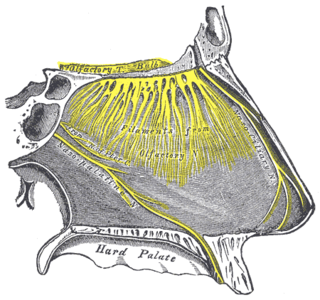
The nasopalatine nerve (also long sphenopalatine nerve) is a nerve of the head. It is a sensory branch of the maxillary nerve (CN V2) that passes through the pterygopalatine ganglion (without synapsing) and then through the sphenopalatine foramen to enter the nasal cavity, and finally out of the nasal cavity through the incisive canal and then the incisive fossa to enter the hard palate. It provides sensory innervation to the posteroinferior part of the nasal septum, and gingiva just posterior to the upper incisor teeth.

The maxillary artery supplies deep structures of the face. It branches from the external carotid artery just deep to the neck of the mandible.

The descending palatine artery is a branch of the third part of the maxillary artery supplying the hard and soft palate.
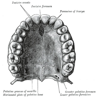
The incisive canals are two bony canals of the anterior hard palate connecting the nasal cavity and the oral cavity. An incisive canal courses through each maxilla. Below, the two incisive canals typically converge medially.
The infraorbital artery is a small artery in the head that arises from the maxillary artery and passes through the inferior orbital fissure to enter the orbit, then passes forward along the floor of the orbit, finally exiting the orbit through the infraorbital foramen to reach the face.

The nerve of the pterygoid canal is formed by the union of the (parasympathetic) greater petrosal nerve and (sympathetic) deep petrosal nerve within the cartilaginous substance filling the foramen lacerum. From the foramen lacerum, the nerve of the pterygoid canal passes through the pterygoid canal to reach the pterygopalatine fossa, ending at the pterygopalatine ganglion.

The palatovaginal canal is a small canal formed between the sphenoidal process of palatine bone, and vaginal process of sphenoid bone. It connects the pterygopalatine fossa and and nasal cavity. It transmits the pharyngeal nerve, and the pharyngeal branch of maxillary artery.

The perpendicular plate of palatine bone is the vertical part of the palatine bone, and is thin, of an oblong form, and presents two surfaces and four borders.

The infraorbital nerve is a branch of the maxillary nerve. It arises in the pterygopalatine fossa. It passes through the inferior orbital fissure to enter the orbit. It travels through the orbit, then enters and traverses the infraorbital canal, exiting the canal at the infraorbital foramen to reach the face. It provides sensory innervation to the skin and mucous membranes around the middle of the face.

In human anatomy of the mouth, the palatine process of maxilla, is a thick, horizontal process of the maxilla. It forms the anterior three quarters of the hard palate, the horizontal plate of the palatine bone making up the rest. It is the most important bone in the midface. It provides structural support for the viscerocranium.

The greater palatine nerve is a branch of the pterygopalatine ganglion. This nerve is also referred to as the anterior palatine nerve, due to its location anterior to the lesser palatine nerve. It carries both general sensory fibres from the maxillary nerve, and parasympathetic fibers from the nerve of the pterygoid canal. It may be anaesthetised for procedures of the mouth and maxillary (upper) teeth.

The greater palatine canal is a passage in the skull that transmits the descending palatine artery, vein, and greater and lesser palatine nerves between the pterygopalatine fossa and the oral cavity.
The lesser palatine arteries are arteries of the head. It is a branch of the descending palatine artery. They supply the palatine tonsils and the soft palate.

The human nose is the first organ of the respiratory system. It is also the principal organ in the olfactory system. The shape of the nose is determined by the nasal bones and the nasal cartilages, including the nasal septum, which separates the nostrils and divides the nasal cavity into two.
References
- 1 2 Elden, Lisa M.; Wetmore, Ralph F.; Potsic, William P. (2012). "Otolaryngologic Disorders". Pediatric Surgery. pp. 707–728. doi:10.1016/B978-0-323-07255-7.00055-6. hdl:1805/23155. ISBN 9780323072557.