Transient synovitis

Last updated
Transient synovitis
Other namesTransitory coxitis, Coxitis fugax, Acute transient epiphysitis, Coxitis serosa seu simplex, Phantom hip disease, Observation hip. [1]
Hip.jpg
The hip joint is formed between the femur and acetabulum of the pelvis.
Specialty Rheumatology   OOjs UI icon edit-ltr-progressive.svg

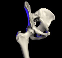
Transient synovitis of hip (also called toxic synovitis; see below for more synonyms) is a self-limiting condition in which there is an inflammation of the inner lining (the synovium) of the capsule of the hip joint. The term irritable hip refers to the syndrome of acute hip pain, joint stiffness, limp or non-weightbearing, indicative of an underlying condition such as transient synovitis or orthopedic infections (like septic arthritis or osteomyelitis). [2] In everyday clinical practice however, irritable hip is commonly used as a synonym for transient synovitis. It should not be confused with sciatica, a condition describing hip and lower back pain much more common to adults than transient synovitis but with similar signs and symptoms.

Contents

Transient synovitis usually affects children between three and ten years old (but it has been reported in a 3-month-old infant and in some adults [3] ). It is the most common cause of sudden hip pain and limp in young children. [4] [5] Boys are affected two to four times as often as girls. [5] [6] [7] The exact cause is unknown. A recent viral infection (most commonly an upper respiratory tract infection) or a trauma have been postulated as precipitating events, although these are reported only in 30% and 5% of cases, respectively. [7]

Transient synovitis is a diagnosis of exclusion. [4] The diagnosis can be made in the typical setting of pain or limp in a young child who is not generally unwell and has no recent trauma. There is a limited range of motion of the hip joint. Nevertheless, children with transient synovitis of the hip can usually weight bear. This is an important clinical differentiating sign from septic arthritis. [8] Blood tests may show mild inflammation. An ultrasound scan of the hip joint can show a fluid collection (effusion). Treatment is with nonsteroidal anti-inflammatory drugs and limited weight-bearing. The condition usually clears by itself within seven to ten days, [5] but a small group of patients will continue to have symptoms for several weeks. The recurrence rate is 4–17%, most of which is in the first six months. [9]

Symptoms and signs

Transient synovitis causes pain in the hip, thigh, groin or knee on the affected side. [5] However, children with transient synovitis of the hip can usually weight bear with varying degrees of limping. There may be a limp (or abnormal crawling in infants) with or without pain. In small infants, the presenting complaint can be unexplained crying (for example, when changing a diaper). The condition is nearly always limited to one side. [5] The pain and limp can range from mild to severe.[ citation needed ]

Some children may have a slightly raised temperature; high fever and general malaise point to other, more serious conditions. On clinical examination, the child typically holds the hip slightly bent, turned outwards and away from the middle line (flexion, external rotation and abduction). [7] Active and passive movements may be limited because of pain, especially abduction and internal rotation. The hip can be tender to palpation. The log roll test involves gently rotating the entire lower limb inwards and outwards with the patient on his back, to check when muscle guarding occurs. The unaffected hip and the knees, ankles, feet and spine are found to be normal. [9]

Complications

In the past, there have been speculations about possible complications after transient synovitis. The current consensus however is that there is no proof of an increased risk of complications after transient synovitis. [10]

One such previously suspected complication was coxa magna, which is an overgrowth of the femoral head and broadening of the femoral neck, accompanied by changes in the acetabulum, which may lead to subluxation of the femur. [9] [11] There was also some controversy about whether continuous high intra-articular pressure in transient synovitis could cause avascular necrosis of the femoral head (Legg-Calvé-Perthes disease), but further studies did not confirm any link between the two conditions. [12]

Diagnosis

There are no set standards for the diagnosis of suspected transient synovitis, so the amount of investigations will depend on the need to exclude other, more serious diseases. [8] It is of great importance to exclude the diagnosis of septic arthritis. This is because if septic arthritis is missed in children, grave complications can occur. The exclusion of septic arthritis is mainly built upon the physician's clinical expertise and is supplemented by basic laboratory test and relevant imaging modalities. [8] Additionally, beware to exclude the diagnosis of acute osteomyelitis, because it not uncommonly cooccurs with septic arthritis of the hip in children. [8]

Inflammatory parameters in the blood may be slightly raised (these include erythrocyte sedimentation rate, C-reactive protein and white blood cell count), but raised inflammatory markers are strong predictors of other more serious conditions such as septic arthritis. [13] [14]

X-ray imaging of the hip is most often unremarkable. Subtle radiographic signs include an accentuated pericapsular shadow, widening of the medial joint space, lateral displacement of the femoral epiphyses with surface flattening (Waldenström sign), prominent obturator shadow, diminution of soft tissue planes around the hip joint or slight demineralisation of the proximal femur. The main reason for radiographic examination is to exclude bony lesions such as occult fractures, slipped upper femoral epiphysis or bone tumours (such as osteoid osteoma). An anteroposterior and frog lateral (Lauenstein) view of the pelvis and both hips is advisable. [15]

An ultrasound scan of the hip can easily demonstrate fluid inside the joint capsule (Fabella sign), although this is not always present in transient synovitis. [7] [16] However, it cannot reliably distinguish between septic arthritis and transient synovitis. [17] [18] If septic arthritis needs to be ruled out, needle aspiration of the fluid can be performed under ultrasound guidance. [19] In transient synovitis, the joint fluid will be clear. [5] In septic arthritis, there will be pus in the joint, which can be sent for bacterial culture and antibiotic sensitivity testing.

More advanced imaging techniques can be used if the clinical picture is unclear; the exact role of different imaging modalities remains uncertain. Some studies have demonstrated findings on magnetic resonance imaging (MRI scan) that can differentiate between septic arthritis and transient synovitis (for example, signal intensity of adjacent bone marrow). [20] [21] [22] Skeletal scintigraphy can be entirely normal in transient synovitis, and scintigraphic findings do not distinguish transient synovitis from other joint conditions in children. [23] CT scanning does not appear helpful.

Differential diagnosis

Pain in or around the hip and/or limp in children can be due to a large number of conditions. Septic arthritis (a bacterial infection of the joint) is the most important differential diagnosis, because it can quickly cause irreversible damage to the hip joint. [8] [4] Fever, raised inflammatory markers on blood tests and severe symptoms (inability to bear weight, pronounced muscle guarding) all point to septic arthritis, [13] [14] but a high index of suspicion remains necessary even if these are not present. [5] [8] Osteomyelitis (infection of the bone tissue) can also cause pain and limp. [8]

Bone fractures, such as a toddler's fracture (spiral fracture of the shin bone), can also cause pain and limp, but are uncommon around the hip joint. Soft tissue injuries can be evident when bruises are present. Muscle or ligament injuries can be contracted during heavy physical activity —however, it is important not to miss a slipped upper femoral epiphysis. Avascular necrosis of the femoral head (Legg-Calvé-Perthes disease) typically occurs in children aged 4–8, and is also more common in boys. There may be an effusion on ultrasound, similar to transient synovitis. [24]

Neurological conditions can also present with a limp. If developmental dysplasia of the hip is missed early in life, it can come to attention later in this way. Pain in the groin can also be caused by diseases of the organs in the abdomen (such as a psoas abscess) or by testicular disease. Rarely, there is an underlying rheumatic condition (juvenile idiopathic arthritis, Lyme arthritis, gonococcal arthritis, ...) or bone tumour.[ citation needed ]

Treatment

Treatment consists of rest, non-weightbearing and painkillers when needed. A small study showed that the nonsteroidal anti-inflammatory drug ibuprofen could shorten the disease course (from 4.5 to 2 days) and provide pain control with minimal side effects (mainly gastrointestinal disturbances). [25] If fever occurs or the symptoms persist, other diagnoses need to be considered. [9]

Related Research Articles

Arthritis Type of joint disorder

Arthritis is a term often used to mean any disorder that affects joints. Symptoms generally include joint pain and stiffness. Other symptoms may include redness, warmth, swelling, and decreased range of motion of the affected joints. In some types of arthritis, other organs are also affected. Onset can be gradual or sudden.

Lemierres syndrome Medical condition

Lemierre's syndrome refers to infectious thrombophlebitis of the internal jugular vein. It most often develops as a complication of a bacterial sore throat infection in young, otherwise healthy adults. The thrombophlebitis is a serious condition and may lead to further systemic complications such as bacteria in the blood or septic emboli.

Tenosynovitis Medical condition

Tenosynovitis is the inflammation of the fluid-filled sheath that surrounds a tendon, typically leading to joint pain, swelling, and stiffness. Tenosynovitis can be either infectious or noninfectious. Common clinical manifestations of noninfectious tenosynovitis include de Quervain tendinopathy and stenosing tenosynovitis

Legg–Calvé–Perthes disease Osteochondrosis that results in death and fracture located in hip joint

Legg–Calvé–Perthes disease (LCPD), is a childhood hip disorder initiated by a disruption of blood flow to the head of the femur. Due to the lack of blood flow, the bone dies and stops growing. Over time, healing occurs by new blood vessels infiltrating the dead bone and removing the necrotic bone which leads to a loss of bone mass and a weakening of the femoral head.

Septic arthritis Medical condition

Acute septic arthritis, infectious arthritis, suppurative arthritis, osteomyelitis, or joint infection is the invasion of a joint by an infectious agent resulting in joint inflammation. Generally speaking, symptoms typically include redness, heat and pain in a single joint associated with a decreased ability to move the joint. Onset is usually rapid. Other symptoms may include fever, weakness and headache. Occasionally, more than one joint may be involved especially in neonates and younger children. In neonates, infants i.e. during the first year of life and toddlers, the signs and symptoms of septic arthritis can be deceptive and mimic other infectious and non-infectious disorders.

Juvenile idiopathic arthritis (JIA), is the most common, chronic rheumatic disease of childhood, affecting approximately one per 1,000 children. Juvenile, in this context, refers to disease onset before 16 years of age, while idiopathic refers to a condition with no defined cause, and arthritis is inflammation within the joint.

Limp Type of asymmetric abnormality of the gait

A limp is a type of asymmetric abnormality of the gait. Limping may be caused by pain, weakness, neuromuscular imbalance, or a skeletal deformity. The most common underlying cause of a painful limp is physical trauma; however, in the absence of trauma, other serious causes, such as septic arthritis or slipped capital femoral epiphysis, may be present. The diagnostic approach involves ruling out potentially serious causes via the use of X-rays, blood tests, and sometimes joint aspiration. Initial treatment involves pain management. A limp is the presenting problem in about 4% of children who visit hospital emergency departments.

Avascular necrosis Death of bone tissue due to interruption of the blood supply

Avascular necrosis (AVN), also called osteonecrosis or bone infarction, is death of bone tissue due to interruption of the blood supply. Early on, there may be no symptoms. Gradually joint pain may develop which may limit the ability to move. Complications may include collapse of the bone or nearby joint surface.

Slipped capital femoral epiphysis Medical condition

Slipped capital femoral epiphysis is a medical term referring to a fracture through the growth plate (physis), which results in slippage of the overlying end of the femur (metaphysis).

Arthropathy Medical condition

An arthropathy is a disease of a joint.

Synovitis is the medical term for inflammation of the synovial membrane. This membrane lines joints that possess cavities, known as synovial joints. The condition is usually painful, particularly when the joint is moved. The joint usually swells due to synovial fluid collection.

Protrusio acetabuli is an uncommon defect of the acetabulum. The acetabulum is the socket that receives the femoral head to make the hip joint. The hip bone of the pelvic bone/girdle is composed of three bones, the ilium, the ischium and the pubis. In protrusio deformity, there is medial displacement of the femoral head in that the medial aspect of the femoral cortex is medial to the ilioischial line. The socket is too deep and may protrude into the pelvis.

Lameness is an abnormal gait or stance of an animal that is the result of dysfunction of the locomotor system. In the horse, it is most commonly caused by pain, but can be due to neurologic or mechanical dysfunction. Lameness is a common veterinary problem in racehorses, sport horses, and pleasure horses. It is one of the most costly health problems for the equine industry, both monetarily for the cost of diagnosis and treatment, and for the cost of time off resulting in loss-of-use.

Hip dysplasia Joint abnormality

Hip dysplasia is an abnormality of the hip joint where the socket portion does not fully cover the ball portion, resulting in an increased risk for joint dislocation. Hip dysplasia may occur at birth or develop in early life. Regardless, it does not typically produce symptoms in babies less than a year old. Occasionally one leg may be shorter than the other. The left hip is more often affected than the right. Complications without treatment can include arthritis, limping, and low back pain.

<i>Kingella kingae</i> Species of bacterium

Kingella kingae is a species of Gram-negative facultative anaerobic β-hemolytic coccobacilli. First isolated in 1960 by Elizabeth O. King, it was not recognized as a significant cause of infection in young children until the 1990s, when culture techniques had improved enough for it to be recognized. It is best known as a cause of septic arthritis, osteomyelitis, spondylodiscitis, bacteraemia, and endocarditis, and less frequently lower respiratory tract infections and meningitis.

Knee pain Medical condition

Knee pain is pain in or around the knee.

Joint effusion Medical condition

A joint effusion is the presence of increased intra-articular fluid. It may affect any joint. Commonly it involves the knee.

The Kocher criteria are a tool useful in the differentiation of septic arthritis from transient synovitis in the child with a painful hip. They are named for Mininder S. Kocher, an orthopaedic surgeon at Boston Children's Hospital and Professor of Orthopaedic Surgery at Harvard Medical School.

Hip pain

Pain in the hip is the experience of pain in the muscles or joints in the hip/ pelvic region, a condition commonly arising from any of a number of factors. Sometimes it is closely associated with lower back pain.

Ultrasound-guided hip joint injection

Ultrasound-guided hip joint injection is a joint injection in the hip, assisted by medical ultrasound. Hip and groin pain often presents a diagnostic and therapeutic challenge. The differential diagnosis is extensive, comprising intra-articular and extra-articular pathology and referred pain from lumbar spine, knee and elsewhere in the pelvis. Various ultrasound-guided techniques have been described in the hip and groin region for diagnostic and therapeutic purposes. Ultrasound has many advantages over other imaging modalities, including portability, lack of ionizing radiation and real-time visualization of soft tissues and neurovascular structures. Many studies have demonstrated the safety, accuracy and efficacy of ultrasound-guided techniques, although there is lack of standardization regarding the injectates used and long-term benefit remains uncertain.

References

  1. Do TT (Feb 2000). "Transient synovitis as a cause of painful limps in children". Current Opinion in Pediatrics. 12 (1): 48–51. doi:10.1097/00008480-200002000-00010. PMID   10676774.
  2. Fischer SU, Beattie TF (Nov 1999). "The limping child: epidemiology, assessment and outcome". J Bone Joint Surg Br. 81 (6): 1029–34. doi:10.1302/0301-620X.81B6.9607. PMID   10615981.
  3. Quintos-Macasa AM, Serebro L, Menon Y (Feb 2006). "Transient synovitis of the hip in an adult". South Med J. 99 (2): 184–5. doi:10.1097/01.smj.0000199746.29009.4c. PMID   16509560. S2CID   26520520.
  4. 1 2 3 Hart JJ (Oct 1996). "Transient synovitis of the hip in children". Am Fam Physician. 54 (5): 1587–91, 1595–6. PMID   8857781.
  5. 1 2 3 4 5 6 7 Scott Moses, MD. "Transient hip tenosynovitis Archived 2007-09-16 at the Wayback Machine ". Family practice notebook. Revision of August 9, 2007. Retrieved December 22, 2007.
  6. Vijlbrief AS, Bruijnzeels MA, van der Wouden JC, van Suijlekom-Smit LW (Oct 1992). "Incidence and management of transient synovitis of the hip: a study in Dutch general practice". The British Journal of General Practice. 42 (363): 426–8. PMC   1372234 . PMID   1466922.
  7. 1 2 3 4 Irritable hip. General Practice Notebook. Retrieved December 22, 2007.
  8. 1 2 3 4 5 6 7 El-Sobky, T; Mahmoud, S (July 2021). "Acute osteoarticular infections in children are frequently forgotten multidiscipline emergencies: beyond the technical skills". EFORT open reviews. 6 (7): 584–592. doi:10.1302/2058-5241.6.200155. PMC   8335954 . PMID   34377550.
  9. 1 2 3 4 ped/1676 at eMedicine
  10. Mattick A, Turner A, Ferguson J, Beattie T, Sharp J (Sep 1999). "Seven year follow up of children presenting to the accident and emergency department with irritable hip". J Accid Emerg Med. 16 (5): 345–7. doi:10.1136/emj.16.5.345. PMC   1347055 . PMID   10505915.
  11. Sharwood PF (Dec 1981). "The irritable hip syndrome in children. A long-term follow-up". Acta Orthop Scand. 52 (6): 633–8. doi:10.3109/17453678108992159. PMID   7331801.
  12. Kallio P, Ryöppy S, Kunnamo I (Nov 1986). "Transient synovitis and Perthes' disease. Is there an aetiological connection?". J Bone Joint Surg Br. 68 (5): 808–11. doi: 10.1302/0301-620X.68B5.3782251 . PMID   3782251.
  13. 1 2 Caird MS, Flynn JM, Leung YL, Millman JE, D'Italia JG, Dormans JP (Jun 2006). "Factors distinguishing septic arthritis from transient synovitis of the hip in children. A prospective study". J Bone Joint Surg Am. 88 (6): 1251–7. doi:10.2106/JBJS.E.00216. PMID   16757758. S2CID   29137759.
  14. 1 2 Kocher MS, Mandiga R, Zurakowski D, Barnewolt C, Kasser JR (Aug 2004). "Validation of a clinical prediction rule for the differentiation between septic arthritis and transient synovitis of the hip in children". J Bone Joint Surg Am. 86-A (8): 1629–35. doi:10.2106/00004623-200408000-00005. PMID   15292409. S2CID   13529642.
  15. Gough-Palmer A, McHugh K (Jun 2007). "Investigating hip pain in a well child". BMJ. 334 (7605): 1216–7. doi:10.1136/bmj.39188.515741.47. PMC   1892599 . PMID   17556478.
  16. Nicola Wright, Vince Choudhery. Ultrasound is better than x-ray at detecting hip effusions in the limping child. BestBETs.org . Retrieved December 22, 2007
  17. Zamzam MM (Nov 2006). "The role of ultrasound in differentiating septic arthritis from transient synovitis of the hip in children". J Pediatr Orthop B. 15 (6): 418–22. doi:10.1097/01.bpb.0000228388.32184.7f. PMID   17001248. S2CID   27006647.
  18. Bienvenu-Perrard M, de Suremain N, Wicart P, et al. (Mar 2007). "[Benefit of hip ultrasound in management of the limping child]" [Benefit of hip ultrasound in management of the limping child]. J Radiol (in French). 88 (3 Pt 1): 377–83. doi:10.1016/S0221-0363(07)89834-9. PMID   17457269.
  19. Skinner J, Glancy S, Beattie TF, Hendry GM (Mar 2002). "Transient synovitis: is there a need to aspirate hip joint effusions?". Eur J Emerg Med. 9 (1): 15–8. doi:10.1097/00063110-200203000-00005. PMID   11989490. S2CID   29742427.
  20. Kwack KS, Cho JH, Lee JH, Cho JH, Oh KK, Kim SY (Aug 2007). "Septic arthritis versus transient synovitis of the hip: gadolinium-enhanced MRI finding of decreased perfusion at the femoral epiphysis". AJR Am J Roentgenol. 189 (2): 437–45. doi:10.2214/AJR.07.2080. PMID   17646472.
  21. Yang WJ, Im SA, Lim GY, et al. (Nov 2006). "MR imaging of transient synovitis: differentiation from septic arthritis". Pediatr Radiol. 36 (11): 1154–8. doi:10.1007/s00247-006-0289-9. PMID   17019590. S2CID   23475331.
  22. Lee SK, Suh KJ, Kim YW, et al. (May 1999). "Septic arthritis versus transient synovitis at MR imaging: preliminary assessment with signal intensity alterations in bone marrow". Radiology. 211 (2): 459–65. doi:10.1148/radiology.211.2.r99ma47459. PMID   10228529.
  23. Connolly LP, Treves ST (Jun 1998). "Assessing the limping child with skeletal scintigraphy". J Nucl Med. 39 (6): 1056–61. PMID   9627343.
  24. Legg-Calve-Perthes Disease at eMedicine
  25. Kermond S, Fink M, Graham K, Carlin JB, Barnett P (Sep 2002). "A randomized clinical trial: should the child with transient synovitis of the hip be treated with nonsteroidal anti-inflammatory drugs?". Annals of Emergency Medicine. 40 (3): 294–9. doi:10.1067/mem.2002.126171. PMID   12192353.

Further reading

Classification
D
External resources