Tubular gland

Last updated
Tubular gland
Gray1054.png
A pyloric gland, from a section of the dog's stomach.
m. Mouth.
n. Neck.
tr. A deep portion of a tubule cut transversely.
Details
Identifiers
Latin glandula tubulosa
TH H2.00.02.0.03021
Anatomical terminology

Tubular glands are glands with a tube-like shape throughout their length, in contrast with alveolar glands, which have a saclike secretory portion. [1] [2]

Contents

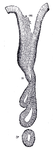
Tubular glands are further classified as one of the following types:

TypeDescriptionLocation
Simple Tubular Gland.png simple tubular
or simple straight tubular [3]
or straight tubular [4]
the gland is a uniform tube Small intestine (Crypts of Lieberkühn), uterine glands
Simple Tubular Coiled Gland.png coiled tubular
or simple coiled tubular [5]
the gland is coiled without losing its tubular form sweat glands
Compound tubular gland.png simple branched tubular [6]
or compound tubular [7]
branching occurs in the tubes pyloric glands of stomach

Additional images

See also

Related Research Articles

<span class="mw-page-title-main">Exocrine gland</span> Gland that secretes substances onto an epithelial surface by way of a duct

Exocrine glands are glands that secrete substances onto an epithelial surface by way of a duct. Examples of exocrine glands include sweat, salivary, mammary, ceruminous, lacrimal, sebaceous, prostate and mucous. Exocrine glands are one of two types of glands in the human body, the other being endocrine glands, which secrete their products directly into the bloodstream. The liver and pancreas are both exocrine and endocrine glands; they are exocrine glands because they secrete products—bile and pancreatic juice—into the gastrointestinal tract through a series of ducts, and endocrine because they secrete other substances directly into the bloodstream. Exocrine sweat glands are part of the integumentary system; they have eccrine and apocrine types.

<span class="mw-page-title-main">Epithelium</span> Tissue lining the surfaces of organs in animals

Epithelium or epithelial tissue is a thin, continuous, protective layer of compactly packed cells with a little intercellular matrix. Epithelial tissues line the outer surfaces of organs and blood vessels throughout the body, as well as the inner surfaces of cavities in many internal organs. An example is the epidermis, the outermost layer of the skin. Epithelial tissue is one of the four basic types of animal tissue, along with connective tissue, muscle tissue and nervous tissue. These tissues also lack blood or lymph supply. The tissue is supplied by nerves.

<span class="mw-page-title-main">Sebaceous hyperplasia</span> Medical condition

Sebaceous hyperplasia is a disorder of the sebaceous glands in which they become enlarged, producing flesh-colored or yellowish, shiny, often umbilicated bumps on the face. Newly formed nodules often swell with sweating, but this diminishes over time.

<span class="mw-page-title-main">Hair follicle</span> Organ found in the dermal layer of the mammalian skin that regulates hair growth

The hair follicle is an organ found in mammalian skin. It resides in the dermal layer of the skin and is made up of 20 different cell types, each with distinct functions. The hair follicle regulates hair growth via a complex interaction between hormones, neuropeptides, and immune cells. This complex interaction induces the hair follicle to produce different types of hair as seen on different parts of the body. For example, terminal hairs grow on the scalp and lanugo hairs are seen covering the bodies of fetuses in the uterus and in some newborn babies. The process of hair growth occurs in distinct sequential stages: anagen is the active growth phase, catagen is the regression of the hair follicle phase, telogen is the resting stage, exogen is the active shedding of hair phase and kenogen is the phase between the empty hair follicle and the growth of new hair.

<span class="mw-page-title-main">Sebaceous gland</span> Gland to lubricate the hair and skin

A sebaceous gland or oil gland is a microscopic exocrine gland in the skin that opens into a hair follicle to secrete an oily or waxy matter, called sebum, which lubricates the hair and skin of mammals. In humans, sebaceous glands occur in the greatest number on the face and scalp, but also on all parts of the skin except the palms of the hands and soles of the feet. In the eyelids, meibomian glands, also called tarsal glands, are a type of sebaceous gland that secrete a special type of sebum into tears. Surrounding the female nipple, areolar glands are specialized sebaceous glands for lubricating the nipple. Fordyce spots are benign, visible, sebaceous glands found usually on the lips, gums and inner cheeks, and genitals.

<span class="mw-page-title-main">Scalp</span> Anatomical area bordered by the face at the front, and by the neck at the sides and back

The scalp is the area of the head where head hair grows. It is made up of skin, layers of connective and fibrous tissues, and the membrane of the skull. Anatomically, the scalp is part of the epicranium, a collection of structures covering the cranium. The scalp is bordered by the face at the front, and by the neck at the sides and back. The scientific study of hair and scalp is called trichology.

<span class="mw-page-title-main">Dermis</span> Layer of skin between the epidermis (with which it makes up the cutis) and subcutaneous tissues

The dermis or corium is a layer of skin between the epidermis and subcutaneous tissues, that primarily consists of dense irregular connective tissue and cushions the body from stress and strain. It is divided into two layers, the superficial area adjacent to the epidermis called the papillary region and a deep thicker area known as the reticular dermis. The dermis is tightly connected to the epidermis through a basement membrane. Structural components of the dermis are collagen, elastic fibers, and extrafibrillar matrix. It also contains mechanoreceptors that provide the sense of touch and thermoreceptors that provide the sense of heat. In addition, hair follicles, sweat glands, sebaceous glands, apocrine glands, lymphatic vessels, nerves and blood vessels are present in the dermis. Those blood vessels provide nourishment and waste removal for both dermal and epidermal cells.

<span class="mw-page-title-main">Sweat gland</span> Small sweat-producing tubular skin structures

Sweat glands, also known as sudoriferous or sudoriparous glands, from Latin sudor 'sweat', are small tubular structures of the skin that produce sweat. Sweat glands are a type of exocrine gland, which are glands that produce and secrete substances onto an epithelial surface by way of a duct. There are two main types of sweat glands that differ in their structure, function, secretory product, mechanism of excretion, anatomic distribution, and distribution across species:

The arrector pili muscles, also known as hair erector muscles, are small muscles attached to hair follicles in mammals. Contraction of these muscles causes the hairs to stand on end, known colloquially as goose bumps (piloerection).

<span class="mw-page-title-main">Esophageal gland</span>

The esophageal glands are glands that are part of the digestive system of various animals, including humans.

<span class="mw-page-title-main">Myoepithelial cell</span>

Myoepithelial cells are cells usually found in glandular epithelium as a thin layer above the basement membrane but generally beneath the luminal cells. These may be positive for alpha smooth muscle actin and can contract and expel the secretions of exocrine glands. They are found in the sweat glands, mammary glands, lacrimal glands, and salivary glands. Myoepithelial cells in these cases constitute the basal cell layer of an epithelium that harbors the epithelial progenitor. In the case of wound healing, myoepithelial cells reactively proliferate. Presence of myoepithelial cells in a hyperplastic tissue proves the benignity of the gland and, when absent, indicates cancer. Only rare cancers like adenoid cystic carcinomas contains myoepithelial cells as one of the malignant components.

<span class="mw-page-title-main">Alveolar gland</span>

Alveolar glands, also called saccular glands, are glands with a saclike secretory portion, in contrast with tubular glands. They typically have an enlarged lumen (cavity), hence the name: they have a shape similar to alveoli, the very small air sacs in the lungs.

<span class="mw-page-title-main">Stratified cuboidal epithelium</span>

Stratified cuboidal epithelium is a type of epithelial tissue composed of multiple layers of cube-shaped cells. Only the most superficial layer is made up of cuboidal cells, and the other layers can be cells of other types. Topmost layer of skin epidermis in frogs, fish is made up of living cuboidal cells.

<span class="mw-page-title-main">Simple cuboidal epithelium</span> Tissue type

Simple cuboidal epithelium is a type of epithelium that consists of a single layer of cuboidal (cube-like) cells which have large, spherical and central nuclei.

An apocrine sweat gland is composed of a coiled secretory portion located at the junction of the dermis and subcutaneous fat, from which a straight portion inserts and secretes into the infundibular portion of the hair follicle. In humans, apocrine sweat glands are found only in certain locations of the body: the axillae (armpits), areola and nipples of the breast, ear canal, eyelids, wings of the nostril, perineal region, and some parts of the external genitalia. Modified apocrine glands include the ciliary glands in the eyelids; the ceruminous glands, which produce ear wax; and the mammary glands, which produce milk. The rest of the body is covered by eccrine sweat glands.

<span class="mw-page-title-main">Metamyelocyte</span>

A metamyelocyte is a cell undergoing granulopoiesis, derived from a myelocyte, and leading to a band cell.

<span class="mw-page-title-main">Uterine gland</span>

Uterine glands or endometrial glands are tubular glands, lined by a simple columnar epithelium, found in the functional layer of the endometrium that lines the uterus. Their appearance varies during the menstrual cycle. During the proliferative phase, uterine glands appear long due to estrogen secretion by the ovaries. During the secretory phase, the uterine glands become very coiled with wide lumens and produce a glycogen-rich secretion known as histotroph or uterine milk. This change corresponds with an increase in blood flow to spiral arteries due to increased progesterone secretion from the corpus luteum. During the pre-menstrual phase, progesterone secretion decreases as the corpus luteum degenerates, which results in decreased blood flow to the spiral arteries. The functional layer of the uterus containing the glands becomes necrotic, and eventually sloughs off during the menstrual phase of the cycle.

<span class="mw-page-title-main">Vermilion border</span> Border between the lips and the rest of the face

The vermilion border, also called margin or zone, is the normally sharp demarcation between the lip and the adjacent normal skin. It represents the change in the epidermis from highly keratinized external skin to less keratinized internal skin. It has no sebaceous glands, sweat glands, or facial hair.

<span class="mw-page-title-main">Syringocystadenoma papilliferum</span> Medical condition

Syringocystadenoma papilliferum is a rare non-malignant adnexal neoplasm that develops from apocrine or eccrine sweat glands and can be identified histologically by cystic, papillary, and ductal invaginations into the dermis lined by double-layered outer cuboidal and luminal high columnar epithelium and connected to the epidermis.

Generalized trichoepitheliomas are characterized histologically by replacement of the hair follicles by trichoepithelioma-like epithelial proliferations associated with hyperplastic sebaceous glands.

References

  1. tubular+gland at eMedicine Dictionary
  2. SIU SOM Histology GI
  3. Histology at KUMC glands-glands04 - "Simple Straight Tubular Gland"
  4. Blue Histology – Epithelia and Glands
  5. Exocrine glands Glandular epithelium
  6. Histology at KUMC glands-glands12
  7. "Epxthelxux C &". Archived from the original on 2008-06-03. Retrieved 2008-08-02.

Additional images