Related Research Articles

An action potential occurs when the membrane potential of a specific cell rapidly rises and falls. This depolarization then causes adjacent locations to similarly depolarize. Action potentials occur in several types of animal cells, called excitable cells, which include neurons, muscle cells, and in some plant cells. Certain endocrine cells such as pancreatic beta cells, and certain cells of the anterior pituitary gland are also excitable cells.

The contraction of cardiac muscle in all animals is initiated by electrical impulses known as action potentials that in the heart are known as cardiac action potentials. The rate at which these impulses fire controls the rate of cardiac contraction, that is, the heart rate. The cells that create these rhythmic impulses, setting the pace for blood pumping, are called pacemaker cells, and they directly control the heart rate. They make up the cardiac pacemaker, that is, the natural pacemaker of the heart. In most humans, the highest concentration of pacemaker cells is in the sinoatrial (SA) node, the natural and primary pacemaker, and the resultant rhythm is a sinus rhythm.
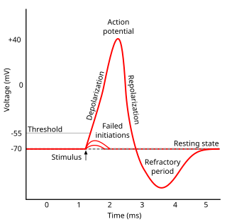
Refractoriness is the fundamental property of any object of autowave nature not responding to stimuli, if the object stays in the specific refractory state. In common sense, refractory period is the characteristic recovery time, a period that is associated with the motion of the image point on the left branch of the isocline .
Hyperpolarization is a change in a cell's membrane potential that makes it more negative. It is the opposite of a depolarization. It inhibits action potentials by increasing the stimulus required to move the membrane potential to the action potential threshold.
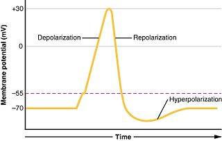
In biology, depolarization or hypopolarization is a change within a cell, during which the cell undergoes a shift in electric charge distribution, resulting in less negative charge inside the cell compared to the outside. Depolarization is essential to the function of many cells, communication between cells, and the overall physiology of an organism.

The cardiac conduction system(CCS) (also called the electrical conduction system of the heart) transmits the signals generated by the sinoatrial node – the heart's pacemaker, to cause the heart muscle to contract, and pump blood through the body's circulatory system. The pacemaking signal travels through the right atrium to the atrioventricular node, along the bundle of His, and through the bundle branches to Purkinje fibers in the walls of the ventricles. The Purkinje fibers transmit the signals more rapidly to stimulate contraction of the ventricles.

The cardiac action potential is a brief change in voltage across the cell membrane of heart cells. This is caused by the movement of charged atoms between the inside and outside of the cell, through proteins called ion channels. The cardiac action potential differs from action potentials found in other types of electrically excitable cells, such as nerves. Action potentials also vary within the heart; this is due to the presence of different ion channels in different cells.
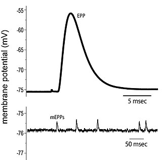
End plate potentials (EPPs) are the voltages which cause depolarization of skeletal muscle fibers caused by neurotransmitters binding to the postsynaptic membrane in the neuromuscular junction. They are called "end plates" because the postsynaptic terminals of muscle fibers have a large, saucer-like appearance. When an action potential reaches the axon terminal of a motor neuron, vesicles carrying neurotransmitters are exocytosed and the contents are released into the neuromuscular junction. These neurotransmitters bind to receptors on the postsynaptic membrane and lead to its depolarization. In the absence of an action potential, acetylcholine vesicles spontaneously leak into the neuromuscular junction and cause very small depolarizations in the postsynaptic membrane. This small response (~0.4mV) is called a miniature end plate potential (MEPP) and is generated by one acetylcholine-containing vesicle. It represents the smallest possible depolarization which can be induced in a muscle.

In neuroscience, the axolemma is the cell membrane of an axon, the branch of a neuron through which signals are transmitted. The axolemma is a three-layered, bilipid membrane. Under standard electron microscope preparations, the structure is approximately 8 nanometers thick.

Guard cells are specialized plant cells in the epidermis of leaves, stems and other organs that are used to control gas exchange. They are produced in pairs with a gap between them that forms a stomatal pore. The stomatal pores are largest when water is freely available and the guard cells become turgid, and closed when water availability is critically low and the guard cells become flaccid. Photosynthesis depends on the diffusion of carbon dioxide (CO2) from the air through the stomata into the mesophyll tissues. Oxygen (O2), produced as a byproduct of photosynthesis, exits the plant via the stomata. When the stomata are open, water is lost by evaporation and must be replaced via the transpiration stream, with water taken up by the roots. Plants must balance the amount of CO2 absorbed from the air with the water loss through the stomatal pores, and this is achieved by both active and passive control of guard cell turgor pressure and stomatal pore size.
The myogenic mechanism is how arteries and arterioles react to an increase or decrease of blood pressure to keep the blood flow constant within the blood vessel. Myogenic response refers to a contraction initiated by the myocyte itself instead of an outside occurrence or stimulus such as nerve innervation. Most often observed in smaller resistance arteries, this 'basal' myogenic tone may be useful in the regulation of organ blood flow and peripheral resistance, as it positions a vessel in a preconstricted state that allows other factors to induce additional constriction or dilation to increase or decrease blood flow.
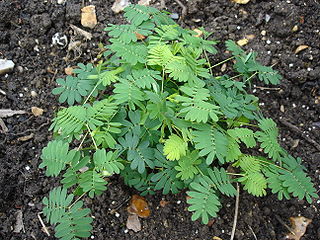
Plant perception is the ability of plants to sense and respond to the environment by adjusting their morphology and physiology. Botanical research has revealed that plants are capable of reacting to a broad range of stimuli, including chemicals, gravity, light, moisture, infections, temperature, oxygen and carbon dioxide concentrations, parasite infestation, disease, physical disruption, sound, and touch. The scientific study of plant perception is informed by numerous disciplines, such as plant physiology, ecology, and molecular biology.
The basal or basic electrical rhythm (BER) or electrical control activity (ECA) is the spontaneous depolarization and repolarization of pacemaker cells known as interstitial cells of Cajal (ICCs) in the smooth muscle of the stomach, small intestine, and large intestine. This electrical rhythm is spread through gap junctions in the smooth muscle of the GI tract. These pacemaker cells, also called the ICCs, control the frequency of contractions in the gastrointestinal tract. The cells can be located in either the circular or longitudinal layer of the smooth muscle in the GI tract; circular for the small and large intestine, longitudinal for the stomach. The frequency of contraction differs at each location in the GI tract beginning with 3 per minute in the stomach, then 12 per minute in the duodenum, 9 per minute in the ileum, and a normally low one contraction per 30 minutes in the large intestines that increases 3 to 4 times a day due to a phenomenon called mass movement. The basal electrical rhythm controls the frequency of contraction but additional neuronal and hormonal controls regulate the strength of each contraction.

In neurophysiology, a dendritic spike refers to an action potential generated in the dendrite of a neuron. Dendrites are branched extensions of a neuron. They receive electrical signals emitted from projecting neurons and transfer these signals to the cell body, or soma. Dendritic signaling has traditionally been viewed as a passive mode of electrical signaling. Unlike its axon counterpart which can generate signals through action potentials, dendrites were believed to only have the ability to propagate electrical signals by physical means: changes in conductance, length, cross sectional area, etc. However, the existence of dendritic spikes was proposed and demonstrated by W. Alden Spencer, Eric Kandel, Rodolfo Llinás and coworkers in the 1960s and a large body of evidence now makes it clear that dendrites are active neuronal structures. Dendrites contain voltage-gated ion channels giving them the ability to generate action potentials. Dendritic spikes have been recorded in numerous types of neurons in the brain and are thought to have great implications in neuronal communication, memory, and learning. They are one of the major factors in long-term potentiation.
Cellular neuroscience is a branch of neuroscience concerned with the study of neurons at a cellular level. This includes morphology and physiological properties of single neurons. Several techniques such as intracellular recording, patch-clamp, and voltage-clamp technique, pharmacology, confocal imaging, molecular biology, two photon laser scanning microscopy and Ca2+ imaging have been used to study activity at the cellular level. Cellular neuroscience examines the various types of neurons, the functions of different neurons, the influence of neurons upon each other, and how neurons work together.
N-(p-Amylcinnamoyl)anthranilic acid (ACA) is a modulator of various ion channels in the heart. ACA is an effective reversible inhibitor of calcium-activated chloride channels and, to a lesser extent, cAMP-activated chloride channels, without affecting L-type calcium channels. Calcium-activated chloride channels are believed to be involved in developing arrhythmia.
In biology, electrotropism, also known as galvanotropism, is a kind of tropism which results in growth or migration of an organism, usually a cell, in response to an exogenous electric field. Several types of cells such as nerve cells, muscle cells, fibroblasts, epithelial cells, green algae, spores, and pollen tubes, among others, have been already reported to respond by either growing or migrating in a preferential direction when exposed to an electric field.
Plants can be exposed to many stress factors such as disease, temperature changes, herbivory, injury and more. Therefore, in order to respond or be ready for any kind of physiological state, they need to develop some sort of system for their survival in the moment and/or for the future. Plant communication encompasses communication using volatile organic compounds, electrical signaling, and common mycorrhizal networks between plants and a host of other organisms such as soil microbes, other plants, animals, insects, and fungi. Plants communicate through a host of volatile organic compounds (VOCs) that can be separated into four broad categories, each the product of distinct chemical pathways: fatty acid derivatives, phenylpropanoids/benzenoids, amino acid derivatives, and terpenoids. Due to the physical/chemical constraints most VOCs are of low molecular mass, are hydrophobic, and have high vapor pressures. The responses of organisms to plant emitted VOCs varies from attracting the predator of a specific herbivore to reduce mechanical damage inflicted on the plant to the induction of chemical defenses of a neighboring plant before it is being attacked. In addition, the host of VOCs emitted varies from plant to plant, where for example, the Venus Fly Trap can emit VOCs to specifically target and attract starved prey. While these VOCs typically lead to increased resistance to herbivory in neighboring plants, there is no clear benefit to the emitting plant in helping nearby plants. As such, whether neighboring plants have evolved the capability to "eavesdrop" or whether there is an unknown tradeoff occurring is subject to much scientific debate. As related to the aspect of meaning-making, the field is also identified as phytosemiotics.
Plants are constantly exposed to different stresses that result in wounding. Plants have adapted to defend themselves against wounding events, like herbivore attacks or environmental stresses. There are many defense mechanisms that plants rely on to help fight off pathogens and subsequent infections. Wounding responses can be local, like the deposition of callose, and others are systemic, which involve a variety of hormones like jasmonic acid and abscisic acid.
Hydraulic signals in plants are detected as changes in the organism's water potential that are caused by environmental stress like drought or wounding. The cohesion and tension properties of water allow for these water potential changes to be transmitted throughout the plant.
References
- 1 2 Stahlberg R, Robert E, Cleland RE, van Volkenburgh E (2006) Slow wave potentials—a propagating electrical signal unique to higher plants.
- ↑ Stankovic´ B, Davies E (1996) Both action potentials and variation potentials induce proteinase inhibitor gene expression in tomato. FEBS Lett 390:275–279.
- ↑ Dziubinska H, Filek M, Koscielniak J, Trebacz K (2003) Variation and action potentials evoked by thermal stimuli accompany enhancement of ethylene emission in distant non-stimulated leaves of Vicia faba minor seedlings. J Plant Physiol 160:1203–1210.
- ↑ Fisahn J, Herde O, Willmitzer L, Pen˜a-Corte´s H (2004) Analysis of the transient increase in cytosolic Ca2? during the action potential of higher plants with high temporal resolution: requirement of Ca2? transients for induction of jasmonic acid biosynthesis and PINII gene expression. Plant Cell Physiol 45:456–459.
- ↑ Hlava´cˇkova´ V, Krchnˇa´k P, Nausˇ J, Nova´k O, Sˇpundova´ M, Strnad M (2006) Electrical and chemical signals involved in short-term systemic photosynthetic responses of tobacco plants to local burning. Planta 225:235–244
- ↑ Koziolek C, Grams TEE, Schreiber U, Matyssek R, Fromm J (2003) Transient knockout of photosynthesis mediated by electrical signals. New Phytol 161:715–722.
- ↑ Filek M, Koscielniak J (1997) The effect of wounding the roots by high temperature on the respiration rate of the shoot and propagation of electric signal in horse bean seedlings (Vicia faba L. minor). Plant Sci 123:39–46.
- ↑ "Result Filters." National Center for Biotechnology Information. U.S. National Library of Medicine, n.d. Web. 10 June 2016.
- ↑ Davies E (2006) Electrical signals in plants: facts and hypotheses. In: Volkov AG (ed) Plant electrophysiology. Theory and methods, Springer-Verlag, Berlin, pp 407–422.
- ↑ Stahlberg, R.; Cosgrove, D. J. (1995). "Comparison of electric and growth responses to excision in cucumber and pea seedlings. II. Long-distance effects are caused by the release of xylem pressure". Plant, Cell & Environment. 18 (1): 33–41. doi:10.1111/j.1365-3040.1995.tb00541.x. ISSN 1365-3040. PMID 11541062.