Vascular anomaly

Last updated

A vascular anomaly is any of a range of lesions from a simple birthmark to a large tumor that may be disfiguring. They are caused by a disorder of the vascular system. [1] A vascular anomaly is a localized defect in blood or lymph vessels. These defects are characterized by an increased number of vessels, and vessels that are both enlarged and sinuous. Some vascular anomalies are congenital, others appear within weeks to years after birth, and others are acquired by trauma or during pregnancy. Inherited vascular anomalies are also described and often present with a number of lesions that increase with age. Vascular anomalies can also be a part of a syndrome.[ citation needed ]

Contents

The estimated prevalence of vascular anomalies is 4.5%. [2] Vascular anomalies can occur throughout the whole body, but in 60% of patients they are localized in the head and neck region. [3] Vascular anomalies can present in various ways, when situated deep below the skin, they appear blue, and are often called cavernous. Superficial vascular anomalies appear as red-coloured stains and are associated with vascular anomalies affecting the dermis. Historically, vascular anomalies have been labeled with descriptive terms, according to the food they resembled (port wine, strawberry, cherry, salmon patch). This imprecise terminology has caused diagnostic confusion, blocked communication and even caused incorrect treatment, as it does not differentiate between various vascular anomalies. [4] However, in 1982, Mulliken introduced a classification that replaced these descriptive terms and gave direction to the management of various vascular anomalies. This classification, based on clinical features, natural history and cellular characteristics, divides vascular anomalies into two groups: vascular tumors and vascular malformations. [5] Although vascular tumors and vascular malformations can resemble each other, there are important differences between both.

Vascular tumors

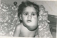
Vascular tumors, include hemangiomas, the most common tumors in infants, occurring in 1-2%, and higher in 10% of premature infants of very low birth weight. [3] Vascular tumors are characterized by an overgrowth of normal vessels, which show increased endothelial proliferation. They are typically present at birth, but can appear within a couple of weeks after birth or during infancy. The four most common types are: infantile hemangioma, congenital hemangioma, kaposiform hemangioendothelioma and pyogenic granuloma.[ citation needed ]

Infantile hemangioma

Infantile hemangioma is the most common vascular tumor. It is a benign tumor, which occurs in 4-5% of Caucasian infants, but rarely in dark skinned infants. [6] It occurs in 20% of low weight premature infants and 2.2 to 4.5 times more frequently in females. [7] IH most commonly presents in the head and neck region (60%), but also involves the trunk and extremities. One third of these lesions is present at birth as a telangiectatic stain or ecchymotic area. During the first four weeks of life, 70% to 90% appear. Lesions that are situated beneath the skin may not appear until 3 to 4 months of age, when the tumor is large enough. During the first 9 months, IH undergoes rapid growth, which is faster than the growth of the child. This is called the proliferating phase. After 9 months, the growth of the tumor will decrease and equal the growth of the child for about 3 months. After 12 months, the tumor will start to involute and might even disappear. Involution occurs in one-third of patient by the age of 3 years, in 50% by the age of 5 years and in 72% by the age of 7 years. [8] Involution may result in residual telangiectasias, pallor, atrophy, textural changes and sometimes fibrofatty residuum. Since 90% of IH is small, localized and asymptomatic, treatment mainly consists of observation and awaiting until involution is complete. IH can be treated with corticosteroids, which accelerate involution: in 95% of patients, growth is stabilized and 75% of tumors decrease in size. Intralesional corticosteroids are most effective, but may require additional injections, as the effect is only temporarily. Systemic corticosteroids may cause a number of side-effects and are only used in problematic IH, which is too large to treat with intralesional injections. During the proliferating phase, the tumor is highly vascular. Patients who undergo operative treatment during this period, are at risk for blood loss. Moreover, surgery during this phase, often leads to an inferior aesthetic outcome. However, patients may require intervention during childhood, because 50% of IH leave residual fibrofatty tissue, redundant skin, or damaged structures after involution. Waiting until involution is completed, ensures that the least amount of fibro fatty residuum and excess skin is resected, giving the smallest possible scar. [6] Another option for treatment is the pulsed-dye laser. After involution residual telangiectasias can be treated with laser therapy.[ citation needed ]

Congenital hemangioma

Congenital hemangioma can be distinguished from infantile hemangioma because it is fully developed at birth. It forms during prenatal life and has reached its maximal size at birth. Congenital hemangioma can even be diagnosed in utero by prenatal ultrasound. Unlike IH, CH is more common in the extremities, has an equal sex distribution, and is solitary, with an average diameter of 5 cm. It commonly presents in the head and neck and in the lower extremities. Congenital hemangioma are divided into 2 subgroups: the rapidly involuting congenital hemangiomas (RICHs) and the non-involuting congenital hemangiomas(NICHs).

The rapidly involuting congenital hemangioma, RICH, presents at birth as a solitary raised tumor with a central depression, scar, or ulceration surrounded by a rim of pallor. It is noted for its involution, which typically begins several weeks after birth and is completed no later than 14 months of age. [9] After regression RICH may cause a residual deformity, such as atrophic skin and subcutaneous tissue. It mainly affects the limbs (52%), but also the head and neck region (42%) and the trunk (6%). [6]

The non-involuting congenital hemangioma, NICH, presents as a solitary, well-circumscribed reddish-pink to purple plaque with central telangiectasia and hypopigmented rim. [9] In contrast to RICH, NICH does not involute and rarely ulcerates. It persists into late childhood and can even mimic a vascular malformation by growing commensurately with the child. Although NICH can resemble RICH in its external appearance, it can be differentiated from RICH by a greater elevation and coarse telangiectases. It mainly affects the head and neck region (43%), but also the limbs (38%) and the trunk (19%).[ citation needed ]

Surgical resection for congenital hemangiomas is rarely needed, because RICH undergoes postnatal regression and NICH is benign and often asymptomatic. Resection may be indicated to improve the appearance of the affected area, as long as the surgical scar is less noticeable than the lesion. Other indications are problematic ulcers with persistent bleeding or chronic infection. Although most NICH lesions are non-problematic and do not cause significant deformity, the threshold for resection of NICH is lower, because it neither involutes, nor responds to pharmacotherapy. RICH tumors are observed until involution is completed. Involuted RICH may leave behind atrophic tissue, which can be reconstructed with autologous grafts. [6] It is often best to postpone excision until regression is complete. There are effective pharmacologic treatments, which include intralesional corticosteroid injection, systemic corticosteroid injection, interferon α-2a or α-2b and angiogenic inhibitors. The use of corticosteroids leads to accelerated regression in 30%, stabilization of growth in 40%, lightening of color and softening of the tumor. However, 30% shows minimal or no response. Another drug treatment is interferon α-2a or α-2b. It is often used for patients who did not respond to corticosteroids. Although the response rate is much slower, it has been successful for 80% of children treated. [10] The most serious side effect of interferon is a spastic diplegia. Other therapeutic options are embolization and pulsed-dye laser, which improves residual telangiectasias in RICH and in NICH.[ citation needed ]

Kaposiform hemangioendothelioma

Kaposiform hemangioendothelioma (KHE) is a rare vascular neoplasm that is locally aggressive but without metastatic potential. It occurs particularly in the skin, deep soft tissue, retroperitoneum, mediastinum, and rarely in bone. Although lesions occur solitary, they often involve large areas of the body, such as the head/neck region (40%), trunk (30%), or extremity (30%). Usually, it is present at birth as a flat, reddish-purple, tense and edematous lesion. Although half of lesions are congenital, 58% of KHE develop during infancy, 32% between age 1 and 10 years (32%) and 10% after 11 years of age. Moreover, adult onset has been described too with mainly males being affected. Both sexes are affected equally in children. Lesions are often greater than 5 cm in diameter and can cause visible deformity and pain. During early childhood, KHE may enlarge and after 2 years of age, it may partially regress. Though, it usually persists longterm. In addition, 50% of patients have coagulopathy due to thrombocytopenia (<25,000/mm3), presenting with petechiae and bleeding. This is called the Kasabach–Merritt syndrome, which is caused by trapping of platelets and other clotting factors within the tumor. Kasabach-Merritt Phenomenon is less likely in patients with lesions less than 8 cm. As two-thirds of adult-onset KHE tumors are less than 2 cm, KHE in adults is rarely associated with Kasabach-Merritt Phenomenon. [6] Patients with KHE and Kasabach-Merritt Phenomenon present with petechiae and ecchymosis. Most KHE tumors are diffuse involving multiple tissue planes and important structures. Resection of KHE is thus often difficult. Treatment of kaposiform hemangioendothelioma is therefore medical. The primary drug is interferon alfa, which is successful in 50% of children. [10] Another option is vincristine, which has many side-effects, but has a response rate of 90%. Drug therapy is often used in shrinking the tumor and treating the coagulopathy. However, many of these kaposiform hemangioendotheliomas do not completely regress and remain as a much smaller asymptomatic tumor. However, KHE still has a high mortality rate of 30%. Although complete surgical removal with a large margin has the best reported outcome, it is usually not done because of the risk of bleeding, extensiveness, and the anatomic site of the lesion. [11] Operative management may be possible for small or localized lesions. Removal of larger areas also may be indicated for symptomatic patients or for patients who have failed pharmacotherapy. Resection is not required for lesions that are not causing functional problems, because KHE is benign and because resection could cause deformity.[ citation needed ]

Pyogenic granuloma

Pyogenic granuloma Pyogenic Granuloma Thumb 1.JPG
Pyogenic granuloma

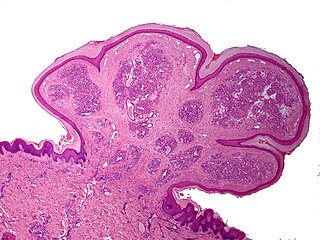
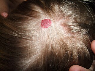
A pyogenic granuloma, is a small benign vascular tumor that primarily involves the skin (88.2%) and mucous membranes. [6] Pyogenic granuloma appears as a red macule that grows rapidly, turns into a papule and eventually becomes pedunculated, being attached to a narrow stalk. [8] The average diameter of these lesions is 6.5 mm. [6] Although these lesions are small, they are often complicated by bleeding, crusting and ulceration. Microscopically, pyogenic granulomas are characterized by vascular proliferation amidst granulation tissue and chronic inflammatory infiltrate. [12]

Pyogenic granulomas are rarely congenital. It commonly develops in infants: 42.1% develops within the first 5 years of life. [6] This vascular tumor is twice as common in males as in females and 25% of lesions seem to be associated with trauma, an underlying cutaneous condition, pregnancy, hormonal alterations and medications. [12] Pyogenic granulomas can also arise within a capillary malformation. Of all pyogenic granulomas, 62% is distributed on the head or neck, occurring mainly on the cheek and in the oral cavity. Lesions on the face may cause visible deformity.[ citation needed ]

Numerous treatment methods have been described for pyogenic granuloma. Lesions involving the reticular dermis, may be out of the reach of pulsed-dye laser, cautery or shave excision and therefore have a recurrence rate of 43.5%. [6] Definitive management requires full-thickness skin excision. Other options are curettage or laser therapy. Furthermore, thorough curettage and cauterization are often used for small lesions and full-thickness excision for larger lesion.[ citation needed ]

Vascular malformations

Vascular malformation is a collective term for different disorders of the vasculature (errors in vascular development). It can be a disorder of the capillaries, arteries, veins and lymphatic vessels or a disorder of a combination of these (lesions are named based on the primary vessel that is malformed). A vascular malformation consists of a cluster of deformed vessels, due to an error in vascular development (dysmorphogenesis). However, endothelial turnover is stable in these defects. Congenital vascular malformations are always already present at birth, although they are not always visible. In contrast to vascular tumors, vascular malformations do not have a growth phase, nor an involution phase. Vascular malformations tend to grow proportionately with the child. [13] Vascular malformations never regress, but persist throughout life. Vascular malformations can be divided into slow-flow, fast-flow and complex-combined types. [14]

Slow-flow vascular malformations

Capillary malformation Myhand.jpg
Capillary malformation
Lymphatic malformation Lymphangioma.jpg
Lymphatic malformation

Fast flow vascular malformations

All fast-flow malformations are malformations involving arteries. They constitute about 14% of all vascular malformations. [2]

Combined-complex vascular malformations

a combination of various vascular malformations. They are 'complex' because they involve a combination of two different types of vessels.

Terminology

Correct termIncorrect terminology commonly used to describe vascular anomalies
HemangiomaStrawberry Hemangioma

Capillary Hemangioma

Cavernous Hemangioma

Kaposiform hemangioendothelioma(Capillary) Hemangioma
Pyogenic granulomaHemangioma
Capillary MalformationPort-wine stain

Capillary Hemangioma

Lymphatic MalformationLymphangioma

Cystic hygroma

Venous MalformationCavernous Hemangioma
Arteriovenous malformationArteriovenous Hemangioma

[2]

Related Research Articles

<span class="mw-page-title-main">Infantile hemangioma</span> Raised red skin lesion that affects infants caused by benign vascular tumor

An infantile hemangioma (IH), sometimes called a strawberry mark due to appearance, is a type of benign vascular tumor or anomaly that affects babies. Other names include capillary hemangioma, strawberry hemangioma, strawberry birthmark and strawberry nevus. and formerly known as a cavernous hemangioma. They appear as a red or blue raised lesion on the skin. Typically, they begin during the first four weeks of life, growing until about five months of life, and then shrinking in size and disappearing over the next few years. Often skin changes remain after they shrink. Complications may include pain, bleeding, ulcer formation, disfigurement, or heart failure. It is the most common tumor of orbit and periorbital areas in childhood. It may occur in the skin, subcutaneous tissues and mucous membranes of oral cavities and lips as well as in extracutaneous locations including the liver and gastrointestinal tract.

<span class="mw-page-title-main">Angioma</span> Medical condition

Angiomas are benign tumors derived from cells of the vascular or lymphatic vessel walls (endothelium) or derived from cells of the tissues surrounding these vessels.

<span class="mw-page-title-main">Hemangioendothelioma</span> Medical condition

Hemangioendotheliomas are a family of vascular neoplasms of intermediate malignancy.

<span class="mw-page-title-main">Lymphangioma</span> Malformations of the lymphatic system characterized by lesions that are thin-walled cysts

Lymphangiomas are malformations of the lymphatic system characterized by lesions that are thin-walled cysts; these cysts can be macroscopic, as in a cystic hygroma, or microscopic. The lymphatic system is the network of vessels responsible for returning to the venous system excess fluid from tissues as well as the lymph nodes that filter this fluid for signs of pathogens. These malformations can occur at any age and may involve any part of the body, but 90% occur in children less than 2 years of age and involve the head and neck. These malformations are either congenital or acquired. Congenital lymphangiomas are often associated with chromosomal abnormalities such as Turner syndrome, although they can also exist in isolation. Lymphangiomas are commonly diagnosed before birth using fetal ultrasonography. Acquired lymphangiomas may result from trauma, inflammation, or lymphatic obstruction.

<span class="mw-page-title-main">Pyogenic granuloma</span> Vascular tumor on both mucosa and skin

A pyogenic granuloma or lobular capillary hemangioma is a vascular tumor that occurs on both mucosa and skin, and appears as an overgrowth of tissue due to irritation, physical trauma, or hormonal factors. It is often found to involve the gums, skin, or nasal septum, and has also been found far from the head, such as in the thigh.

<span class="mw-page-title-main">Focal nodular hyperplasia</span> Medical condition

Focal nodular hyperplasia (FNH) is a benign tumor of the liver, which is the second most prevalent tumor of the liver. It is usually asymptomatic, rarely grows or bleeds, and has no malignant potential. This tumour was once often resected because it was difficult to distinguish from hepatic adenoma, but with modern multiphase imaging is usually now diagnosed by strict imaging criteria and not resected.

<span class="mw-page-title-main">Kasabach–Merritt syndrome</span> Medical condition

Kasabach–Merritt syndrome, also known as hemangioma with thrombocytopenia, is a rare disease, usually of infants, in which a vascular tumor leads to decreased platelet counts and sometimes other bleeding problems, which can be life-threatening. It is also known as hemangioma thrombocytopenia syndrome. It is named after Haig Haigouni Kasabach and Katharine Krom Merritt, the two pediatricians who first described the condition in 1940.

<span class="mw-page-title-main">Vascular tumor</span> Medical condition

A vascular tumor is a tumor of vascular origin; a soft tissue growth that can be either benign or malignant, formed from blood vessels or lymph vessels. Examples of vascular tumors include hemangiomas, lymphangiomas, hemangioendotheliomas, Kaposi's sarcomas, angiosarcomas, and hemangioblastomas. An angioma refers to any type of benign vascular tumor.

<span class="mw-page-title-main">Vascular disease</span> Medical condition

Vascular disease is a class of diseases of the vessels of the circulatory system in the body, including blood vessels – the arteries and veins, and the lymphatic vessels. Vascular disease is a subgroup of cardiovascular disease. Disorders in this vast network of blood and lymph vessels can cause a range of health problems that can sometimes become severe, and fatal. Coronary heart disease for example, is the leading cause of death for men and women in the United States.

<span class="mw-page-title-main">Cutis marmorata telangiectatica congenita</span> Medical condition

Cutis marmorata telangiectatica congenita is a rare congenital vascular disorder that usually manifests in affecting the blood vessels of the skin. The condition was first recognised and described in 1922 by Cato van Lohuizen, a Dutch pediatrician whose name was later adopted in the other common name used to describe the condition – Van Lohuizen Syndrome. CMTC is also used synonymously with congenital generalized phlebectasia, nevus vascularis reticularis, congenital phlebectasia, livedo telangiectatica, congenital livedo reticularis and Van Lohuizen syndrome.

Pediatric plastic surgery is plastic surgery performed on children. Its procedures are most often conducted for reconstructive or cosmetic purposes. In children, this line is often blurred, as many congenital deformities impair physical function as well as aesthetics.

A vascular malformation is a blood vessel or lymph vessel abnormality. Vascular malformations are one of the classifications of vascular anomalies, the other grouping is vascular tumors. They may cause aesthetic problems as they have a growth cycle, and can continue to grow throughout life.

<span class="mw-page-title-main">Bonnet–Dechaume–Blanc syndrome</span> Medical condition

Bonnet–Dechaume–Blanc syndrome, also known as Wyburn-Mason syndrome, is a rare congenital disorder characterized by arteriovenous malformations of the brain, retina or facial nevi. The syndrome has a number of possible symptoms and can, more rarely, affect the skin, bones, kidneys, muscles, and gastrointestinal tract. When the syndrome affects the brain, people can experience severe headaches, seizures, acute stroke, meningism, and progressive neurological deficits due to acute or chronic ischaemia caused by arteriovenous shunting.

Sinus pericranii (SP) is a rare disorder characterized by a congenital epicranial venous malformation of the scalp. Sinus pericranii is an abnormal communication between the intracranial and extracranial venous drainage pathways. Treatment of this condition has mainly been recommended for aesthetic reasons and prevention of bleeding.

Diffuse neonatal hemangiomatosis (DNH) is a potentially fatal disorder where multiple benign (non-cancerous) blood vessel tumors (hemangiomas) are present in the skin and other organs. The mortality rate of diffuse neonatal hemangiomatosis is 50-90%. This disease is normally found in female Caucasian infants. The most common site of internal organ damage, or lesions, is the liver, which can redirect blood away from the heart and cause arteriovenous shunting. This can cause high cardiac output, leading to further complications such as congestive heart failure. This condition affecting the liver is also known as infantile hepatic hemangioma (IHH). Other sites of internal organ damage can include the intestines, nervous system, lungs, and sometimes the skeletal system. Early detection and treatment with steroids results in most newborn babies with this disease remaining healthy, with serious problems developing for some individuals during the hemangioma's growth phase.

<span class="mw-page-title-main">Cavernous hemangioma</span> Human disease

Cavernous hemangioma, also called cavernous angioma, venous malformation, or cavernoma, is a type of venous malformation due to endothelial dysmorphogenesis from a lesion which is present at birth. A cavernoma in the brain is called a cerebral cavernous malformation or CCM. Despite its designation as a hemangioma, a cavernous hemangioma is not a tumor as it does not display endothelial hyperplasia. The abnormal tissue causes a slowing of blood flow through the cavities, or "caverns". The blood vessels do not form the necessary junctions with surrounding cells, and the structural support from the smooth muscle is hindered, causing leakage into the surrounding tissue. It is the leakage of blood, referred to as hemorrhage, that causes a variety of symptoms known to be associated with the condition.

Fibro-adipose vascular anomaly, also known as FAVA, is a type of vascular anomaly that is both rare and painful. FAVA is characterized by tough fibrofatty tissue taking over portions of muscle, most often contained within a single limb. FAVA also causes venous and/or lymphatic abnormalities.

<span class="mw-page-title-main">CLOVES syndrome</span> Medical condition

CLOVES syndrome is a rare overgrowth syndrome with complex vascular anomalies. CLOVES syndrome affects people with various symptoms, ranging from mild fatty soft-tissue tumors to vascular malformations encompassing the spine or internal organs.

<span class="mw-page-title-main">Hemangioma</span> Medical condition

A hemangioma or haemangioma is a usually benign vascular tumor derived from blood vessel cell types. The most common form, seen in infants, is an infantile hemangioma, known colloquially as a "strawberry mark", most commonly presenting on the skin at birth or in the first weeks of life. A hemangioma can occur anywhere on the body, but most commonly appears on the face, scalp, chest or back. They tend to grow for up to a year before gradually shrinking as the child gets older. A hemangioma may need to be treated if it interferes with vision or breathing or is likely to cause long-term disfigurement. In rare cases internal hemangiomas can cause or contribute to other medical problems. Most of the time they tend to disappear in 10 years. The first line treatment option is beta blockers, which are highly effective in the majority of cases. Ones that form at birth are called congenital hemangiomas while ones that form later in life are called infantile hemangiomas.

Diffuse capillary malformation with overgrowth (DCMO) is a subset of capillary malformations (CM) associated with hypertrophy, i.e. increased size of body structures. CM can be considered an umbrella term for various vascular anomalies caused by increased diameter or number of capillary blood vessels. It is commonly referred to as "port-wine stain", and is thought to affect approximately 0.5% of the population. Typically capillaries in the papillary dermis are involved, and this gives rise to pink or violaceous colored lesions. The majority of DCMO lesions are diffuse, reticulated pale-colored stains.

References

  1. Steiner, JE; Drolet, BA (September 2017). "Classification of Vascular Anomalies: An Update". Seminars in Interventional Radiology. 34 (3): 225–232. doi:10.1055/s-0037-1604295. PMC   5615389 . PMID   28955111.
  2. 1 2 3 4 5 6 Greene, AK (January 2011). "Vascular anomalies: current overview of the field". Clinics in Plastic Surgery. 38 (1): 1–5. doi: 10.1016/j.cps.2010.08.004 . PMID   21095467.
  3. 1 2 3 4 5 6 Ernemann, U; Kramer, U; Miller, S; Bisdas, S; Rebmann, H; Breuninger, H; Zwick, C; Hoffmann, J (July 2010). "Current concepts in the classification, diagnosis and treatment of vascular anomalies". European Journal of Radiology. 75 (1): 2–11. doi:10.1016/j.ejrad.2010.04.009. PMID   20466500.
  4. Hassanein, AH; Mulliken, JB; Fishman, SJ; Greene, AK (January 2011). "Evaluation of terminology for vascular anomalies in current literature". Plastic and Reconstructive Surgery. 127 (1): 347–51. doi:10.1097/PRS.0b013e3181f95b83. PMID   21200229. S2CID   21459193.
  5. Mulliken, JB; Glowacki, J (March 1982). "Hemangiomas and vascular malformations in infants and children: a classification based on endothelial characteristics". Plastic and Reconstructive Surgery. 69 (3): 412–22. doi:10.1097/00006534-198203000-00002. PMID   7063565. S2CID   21584838.
  6. 1 2 3 4 5 6 7 8 9 Greene, AK (January 2011). "Management of hemangiomas and other vascular tumors". Clinics in Plastic Surgery. 38 (1): 45–63. doi: 10.1016/j.cps.2010.08.001 . PMID   21095471.
  7. Kim, LH; Hogeling, M; Wargon, O; Jiwane, A; Adams, S (April 2011). "Propranolol: useful therapeutic agent for the treatment of ulcerated infantile hemangiomas". Journal of Pediatric Surgery. 46 (4): 759–63. doi:10.1016/j.jpedsurg.2011.01.012. PMID   21496551.
  8. 1 2 Van Aalst, JA; Bhuller, A; Sadove, AM (July 2003). "Pediatric vascular lesions". The Journal of Craniofacial Surgery. 14 (4): 566–83. doi:10.1097/00001665-200307000-00032. PMID   12867875.
  9. 1 2 Beck, DO; Gosain, AK (June 2009). "The presentation and management of hemangiomas". Plastic and Reconstructive Surgery. 123 (6): 181e–91e. doi:10.1097/PRS.0b013e3181a65c59. PMID   19483535. S2CID   44534389.
  10. 1 2 Hentz, Ed. Vincent R. (2006). "209: Vascular Anomalies of the Upper Extremity". The hand and upper limb; part 2 (2. ed.). Philadelphia, Pa.: Saunders Elsevier. ISBN   978-0-7216-8819-0.
  11. Hermans DJ, van Beynum IM, van der Vijver RJ, Kool LJ, de Blaauw I, van der Vleuten CJ (May 2011). "Kaposiform hemangioendothelioma with Kasabach-Merritt syndrome: a new indication for propranolol treatment". Journal of Pediatric Hematology/Oncology. 33 (4): e171-3. doi:10.1097/MPH.0b013e3182152e4e. PMID   21516018. S2CID   45058591.
  12. 1 2 Gupta, A; Kozakewich, H (January 2011). "Histopathology of vascular anomalies". Clinics in Plastic Surgery. 38 (1): 31–44. doi:10.1016/j.cps.2010.08.007. PMID   21095470.
  13. 1 2 3 Chim, H; Drolet, B; Duffy, K; Koshima, I; Gosain, AK (August 2010). "Vascular anomalies and lymphedema". Plastic and Reconstructive Surgery. 126 (2): 55e–69e. doi:10.1097/PRS.0b013e3181df803d. PMID   20679788. S2CID   30865392.
  14. 1 2 3 Enjolras, O (2007). "Introduction: ISSVA Classification". Color atlas of vascular tumors and vascular malformations. Cambridge [u.a.]: Cambridge University Press. ISBN   978-0-521-84851-0.
  15. Perkins JA, Manning SC, Tempero RM, Cunningham MJ, Edmonds JL, Hoffer FA, Egbert MA (June 2010). "Lymphatic malformations: current cellular and clinical investigations". Otolaryngology–Head and Neck Surgery. 142 (6): 789–94. doi:10.1016/j.otohns.2010.02.025. PMID   20493347. S2CID   33570025.