Angiolymphoid hyperplasia with eosinophilia

Last updated
Angiolymphoid hyperplasia with eosinophilia
Other namesEpithelioid hemangioma
SkinTumors-PA090956.JPG
Angiolymphoid hyperplasia with eosinophilia
Specialty Dermatology   OOjs UI icon edit-ltr-progressive.svg

Angiolymphoid hyperplasia with eosinophilia (also known as: [1] "Epithelioid hemangioma," "Histiocytoid hemangioma," "Inflammatory angiomatous nodule," "Intravenous atypical vascular proliferation," "Papular angioplasia," "Inflammatory arteriovenous hemangioma," and "Pseudopyogenic granuloma") usually presents with pink to red-brown, dome-shaped, dermal papules or nodules of the head or neck, especially about the ears and on the scalp. [2]

Contents

It, or a similar lesion, has been suggested as a feature of IgG4-related skin disease, which is the name used for skin manifestations of IgG4-related disease. [3] [4]

Signs and symptoms

Angiolymphoid hyperplasia with eosinophilia is characterized by papulonodular lesions on the head and neck that are violaceous or erythematous. [5] Lesions on the genital area, upper limbs, and trunk are uncommon. The lesions could be painful, pruriginous, or asymptomatic. Peripheral eosinophilia and lymphadenomegaly could be present. [6]

Causes

Currently, the cause of angiolymphoid hyperplasia with eosinophilia is unknown. Several theories have been proposed, but none of them have proven to be conclusive or definitive. These include reactive processes, neoplastic processes, and infectious mechanisms that may be connected to the human immunodeficiency virus. [7]

Though its significance in the etiology of angiolymphoid hyperplasia with eosinophilia remains unclear, the distinctive inflammatory infiltrate in angiolymphoid hyperplasia with eosinophilia seems to be a crucial aspect of this illness. [7] Vascular proliferation may be explained by the endothelial cells' response to proliferative stimuli produced by the inflammatory cells and immunologic allergic reaction that surround them. [8] [9] Elevated serum estrogen levels, [8] [10] local trauma, [9] [11] and arteriovenous shunting [8] [12] are most likely contributing factors. [7]

The TEK gene, which codes for the endothelial cell tyrosine kinase receptor Tie-2, was found to contain a mutation in a case of dermal angiolymphoid hyperplasia with eosinophilia, suggesting that specific molecular changes may play a role in the pathophysiology of this condition. [13]

While angiolymphoid hyperplasia with eosinophilia is thought to be a benign tumefaction, it has been linked to a number of lymphoproliferative disorders, which lends credence to the argument put forth by some that angiolymphoid hyperplasia with eosinophilia could occasionally be a monoclonal T-cell process. [14] [15] Follic mucinosis was found to be present concurrently in a small number of angiolymphoid hyperplasia with eosinophilia cases. [16] A clear correlation between angiolymphoid hyperplasia with eosinophilia and mycosis fungoides has not yet been documented, and this association is fairly ambiguous. [7] Remarkably, a patient with angiolymphoid hyperplasia with eosinophilia has been documented to develop peripheral T-cell lymphoma. [17] T-cell receptor gene (TCR) rearrangement and monoclonality have also been found in angiolymphoid hyperplasia with eosinophilia cases. [14] [15] The same monoclonal TCR gene rearrangement was found in both lesions in a patient, who had both peripheral T-cell lymphoma and angiolymphoid hyperplasia with eosinophilia. [15]

Diagnosis

A complete blood count, which is part of the laboratory analysis, indicates peripheral blood eosinophilia in about 20% of the cases. [8]

To make the diagnosis, a biopsy of the lesions is needed. Strong reactivity for CD31 and lower reactivity for CD34 and factor VIII-related antigen is shown by an immunohistochemical analysis. [18]

When angiolymphoid hyperplasia with eosinophilia lesions are examined under a microscope, a polymorphous vascular pattern made up of uniformly spaced linear and dotted vessels is visible over a background that ranges in color from pink to red. [19]

Treatment

The preferred course of treatment is surgical excision; after being fully removed, angiolymphoid hyperplasia with eosinophilia rarely returns. [20] Other treatments include cryosurgery,[ citation needed ] carbon dioxide laser, [21] pulsed-dye laser, [22] and Mohs micrographic surgery. [23]

See also

Related Research Articles

<span class="mw-page-title-main">Cherry angioma</span> Small bright red dome-shaped bump on the skin

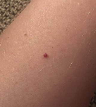
Cherry angioma, also called cherry hemangioma or Campbell de Morgan Spot, is a small bright red dome-shaped bump on the skin. It ranges between 0.5 – 6 mm in diameter and usually several are present, typically on the chest and arms, and increasing in number with age. If scratched, they may bleed.

<span class="mw-page-title-main">Pyogenic granuloma</span> Vascular tumor on both mucosa and skin

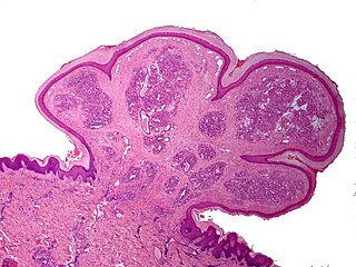
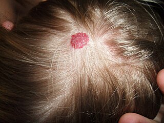
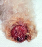
A pyogenic granuloma or lobular capillary hemangioma is a vascular tumor that occurs on both mucosa and skin, and appears as an overgrowth of tissue due to irritation, physical trauma, or hormonal factors. It is often found to involve the gums, skin, or nasal septum, and has also been found far from the head, such as in the thigh.

<span class="mw-page-title-main">Focal nodular hyperplasia</span> Medical condition

Focal nodular hyperplasia is a benign tumor of the liver, which is the second most prevalent tumor of the liver after hepatic hemangioma. It is usually asymptomatic, rarely grows or bleeds, and has no malignant potential. This tumour was once often resected because it was difficult to distinguish from hepatic adenoma, but with modern multiphase imaging it is usually now diagnosed by strict imaging criteria and not resected.

<span class="mw-page-title-main">Vascular tumor</span> Medical condition

A vascular tumor is a tumor of vascular origin; a soft tissue growth that can be either benign or malignant, formed from blood vessels or lymph vessels. Examples of vascular tumors include hemangiomas, lymphangiomas, hemangioendotheliomas, Kaposi's sarcomas, angiosarcomas, and hemangioblastomas. An angioma refers to any type of benign vascular tumor.

<span class="mw-page-title-main">Amelanotic melanoma</span> Medical condition

Amelanotic melanoma is a type of skin cancer in which the cells do not make any melanin. They can be pink, red, purple or of normal skin color, and are therefore difficult to diagnose correctly. They can occur anywhere on the body, just as a typical melanoma can.

<span class="mw-page-title-main">Kimura's disease</span> Medical condition

Kimura's disease is a benign rare chronic inflammatory disorder. Its primary symptoms are subdermal lesions in the head or neck or painless unilateral inflammation of cervical lymph nodes.

Lupus erythematosus panniculitis presents with subcutaneous nodules that are commonly firm, sharply defined and nontender.

Anetoderma is a benign but uncommon disorder that causes localized areas of flaccid or herniated sac-like skin due to a focal reduction of dermal elastic tissue. Anetoderma is subclassified as primary anetoderma, secondary anetoderma, iatrogenic anetoderma of prematurity, congenital anetoderma, familial anetoderma, and drug-induced anetoderma.

Palisaded neutrophilic and granulomatous dermaititis (PNGS) is usually associated with a well-defined connective tissue disease, lupus erythematosus or rheumatoid arthritis most commonly, and often presents with eroded or ulcerated symmetrically distributed umbilicated papules or nodules on the elbows.

<span class="mw-page-title-main">Chondrodermatitis nodularis chronica helicis</span> Medical condition

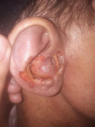
Chondrodermatitis nodularis chronica helicis(CNCH) is a small, nodular, tender, chronic inflammatory lesion occurring on the helix of the ear, most often in men. it often presents as a benign painful erythematous nodule fixed to the cartilage of the helix or antihelix of the external ear. Although the exact cause of the condition is unknown, it has been linked to head pressure on the ear while sleeping. Treatment options include the use of pressure-relieving devices or, in cases where that is not an option, surgery.

<span class="mw-page-title-main">Linear IgA bullous dermatosis</span> Medical condition

Linear IgA bullous dermatosis is a rare immune-mediated blistering skin disease frequently associated with medication exposure, especially vancomycin, with men and women being equally affected. It was first described by Tadeusz Chorzelski in 1979 and may be divided into two types:

Neutrophilic dermatosis of the dorsal hands (NDDH) is a skin condition that presents with edematous pustular or ulcerative nodules or plaques localized to the dorsal hands.

Self-healing juvenile cutaneous mucinosis is a skin condition caused by fibroblasts producing abnormally large amounts of mucopolysaccharides, and is characterized by the sudden onset of skin lesions and polyarthritis.

<span class="mw-page-title-main">Trigeminal trophic syndrome</span> Medical condition

Trigeminal trophic syndrome is a rare disease caused by the interruption of peripheral or central sensory pathways of the trigeminal nerve. A slowly enlarging, uninflammed ulcer can occur in the area that has had trigeminal nerve damage; including but not limited to the cheek beside the ala nasi.

<span class="mw-page-title-main">Generalized granuloma annulare</span> Medical condition

Generalized granuloma annulare is a skin condition of unknown cause, tending to affect women in the fifth and sixth decades, presenting as a diffuse but symmetrical, papular or annular eruption of more than ten skin lesions, and often hundreds.

<span class="mw-page-title-main">Pagetoid reticulosis</span> Medical condition

Pagetoid reticulosis is a cutaneous condition, an uncommon lymphoproliferative disorder, sometimes considered a form of mycosis fungoides.

Primary cutaneous anaplastic large cell lymphoma belongs to the group of cutaneous processes that are CD30+ lymphoproliferative and are characterized by autoregressive, recurrent, single or multifocal ulcerating nodules. Single or localized nodules, papules, or plaques are present in the majority of patients. However, a patient may have more than one lesion in up to 20% of cases.

<span class="mw-page-title-main">Airbag dermatitis</span> Skin irritation caused by airbag deployment

Airbag dermatitis is skin irritation secondary to the deployment of airbags. The diagnosis of "air bag dermatitis" is relatively recent; the first case was reported in 1994.

Rheumatoid neutrophilic dermatitis, also known as rheumatoid neutrophilic dermatosis, is a cutaneous condition associated with rheumatoid arthritis.

IgG4-related skin disease is the recommended name for skin manifestations in IgG4-related disease (IgG4-RD). Multiple different skin manifestations have been described.

References

  1. Rapini, Ronald P.; Bolognia, Jean L.; Jorizzo, Joseph L. (2007). Dermatology: 2-Volume Set. St. Louis: Mosby. ISBN   978-1-4160-2999-1.
  2. James, William; Berger, Timothy; Elston, Dirk (2005). Andrews' Diseases of the Skin: Clinical Dermatology. (10th ed.). Saunders. ISBN   0-7216-2921-0.
  3. Yoshiki Tokura; Hiroaki Yagi; H. Yanaguchi; Yuta Majima; Akira Kasuya; Taisuke Ito; M Maekawa; Hideo Hashizume (November 2014). "IgG4-related skin disease". British Journal of Dermatology . 171 (5): 959–967. doi:10.1111/bjd.13296. PMID   25065694. S2CID   5374017.
  4. Yasuhito Hamaguchi; Manabu Fujimoto; Yukiyo Matsushita; Seiko Kitamura-Sawada; Mitsuhiro Kawano; Kazuhiko Takehara (2011). "IgG4-related skin disease, a mimic of angiolymphoid hyperplasia with eosinophilia". Dermatology . 223 (4): 301–305. doi:10.1159/000335372. PMID   22269779. S2CID   22928854.
  5. Nogueira, Andreia; Accioly Filho, José Wilson; Castro, Maria Cristina Ribeiro de; Maceira, Juan Piñero; Ramos-e-Silva, Marcia (2003). "Hiperplasia angiolinfóide com eosinofilia: relato de dois casos". Anais Brasileiros de Dermatologia. FapUNIFESP (SciELO). 78 (1): 79–85. doi: 10.1590/s0365-05962003000100008 . ISSN   0365-0596.
  6. Esteves, Paola; Barbalho, Marcella; Lima, Tiago; Quintella, Leonardo; Niemeyer-Corbellini, João Paulo; Ramos-e-Silva, Marcia (May 29, 2015). "Angiolymphoid Hyperplasia with Eosinophilia: A Case Report". Case Reports in Dermatology. S. Karger AG. 7 (2): 113–116. doi:10.1159/000381843. ISSN   1662-6567. PMC   4478319 . PMID   26120306.
  7. 1 2 3 4 Guo, Ruifeng; Gavino, Alde Carlo P. (May 1, 2015). "Angiolymphoid Hyperplasia With Eosinophilia". Archives of Pathology & Laboratory Medicine. 139 (5): 683–686. doi:10.5858/arpa.2013-0334-rs. ISSN   1543-2165. PMID   25927152.
  8. 1 2 3 4 Olsen, Thomas G.; Helwig, Elson B. (1985). "Angiolymphoid hyperplasia with eosinophilia". Journal of the American Academy of Dermatology. Elsevier BV. 12 (5): 781–796. doi:10.1016/s0190-9622(85)70098-9. ISSN   0190-9622. PMID   4008683.
  9. 1 2 Stewart, Nicholas; Zagarella, Samuel; Mann, Steve (2013). "Angiolyphoid hyperplasia with eosinophilia occurring after venipuncture trauma". The Journal of Dermatology. 40 (5): 393–395. doi:10.1111/1346-8138.12106. ISSN   0385-2407. PMID   23488565. S2CID   20965967.
  10. Moy, Ronald L. (1992-06-01). "Estrogen Receptors and the Response to Sex Hormones in Angiolymphoid Hyperplasia With Eosinophilia". Archives of Dermatology. 128 (6): 825–828. doi:10.1001/archderm.1992.01680160109016. ISSN   0003-987X. PMID   1599273.
  11. Fetsch, J F; Weiss, S W (July 1991). "Observations concerning the pathogenesis of epithelioid hemangioma (angiolymphoid hyperplasia)". Modern Pathology. 4 (4): 449–455. PMID   1924276.
  12. Moesner, Jørgen (1981-10-01). "Angiolymphoid Hyperplasia With Eosinophilia (Kimura's Disease): A Case With Dermal Lesions in the Knee Region and a Popliteal Arteriovenous Fistula". Archives of Dermatology. 117 (10): 650. doi:10.1001/archderm.1981.01650100052028. ISSN   0003-987X.
  13. Ye, Caisheng; Pan, Lingxiao; Huang, Yongbo; Ye, Runyi; Han, Anjia; Li, Songqi; Li, Xiaoxi; Wang, Shenming (2011). "Somatic mutations in exon 17 of the TEK gene in vascular tumors and vascular malformations". Journal of Vascular Surgery. Elsevier BV. 54 (6): 1760–1768.e3. doi: 10.1016/j.jvs.2011.06.098 . ISSN   0741-5214. PMID   21962923.
  14. 1 2 Kempf, Werner; Haeffner, Andreas C.; Zepter, Karoline; Sander, Christian A.; Flaig, Michael J.; Mueller, Beatrix; Panizzon, Renato G.; Hardmeier, Thomas; Adams, Volker; Burg, Günter (2002). "Angiolymphoid hyperplasia with eosinophilia: Evidence for a T-cell lymphoproliferative origin". Human Pathology. Elsevier BV. 33 (10): 1023–1029. doi:10.1053/hupa.2002.128247. ISSN   0046-8177. PMID   12395376.
  15. 1 2 3 Gonzalez-Cuyar, Luis F; Tavora, Fabio; Zhao, X Frank; Wang, Guanghua; Auerbach, Aaron; Aguilera, Nadine; Burke, Allen P (2008). "Angiolymphoid hyperplasia with eosinophilia developing in a patient with history of peripheral T-cell lymphoma: evidence for multicentric T-cell lymphoproliferative process". Diagnostic Pathology. 3 (1): 22. doi: 10.1186/1746-1596-3-22 . ISSN   1746-1596. PMC   2427016 . PMID   18510751.
  16. Joshi, Rajiv (2007). "Angiolymphoid hyperplasia with follicular mucinosis". Indian Journal of Dermatology, Venereology and Leprology. Scientific Scholar. 73 (5): 346–347. doi: 10.4103/0378-6323.35739 . ISSN   0378-6323. PMID   17921619.
  17. Andreae, J.; Galle, C.; Magdorf, K.; Staab, D.; Meyer, L.; Goldman, M.; Querfeld, U. (2005). "Severe atherosclerosis of the aorta and development of peripheral T-cell lymphoma in an adolescent with angiolymphoid hyperplasia with eosinophilia". British Journal of Dermatology. 152 (5): 1033–1038. doi:10.1111/j.1365-2133.2005.06421.x. ISSN   0007-0963. PMID   15888166. S2CID   7040587.
  18. Fetsch, John F; Sesterhenn, Isabell A; Miettinen, Markku; Davis, Charles J (2004). "Epithelioid Hemangioma of the Penis". The American Journal of Surgical Pathology. Ovid Technologies (Wolters Kluwer Health). 28 (4): 523–533. doi:10.1097/00000478-200404000-00012. ISSN   0147-5185. PMID   15087672. S2CID   33137366.
  19. Rodríguez-Lomba, Enrique; Avilés-Izquierdo, Jose Antonio; Molina-López, Irene; Parra-Blanco, Verónica; Lázaro-Ochaita, Pablo; Suárez-Fernández, Ricardo (2016). "Dermoscopic features in 2 cases of angiolymphoid hyperplasia with eosinophilia". Journal of the American Academy of Dermatology. Elsevier BV. 75 (1): e19–e21. doi:10.1016/j.jaad.2016.02.1145. ISSN   0190-9622. PMID   27317536.
  20. Baghestani, Shahram; Firooz, Alireza; Ghazisaidi, Mohammad Reza (2011-02-01). "A refractory case of angiolymphoid hyperplasia with eosinophilia successfully treated by surgery". Journal of Dermatological Treatment. 22 (1): 49–51. doi:10.3109/09546630903440072. ISSN   0954-6634. PMID   20524877. S2CID   30142901.
  21. Kaur, T; Sandhu, K; Gupta, S; Kanwar, Aj; Kumar, B (2004-09-01). "Treatment of angiolymphoid hyperplasia with eosinophilia with the carbon dioxide laser". Journal of Dermatological Treatment. 15 (5): 328–330. doi:10.1080/09546630410015547. ISSN   0954-6634. PMID   15370402. S2CID   36522330.
  22. Abrahamson, Timothy G; Davis, David A (2003). "Angiolymphoid hyperplasia with eosinophilia responsive to pulsed dye laser". Journal of the American Academy of Dermatology. Elsevier BV. 49 (2): 195–196. doi:10.1067/mjd.2003.314. ISSN   0190-9622. PMID   12894122.
  23. Miller, Christopher J.; Ioffreda, Michael D.; Ammirati, Christie T. (2004). "Mohs Micrographic Surgery for Angiolymphoid Hyperplasia with Eosinophilia". Dermatologic Surgery. 30 (8): 1169–1173. doi:10.1111/j.1524-4725.2004.30349.x. ISSN   1076-0512. PMID   15274714. S2CID   37140412.

Further reading