Febrile neutrophilic dermatosis

Last updated
Sweet syndrome
Sweet-syndrome skin.png
Sweet syndrome lesions with the classical form of the dermatosis.
Specialty Dermatology   OOjs UI icon edit-ltr-progressive.svg

Sweet syndrome (SS), or acute febrile neutrophilic dermatosis, [1] [2] is a skin disease characterized by the sudden onset of fever, an elevated white blood cell count, and tender, red, well-demarcated papules and plaques that show dense infiltrates by neutrophil granulocytes on histologic examination.

Contents

The syndrome was first described in 1964 by Robert Douglas Sweet. It was also known as Gomm–Button disease in honour of the first two patients Sweet diagnosed with the condition. [3] [4] [5]

Signs and symptoms

Pustular lesions with central necrosis on the left leg of a patient with Sweet's syndrome associated with Crohn's disease. Sweet's syndrome Crohn's disease.gif
Pustular lesions with central necrosis on the left leg of a patient with Sweet's syndrome associated with Crohn's disease.
Punch biopsy of a skin lesion showing neutrophilic infiltration in the dermis, with no evidence of vasculitis (same patient with Crohn's disease). Sweet's syndrome pathology.jpg
Punch biopsy of a skin lesion showing neutrophilic infiltration in the dermis, with no evidence of vasculitis (same patient with Crohn's disease).

Acute, tender, erythematous plaques, nodes, pseudovesicles and, occasionally, blisters with an annular or arciform pattern occur on the head, neck, legs, and arms, particularly the back of the hands and fingers. The trunk is rarely involved. Fever (50%); arthralgia or arthritis (62%); eye involvement, most frequently conjunctivitis or iridocyclitis (38%); and oral aphthae (13%) are associated features.[ citation needed ]

Cause

SS can be classified based upon the clinical setting in which it occurs: classical or idiopathic SS, malignancy-associated SS, and drug-induced SS. [5]

Systemic diseases

SS is a reactive phenomenon and should be considered a cutaneous marker of systemic disease. [5] Careful systemic evaluation is indicated, especially when cutaneous lesions are severe or hematologic values are abnormal. Approximately 20% of cases are associated with malignancy, predominantly hematological, especially acute myelogenous leukemia (AML). An underlying condition (streptococcal infection, inflammatory bowel disease, nonlymphocytic leukemia and other hematologic malignancies, solid tumors, pregnancy) is found in up to 50% of cases. Attacks of SS may precede the hematologic diagnosis by 3 months to 6 years, so that close evaluation of patients in the “idiopathic” group is required.[ citation needed ]

There is now good evidence that treatment with hematopoietic growth factors—including granulocyte colony-stimulating factor (G-CSF), which is used to treat neutropenia, and granulocyte-macrophage colony-stimulating factor—can cause SS.[ citation needed ] Lesions typically occur when the patient has leukocytosis and neutrophilia but not when the patient is neutropenic. However, G-CSF may cause SS in neutropenic patients because of the induction of stem cell proliferation, the differentiation of neutrophils, and the prolongation of neutrophil survival.[ citation needed ]

Associations

Although it may occur in the absence of other known disease, SS is often associated with hematologic disease (including leukemia, most commonly acute myelogenous leukemia), solid cancers (especially genitourinary, gastrointestinal and breast cancer) and immunologic diseases including rheumatoid arthritis, inflammatory bowel disease, Behçet's syndrome. [5]

Pregnancy associated Sweet syndrome typically present in first or second trimester. It may recur with subsequent pregnancy, but there seems to be no risk to the fetus.[ citation needed ]

A genetic association has been suggested, [6] but no specific genetic link has been identified.

In SS, inflammatory lesions are known to occur not only on the skin but also in various organs. When complications such as encephalitis or meningitis occur, it is referred to as neuro-Sweet disease. [7] It is known that treatment with corticosteroids often leads to favorable outcomes, and the frequencies of human leukocyte antigen (HLA) types B54 and Cw1 are notably high, indicating their association with the condition. However, it is suggested that multiple risk factors, including these, are involved in the onset of the disease. While the more frequent HLA type associated with a related disorder, neuro-Behçet's disease, is different (B51), it is believed to form a spectrum of disorders with other shared predisposing factors. [8]

Diagnosis

The clinical differential diagnosis includes pyoderma gangrenosum, infection, erythema multiforme, adverse drug reactions, and urticaria.[ citation needed ] Recurrences are common and affect up to one third of patients.

Laboratory studies

Studies show a moderate neutrophilia (less than 50%), elevated ESR (greater than 30 mm/h) (90%), and a slight increase in alkaline phosphatase (83%). Skin biopsy shows a papillary and mid-dermal mixed infiltrate of polymorphonuclear leukocytes with nuclear fragmentation and histiocytic cells. The infiltrate is predominantly perivascular with endothelial-cell swelling in some vessels, but vasculitic changes (blood clots; deposition of fibrin, complement, or immunoglobulins within the vessel walls; red blood cell extravasation;inflammatory infiltration of vascular walls) are absent in early lesions.Perivasculitis occurs secondarily, because of cytokines released by the lesional neutrophils. True transmural vasculitis is not an expected finding histopathologically in SS.[ citation needed ]

Definition

Sweet described a disease with four features: fever; leukocytosis; acute, tender, red plaques; and a papillary dermal infiltrate of neutrophils. This led to the name acute febrile neutrophilic dermatosis. Larger series of patients showed that fever and neutrophilia are not consistently present.[ citation needed ] The diagnosis is based on the two constant features, a typical eruption and the characteristic histologic features;[ citation needed ] thus the eponym "Sweet's syndrome" is used.

Treatment

Systemic corticosteroids such as (prednisone) can produce rapid improvement and are the “gold standard” for treatment.[ citation needed ] The temperature, white blood cell count, and eruption improve within 72 hours. The skin lesions clear within 3 to 9 days. Abnormal laboratory values rapidly return to normal. There are, however, frequent recurrences. Corticosteroids are tapered within 2 to 6 weeks to zero.Resolution of the eruption is occasionally followed by milia and scarring. The disease clears spontaneously in some patients. Topical and/or intralesional corticosteroids may be effective as either monotherapy or adjuvant therapy. Oral potassium iodide or colchicine may induce rapid resolution.Patients who have a potential systemic infection or in whom corticosteroids are contraindicated can use these agents as a first-line therapy. In one study, indomethacin, 150 mg per day, was given for the first week, and 100 mg per day was given for 2 additional weeks. Seventeen of 18 patients had a good initial response; fever and arthralgias were markedly attenuated within 48 hours, and eruptions cleared between 7 and 14 days. Patients whose cutaneous lesions continued to develop were successfully treated with prednisone (1 mg/kg per day). No patient had a relapse after discontinuation of indomethacin. Other alternatives to corticosteroid treatment include dapsone, doxycycline, clofazimine, and cyclosporine. All of these drugs influence migration and other functions of neutrophils.[ citation needed ]

See also

Related Research Articles

<span class="mw-page-title-main">Neutropenia</span> Abnormally low concentration of neutrophils (a type of white blood cell) in the blood

Neutropenia is an abnormally low concentration of neutrophils in the blood. Neutrophils make up the majority of circulating white blood cells and serve as the primary defense against infections by destroying bacteria, bacterial fragments and immunoglobulin-bound viruses in the blood. People with neutropenia are more susceptible to bacterial infections and, without prompt medical attention, the condition may become life-threatening.

<span class="mw-page-title-main">Eosinophilia</span> Blood condition

Eosinophilia is a condition in which the eosinophil count in the peripheral blood exceeds 5×10⁸/L (500/μL). Hypereosinophilia is an elevation in an individual's circulating blood eosinophil count above 1.5 × 109/L (i.e. 1,500/μL). The hypereosinophilic syndrome is a sustained elevation in this count above 1.5 × 109/L (i.e. 1,500/μL) that is also associated with evidence of eosinophil-based tissue injury.

Leukocytosis is a condition in which the white cell (leukocyte) count is above the normal range in the blood. It is frequently a sign of an inflammatory response, most commonly the result of infection, but may also occur following certain parasitic infections or bone tumors as well as leukemia. It may also occur after strenuous exercise, convulsions such as epilepsy, emotional stress, pregnancy and labor, anesthesia, as a side effect of medication, and epinephrine administration. There are five principal types of leukocytosis:

  1. Neutrophilia
  2. Lymphocytosis
  3. Monocytosis
  4. Eosinophilia
  5. Basophilia
<span class="mw-page-title-main">Cryoglobulinemia</span> Medical condition

Cryoglobulinemia is a medical condition in which the blood contains large amounts of pathological cold sensitive antibodies called cryoglobulins – proteins that become insoluble at reduced temperatures. This should be contrasted with cold agglutinins, which cause agglutination of red blood cells.

<span class="mw-page-title-main">Pyoderma gangrenosum</span> Medical condition

Pyoderma gangrenosum is a rare, inflammatory skin disease where painful pustules or nodules become ulcers that progressively grow. Pyoderma gangrenosum is not infectious.

Fever of unknown origin (FUO) refers to a condition in which the patient has an elevated temperature (fever) but, despite investigations by one or more qualified physicians, no explanation is found.

<span class="mw-page-title-main">Promyelocyte</span> Granulocyte precursor cell

A promyelocyte is a granulocyte precursor, developing from the myeloblast and developing into the myelocyte. Promyelocytes measure 12–20 microns in diameter. The nucleus of a promyelocyte is approximately the same size as a myeloblast but their cytoplasm is much more abundant. They also have less prominent nucleoli than myeloblasts and their chromatin is more coarse and clumped. The cytoplasm is basophilic and contains primary red/purple granules.

A genital ulcer is an open sore located on the genital area, which includes the vulva, penis, perianal region, or anus. Genital ulcers are most commonly caused by infectious agents. However, this is not always the case, as a genital ulcer may have noninfectious causes as well.

Chronic recurrent multifocal osteomyelitis (CRMO) is a rare condition (1:1,000,000), in which the bones have lesions, inflammation, and pain. It is called multifocal because it can appear in different parts of the body, primarily bones, and osteomyelitis because it is very similar to that disease, although CRMO appears to be without any infection.

Neutrophilic eccrine hidradenitis (NEH) usually is a cutaneous complication of chemotherapy, but it can also occur for other reasons. It consists of fever and non specific skin lesions. It is rare, and self-limited.

<span class="mw-page-title-main">Acute generalized exanthematous pustulosis</span> Medical condition

Acute generalized exanthematous pustulosis (AGEP) is a rare skin reaction that in 90% of cases is related to medication.

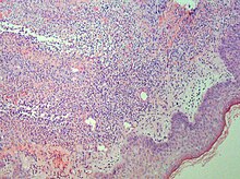
Reactive neutrophilic dermatoses are a spectrum of conditions mediated by neutrophils, and typically associated with underlying diseases, such as inflammatory bowel disease and hematologic malignancy.

Neutrophilic dermatosis of the dorsal hands (NDDH) is a skin condition that presents with edematous pustular or ulcerative nodules or plaques localized to the dorsal hands.

Behçet's disease is recognized as a disease that cause inflammatory perivasculitis, inflammation of the tissue around a blood or lymph vessel, in practically any tissue in the body. Usually, prevalent symptoms include canker sores or ulcers in the mouth and on the genitals, and inflammation in parts of the eye. In addition, patients experience severe headache and papulopustular skin lesions as well. The disease was first described in 1937 by a Turkish dermatologist, Dr. Hulusi Behçet. Behçet's disease is most prevalent in the Middle East and the Far East regions; however, it is rare in America regions.

<span class="mw-page-title-main">White blood cell</span> Type of cells of the immunological system

White blood cells, also called immune cells or immunocytes, are cells of the immune system that are involved in protecting the body against both infectious disease and foreign invaders. White blood cells include three main subtypes: granulocytes, lymphocytes and monocytes.

<span class="mw-page-title-main">Bowel-associated dermatosis–arthritis syndrome</span> Medical condition

Bowel-associated dermatosis–arthritis syndrome (BADAS), is a complication of jejunoileal bypass surgery consisting of flu-like symptoms, multiple painful joints (polyarthralgia), muscle aches (myalgia) and skin changes. It has been reported to occur in up to 20% of patients who had jejunoileal bypass surgery, a form of obesity surgery that is rarely performed today.

<span class="mw-page-title-main">Behçet's disease</span> Inflammatory disorder

Behçet's disease (BD) is a type of inflammatory disorder which affects multiple parts of the body. The most common symptoms include painful sores on the mucous membranes of the mouth and other parts of the body, inflammation of parts of the eye, and arthritis. The sores can last from a few days, up to a week or more. Less commonly there may be inflammation of the brain or spinal cord, blood clots, aneurysms, or blindness. Often, the symptoms come and go.

Cutaneous manifestations of COVID-19 are characteristic signs or symptoms of the Coronavirus disease 2019 that occur in the skin. The American Academy of Dermatology reports that skin lesions such as morbilliform, pernio, urticaria, macular erythema, vesicular purpura, papulosquamous purpura and retiform purpura are seen in people with COVID-19. Pernio-like lesions were more common in mild disease while retiform purpura was seen only in critically ill patients. The major dermatologic patterns identified in individuals with COVID-19 are urticarial rash, confluent erythematous/morbilliform rash, papulovesicular exanthem, chilbain-like acral pattern, livedo reticularis and purpuric "vasculitic" pattern. Chilblains and Multisystem inflammatory syndrome in children are also cutaneous manifestations of COVID-19.

Autoinflammatory diseases (AIDs) are a group of rare disorders caused by dysfunction of the innate immune system. These responses are characterized by periodic or chronic systemic inflammation, usually without the involvement of adaptive immunity.

References

  1. Mustafa NM, Lavizzo M (2008). "Sweet syndrome in a patient with Crohn disease: a case report". J Med Case Rep. 2: 221. doi: 10.1186/1752-1947-2-221 . PMC   2503996 . PMID   18588703.
  2. James, W; Berger, T; Elston D (2005). Andrews Diseases of the Skin: Clinical Dermatology (10th ed.). Saunders. p. 145. ISBN   978-0-7216-2921-6.
  3. synd/3019 at Who Named It?
  4. Sweet RD (1964). "An acute febrile neutrophilic dermatosis". Br. J. Dermatol. 76 (8–9): 349–56. doi:10.1111/j.1365-2133.1964.tb14541.x. PMID   14201182. S2CID   53772268.
  5. 1 2 3 4 Cohen, Philip R (December 2007). "Sweet's syndrome – a comprehensive review of an acute febrile neutrophilic dermatosis". Orphanet Journal of Rare Diseases. 2 (1). doi: 10.1186/1750-1172-2-34 .
  6. Parsapour K, Reep MD, Gohar K, Shah V, Church A, Shwayder TA (July 2003). "Familial Sweet's syndrome in 2 brothers, both seen in the first 2 weeks of life". J. Am. Acad. Dermatol. 49 (1): 132–8. doi:10.1067/mjd.2003.328. PMID   12833027.
  7. Hisanaga K, Iwasaki Y, Itoyama Y, the neuro-Sweet disease study group (2005). “Neuro-Sweet disease": Clinical manifestations and criteria for diagnosis. Neurology. 64: 1756-1761.
  8. Hisanaga K (2022). “Neuro-Behçet disease, neuro-Sweet disease, and spectrum disorders.” Intern Med. 61: 447-450.