Vascular endothelial growth factor C

Last updated
VEGFC
Protein VEGFC PDB 2X1W.png
Available structures
PDB Ortholog search: PDBe RCSB
Identifiers
Aliases VEGFC , Flt4-L, LMPH1D, VRP, vascular endothelial growth factor C, LMPHM4
External IDs OMIM: 601528 MGI: 109124 HomoloGene: 3962 GeneCards: VEGFC
Orthologs
SpeciesHumanMouse
Entrez
Ensembl
UniProt
RefSeq (mRNA)

NM_005429

NM_009506

RefSeq (protein)

NP_005420

NP_033532

Location (UCSC) Chr 4: 176.68 – 176.79 Mb Chr 8: 54.53 – 54.64 Mb
PubMed search [3] [4]
Wikidata
View/Edit Human View/Edit Mouse

Vascular endothelial growth factor C (VEGF-C) is a protein that is a member of the platelet-derived growth factor / vascular endothelial growth factor (PDGF/VEGF) family. It is encoded in humans by the VEGFC gene, which is located on chromosome 4q34. [5]

Contents

Functions

The main function of VEGF-C is to promote the growth of lymphatic vessels (lymphangiogenesis). It acts on lymphatic endothelial cells (LECs) primarily via its receptor VEGFR-3 promoting survival, growth and migration. It was discovered in 1996 as a ligand for the orphan receptor VEGFR-3. [6] Soon thereafter, it was shown to be a specific growth factor for lymphatic vessels in a variety of models. [7] [8] However, in addition to its effect on lymphatic vessels, it can also promote the growth of blood vessels and regulate their permeability. The effect on blood vessels can be mediated via its primary receptor VEGFR-3 [9] or its secondary receptor VEGFR-2. Apart from vascular targets, VEGF-C is also important for neural development [10] and blood pressure regulation. [11]

Biosynthesis

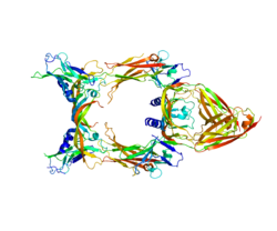
VEGF-C is a dimeric, secreted protein, which undergoes a complex proteolytic maturation resulting in multiple processed forms. After translation, VEGF-C consists of three domains: the central VEGF homology domain (VHD), the N-terminal domain (propeptide) and a C-terminal domain (propeptide). [12] It is referred to as "uncleaved VEGF-C" and has a size of approximately 58 kDa. The first cleavage (which happens already before secretion) occurs between the VHD and the C-terminal domain and is mediated by proprotein convertases. [13] However, the resulting protein is still held together by disulfide bonds and remains inactive (although it can bind already VEGFR-3). [14] This form is referred to as "intermediate form" or pro-VEGF-C and it consists of two polypeptide chains of 29 and 31 kDa. In order to activate VEGF-C, a second cleavage has to occur between the N-terminal propeptide and the VHD. This cleavage can be performed either by ADAMTS3, [14] plasmin, [15] KLK3/PSA or cathepsin D. [16] With progressing maturation, the affinity of VEGF-C for both VEGFR-2 and VEGFR-3 increases and only the fully processed, mature forms of VEGF-C have a significant affinity for VEGFR-2. [12]

Relationship to VEGF-D

The closest structural and functional relative of VEGF-C is VEGF-D. [17] However, at least in mice, VEGF-C is absolutely essential for the development of the lymphatic system, [18] whereas VEGF-D appears to be unnecessary. [19] Whether this holds true for humans is unknown, because there are major differences between human and mouse VEGF-D. [20]

Disease relevance

In a minority of lymphedema patients, the condition is caused by mutations in the VEGFC gene [21] and VEGF-C is a potential treatment for lymphedema, [22] [23] even though the underlying molecular cause appears more often in the VEGF-Receptor-3 instead of VEGF-C itself. [24] Because in Milroy's disease (Hereditary lymphedema type I), only one allele is mutated, not all VEGFR-3 molecules are non-functional and it is thought, that high amounts of VEGF-C can compensate for the mutated, nonfunctional receptors by increasing the signaling levels of the remaining functional receptors. [25] Therefore, VEGF-C is developed as a lymphedema drug under the name of Lymfactin. [26] Also indirectly VEGF-C can be responsible for hereditary lymphedema: The rare Hennekam syndrome can result from the inability of the mutated CCBE1 to assist the ADAMTS3 protease in activating VEGF-C. [14] While lack of VEGF-C results in lymphedema, VEGF-C production is implicated in tumor lymphangiogenesis and metastasis. Expression of VEGF-C by tumors induces peri-tumoral and intratumoral lymphangiogenesis what potently promotes metastatic dissemination of tumor cells. [27] [28] VEGF-C primarily stimulates lymphangiogenesis by activating VEGFR-3, yet under certain conditions it can also act directly on blood vessels to promote tumor angiogenesis. [9] [29]

Evolution

The PDGF family is so closely related to the VEGF family that the two are sometimes grouped together as the PDGF/VEGF family. In invertebrates, molecules from this families are not easily distinguished from each other and are collectively referred to as PVFs (PDGF/VEGF-like growth factors. [30] The comparison of human VEGFs with these PVFs allows conclusions on the structure of the ancestral molecules, which appear more closely related to today's lymphangiogenic VEGF-C than to the other members of the VEGF family and despite their large evolutionary distance are still able to interact with human VEGF receptors. The PVFs in Drosophila melanogaster have functions for the migration of hemocytes [31] and the PVFs in the jellyfish Podocoryne carnea for the development of the tentacles and the gastrovascular apparatus. [32] However, the function of the PVF-1 of the nematode Caenorhabditis elegans is unknown [30]

Related Research Articles

<span class="mw-page-title-main">Angiogenesis</span> Blood vessel formation, when new vessels emerge from existing vessels

Angiogenesis is the physiological process through which new blood vessels form from pre-existing vessels, formed in the earlier stage of vasculogenesis. Angiogenesis continues the growth of the vasculature mainly by processes of sprouting and splitting, but processes such as coalescent angiogenesis, vessel elongation and vessel cooption also play a role. Vasculogenesis is the embryonic formation of endothelial cells from mesoderm cell precursors, and from neovascularization, although discussions are not always precise. The first vessels in the developing embryo form through vasculogenesis, after which angiogenesis is responsible for most, if not all, blood vessel growth during development and in disease.

<span class="mw-page-title-main">Lymphatic vessel</span> Tubular vessels that are involved in the transport of lymph and lymphocytes

The lymphatic vessels are thin-walled vessels (tubes), structured like blood vessels, that carry lymph. As part of the lymphatic system, lymph vessels are complementary to the cardiovascular system. Lymph vessels are lined by endothelial cells, and have a thin layer of smooth muscle, and adventitia that binds the lymph vessels to the surrounding tissue. Lymph vessels are devoted to the propulsion of the lymph from the lymph capillaries, which are mainly concerned with the absorption of interstitial fluid from the tissues. Lymph capillaries are slightly bigger than their counterpart capillaries of the vascular system. Lymph vessels that carry lymph to a lymph node are called afferent lymph vessels, and those that carry it from a lymph node are called efferent lymph vessels, from where the lymph may travel to another lymph node, may be returned to a vein, or may travel to a larger lymph duct. Lymph ducts drain the lymph into one of the subclavian veins and thus return it to general circulation.

<span class="mw-page-title-main">Platelet-derived growth factor</span> Signaling glycoprotein regulating cell proliferation

Platelet-derived growth factor (PDGF) is one among numerous growth factors that regulate cell growth and division. In particular, PDGF plays a significant role in blood vessel formation, the growth of blood vessels from already-existing blood vessel tissue, mitogenesis, i.e. proliferation, of mesenchymal cells such as fibroblasts, osteoblasts, tenocytes, vascular smooth muscle cells and mesenchymal stem cells as well as chemotaxis, the directed migration, of mesenchymal cells. Platelet-derived growth factor is a dimeric glycoprotein that can be composed of two A subunits (PDGF-AA), two B subunits (PDGF-BB), or one of each (PDGF-AB).

Lymphangiogenesis is the formation of lymphatic vessels from pre-existing lymphatic vessels in a method believed to be similar to angiogenesis.

Vascular endothelial growth factor, originally known as vascular permeability factor (VPF), is a signal protein produced by many cells that stimulates the formation of blood vessels. To be specific, VEGF is a sub-family of growth factors, the platelet-derived growth factor family of cystine-knot growth factors. They are important signaling proteins involved in both vasculogenesis and angiogenesis.

<span class="mw-page-title-main">Endothelial stem cell</span> Stem cell in bone marrow that gives rise to endothelial cells

Endothelial stem cells (ESCs) are one of three types of stem cells found in bone marrow. They are multipotent, which describes the ability to give rise to many cell types, whereas a pluripotent stem cell can give rise to all types. ESCs have the characteristic properties of a stem cell: self-renewal and differentiation. These parent stem cells, ESCs, give rise to progenitor cells, which are intermediate stem cells that lose potency. Progenitor stem cells are committed to differentiating along a particular cell developmental pathway. ESCs will eventually produce endothelial cells (ECs), which create the thin-walled endothelium that lines the inner surface of blood vessels and lymphatic vessels. The lymphatic vessels include things such as arteries and veins. Endothelial cells can be found throughout the whole vascular system and they also play a vital role in the movement of white blood cells

<span class="mw-page-title-main">Receptor tyrosine kinase</span> Class of enzymes

Receptor tyrosine kinases (RTKs) are the high-affinity cell surface receptors for many polypeptide growth factors, cytokines, and hormones. Of the 90 unique tyrosine kinase genes identified in the human genome, 58 encode receptor tyrosine kinase proteins. Receptor tyrosine kinases have been shown not only to be key regulators of normal cellular processes but also to have a critical role in the development and progression of many types of cancer. Mutations in receptor tyrosine kinases lead to activation of a series of signalling cascades which have numerous effects on protein expression. Receptor tyrosine kinases are part of the larger family of protein tyrosine kinases, encompassing the receptor tyrosine kinase proteins which contain a transmembrane domain, as well as the non-receptor tyrosine kinases which do not possess transmembrane domains.

<span class="mw-page-title-main">Angiopoietin</span> Protein family

Angiopoietin is part of a family of vascular growth factors that play a role in embryonic and postnatal angiogenesis. Angiopoietin signaling most directly corresponds with angiogenesis, the process by which new arteries and veins form from preexisting blood vessels. Angiogenesis proceeds through sprouting, endothelial cell migration, proliferation, and vessel destabilization and stabilization. They are responsible for assembling and disassembling the endothelial lining of blood vessels. Angiopoietin cytokines are involved with controlling microvascular permeability, vasodilation, and vasoconstriction by signaling smooth muscle cells surrounding vessels. There are now four identified angiopoietins: ANGPT1, ANGPT2, ANGPTL3, ANGPT4.

<span class="mw-page-title-main">VEGF receptor</span> Protein family

VEGF receptors (VEGFRs) are receptors for vascular endothelial growth factor (VEGF). There are three main subtypes of VEGFR, numbered 1, 2 and 3. Depending on alternative splicing, they may be membrane-bound (mbVEGFR) or soluble (sVEGFR).

<span class="mw-page-title-main">VEGFR1</span> Protein-coding gene in the species Homo sapiens

Vascular endothelial growth factor receptor 1 is a protein that in humans is encoded by the FLT1 gene.

<span class="mw-page-title-main">Kinase insert domain receptor</span> Protein-coding gene in the species Homo sapiens

Kinase insert domain receptor also known as vascular endothelial growth factor receptor 2 (VEGFR-2) is a VEGF receptor. KDR is the human gene encoding it. KDR has also been designated as CD309. KDR is also known as Flk1.

<span class="mw-page-title-main">C-fos-induced growth factor</span> Mammalian protein found in humans

C-fos-induced growth factor (FIGF) is a vascular endothelial growth factor that in humans is encoded by the FIGF gene.

<span class="mw-page-title-main">Vascular endothelial growth factor B</span> Protein-coding gene in the species Homo sapiens

Vascular endothelial growth factor B also known as VEGF-B is a protein that, in humans, is encoded by the VEGF-B gene. VEGF-B is a growth factor that belongs to the vascular endothelial growth factor family, of which VEGF-A is the best-known member.

<span class="mw-page-title-main">ADAMTS3</span> Protein-coding gene in the species Homo sapiens

A disintegrin and metalloproteinase with thrombospondin motifs 3 is an enzyme that in humans is encoded by the ADAMTS3 gene. The protein encoded by this gene is the major procollagen II N-propeptidase.

<span class="mw-page-title-main">FLT4</span> Protein-coding gene in the species Homo sapiens

Fms-related tyrosine kinase 4, also known as FLT4, is a protein which in humans is encoded by the FLT4 gene.

The lymphatic endothelium refers to a specialized subset of endothelial cells located in the sinus systems of draining lymph nodes. Specifically, these endothelial cells line the branched sinus systems formed by afferent lymphatic vessels, forming a single-cell layer which functions in a variety of critical physiological processes. These lymphatic endothelial cells contribute directly to immune function and response modulation, provide transport selectivity, and demonstrate orchestration of bidirectional signaling cascades. Additionally, lymphatic endothelial cells may be implicated in downstream immune cell development as well as lymphatic organogenesis. Until recently, lymphatic endothelial cells have not been characterized to their optimal potential. This system is very important in the function of continuous removal of interstitial fluid and proteins, while also having a significant function of entry for leukocytes and tumor cells. This leads to further research that is being developed on the relationship between lymphatic endothelium and metastasis of tumor cells . The lymphatic capillaries are described to be blind ended vessels, and they are made up of a single non-fenestrated layer of endothelial cells; The lymph capillaries function to aid in the uptake of fluids, macromolecules, and cells. Although they are generally similar to blood capillaries, the lymph capillaries have distinct structural differences. Lymph capillaries consist of a more wide and irregular lumen, and the endothelium in lymph capillaries is much thinner as well. Their origin has been speculated to vary based on them being dependent on specific tissue environments, and powered by organ-specific signals.(L. Gutierrez-Miranda, K. Yaniv, 2020). A lymph capillary endothelial cell is distinct from other endothelial cells in that collagen fibers are directly attached to its plasma membrane.

<span class="mw-page-title-main">Hennekam syndrome</span> Medical condition

Hennekam syndrome also known as intestinal lymphagiectasia–lymphedema–mental retardation syndrome, is an autosomal recessive disorder consisting of intestinal lymphangiectasia, facial anomalies, peripheral lymphedema, and mild to moderate levels of growth and intellectual disability.

<span class="mw-page-title-main">CCBE1</span> Protein-coding gene in the species Homo sapiens

Collagen and calcium-binding EGF domain-containing protein 1 is a protein that in humans is encoded by the CCBE1 gene.

<span class="mw-page-title-main">Kari Alitalo</span> Finnish MD and a medical researcher

Kari Kustaa Alitalo is a Finnish MD and a medical researcher. He is a foreign associated member of the National Academy of Sciences of the US. He became famous for his discoveries of several receptor tyrosine kinases (RTKs) and the first growth factor capable of inducing lymphangiogenesis: vascular endothelial growth factor C (VEGF-C). In the years 1996–2007 he was Europe's second most cited author in the field of cell biology. Alitalo is currently serving as an Academy Professor for the Academy of Finland.

Michael Jeltsch is a German-Finnish researcher in the field of Biochemistry. He is an associate professor at the University of Helsinki, Finland. He has more than 70 publications. Jeltsch was the first to show that VEGF-C and VEGF-D are the principal growth factors for the lymphatic vasculature and his research focuses on cancer drug targets and lymphangiogenesis. He has also contributed to other seminal publications in cell biology with transgenesis, protein engineering, recombinant production and purification. In 2006, he developed a synthetic super-VEGF, using a library of VEGF hybrid molecules using a novel, non-random DNA family shuffling method.

References

  1. 1 2 3 GRCh38: Ensembl release 89: ENSG00000150630 - Ensembl, May 2017
  2. 1 2 3 GRCm38: Ensembl release 89: ENSMUSG00000031520 - Ensembl, May 2017
  3. "Human PubMed Reference:". National Center for Biotechnology Information, U.S. National Library of Medicine.
  4. "Mouse PubMed Reference:". National Center for Biotechnology Information, U.S. National Library of Medicine.
  5. Paavonen K, Horelli-Kuitunen N, Chilov D, Kukk E, Pennanen S, Kallioniemi OP, et al. (March 1996). "Novel human vascular endothelial growth factor genes VEGF-B and VEGF-C localize to chromosomes 11q13 and 4q34, respectively". Circulation. 93 (6): 1079–1082. doi:10.1161/01.CIR.93.6.1079. PMID   8653826.
  6. Joukov V, Pajusola K, Kaipainen A, Chilov D, Lahtinen I, Kukk E, et al. (January 1996). "A novel vascular endothelial growth factor, VEGF-C, is a ligand for the Flt4 (VEGFR-3) and KDR (VEGFR-2) receptor tyrosine kinases". The EMBO Journal. 15 (2): 290–298. doi:10.1002/j.1460-2075.1996.tb00359.x. PMC   449944 . PMID   8617204.
  7. Oh SJ, Jeltsch MM, Birkenhäger R, McCarthy JE, Weich HA, Christ B, et al. (August 1997). "VEGF and VEGF-C: specific induction of angiogenesis and lymphangiogenesis in the differentiated avian chorioallantoic membrane". Developmental Biology. 188 (1): 96–109. doi: 10.1006/dbio.1997.8639 . PMID   9245515.
  8. Jeltsch M, Kaipainen A, Joukov V, Meng X, Lakso M, Rauvala H, et al. (May 1997). "Hyperplasia of lymphatic vessels in VEGF-C transgenic mice". Science. 276 (5317): 1423–1425. doi:10.1126/science.276.5317.1423. PMID   9162011. S2CID   21835142.
  9. 1 2 Tammela T, Zarkada G, Wallgard E, Murtomäki A, Suchting S, Wirzenius M, et al. (July 2008). "Blocking VEGFR-3 suppresses angiogenic sprouting and vascular network formation". Nature. 454 (7204): 656–660. Bibcode:2008Natur.454..656T. doi:10.1038/nature07083. PMID   18594512. S2CID   2251527.
  10. Le Bras B, Barallobre MJ, Homman-Ludiye J, Ny A, Wyns S, Tammela T, et al. (March 2006). "VEGF-C is a trophic factor for neural progenitors in the vertebrate embryonic brain". Nature Neuroscience. 9 (3): 340–348. doi:10.1038/nn1646. PMID   16462734. S2CID   24197350.
  11. Machnik A, Neuhofer W, Jantsch J, Dahlmann A, Tammela T, Machura K, et al. (May 2009). "Macrophages regulate salt-dependent volume and blood pressure by a vascular endothelial growth factor-C-dependent buffering mechanism". Nature Medicine. 15 (5): 545–552. doi:10.1038/nm.1960. PMID   19412173. S2CID   10526891.
  12. 1 2 Joukov V, Sorsa T, Kumar V, Jeltsch M, Claesson-Welsh L, Cao Y, et al. (July 1997). "Proteolytic processing regulates receptor specificity and activity of VEGF-C". The EMBO Journal. 16 (13): 3898–3911. doi:10.1093/emboj/16.13.3898. PMC   1170014 . PMID   9233800.
  13. Siegfried G, Basak A, Cromlish JA, Benjannet S, Marcinkiewicz J, Chrétien M, et al. (June 2003). "The secretory proprotein convertases furin, PC5, and PC7 activate VEGF-C to induce tumorigenesis". The Journal of Clinical Investigation. 111 (11): 1723–1732. doi:10.1172/JCI17220. PMC   156106 . PMID   12782675.
  14. 1 2 3 Jeltsch M, Jha SK, Tvorogov D, Anisimov A, Leppänen VM, Holopainen T, et al. (May 2014). "CCBE1 enhances lymphangiogenesis via A disintegrin and metalloprotease with thrombospondin motifs-3-mediated vascular endothelial growth factor-C activation". Circulation. 129 (19): 1962–1971. doi: 10.1161/CIRCULATIONAHA.113.002779 . PMID   24552833.
  15. McColl BK, Baldwin ME, Roufail S, Freeman C, Moritz RL, Simpson RJ, et al. (September 2003). "Plasmin activates the lymphangiogenic growth factors VEGF-C and VEGF-D". The Journal of Experimental Medicine. 198 (6): 863–868. doi:10.1084/jem.20030361. PMC   2194198 . PMID   12963694.
  16. Jha SK, Rauniyar K, Chronowska E, Mattonet K, Maina EW, Koistinen H, et al. (May 2019). "KLK3/PSA and cathepsin D activate VEGF-C and VEGF-D". eLife. 8: 44478. doi: 10.7554/eLife.44478 . PMC   6588350 . PMID   31099754.
  17. Achen MG, Jeltsch M, Kukk E, Mäkinen T, Vitali A, Wilks AF, et al. (January 1998). "Vascular endothelial growth factor D (VEGF-D) is a ligand for the tyrosine kinases VEGF receptor 2 (Flk1) and VEGF receptor 3 (Flt4)". Proceedings of the National Academy of Sciences of the United States of America. 95 (2): 548–553. Bibcode:1998PNAS...95..548A. doi: 10.1073/pnas.95.2.548 . PMC   18457 . PMID   9435229.
  18. Karkkainen MJ, Haiko P, Sainio K, Partanen J, Taipale J, Petrova TV, et al. (January 2004). "Vascular endothelial growth factor C is required for sprouting of the first lymphatic vessels from embryonic veins". Nature Immunology. 5 (1): 74–80. doi:10.1038/ni1013. PMID   14634646. S2CID   22078757.
  19. Baldwin ME, Halford MM, Roufail S, Williams RA, Hibbs ML, Grail D, et al. (March 2005). "Vascular endothelial growth factor D is dispensable for development of the lymphatic system". Molecular and Cellular Biology. 25 (6): 2441–2449. doi:10.1128/MCB.25.6.2441-2449.2005. PMC   1061605 . PMID   15743836.
  20. Baldwin ME, Catimel B, Nice EC, Roufail S, Hall NE, Stenvers KL, et al. (June 2001). "The specificity of receptor binding by vascular endothelial growth factor-d is different in mouse and man". The Journal of Biological Chemistry. 276 (22): 19166–19171. doi: 10.1074/jbc.M100097200 . PMID   11279005. S2CID   41677159.
  21. Balboa-Beltran E, Fernández-Seara MJ, Pérez-Muñuzuri A, Lago R, García-Magán C, Couce ML, et al. (July 2014). "A novel stop mutation in the vascular endothelial growth factor-C gene (VEGFC) results in Milroy-like disease". Journal of Medical Genetics. 51 (7): 475–478. doi:10.1136/jmedgenet-2013-102020. PMID   24744435. S2CID   6613861.
  22. Enholm B, Karpanen T, Jeltsch M, Kubo H, Stenback F, Prevo R, et al. (March 2001). "Adenoviral expression of vascular endothelial growth factor-C induces lymphangiogenesis in the skin". Circulation Research. 88 (6): 623–629. doi: 10.1161/01.RES.88.6.623 . PMID   11282897. S2CID   28806663.
  23. Honkonen KM, Visuri MT, Tervala TV, Halonen PJ, Koivisto M, Lähteenvuo MT, et al. (May 2013). "Lymph node transfer and perinodal lymphatic growth factor treatment for lymphedema". Annals of Surgery. 257 (5): 961–7. doi:10.1097/SLA.0b013e31826ed043. PMID   23013803. S2CID   2042145.
  24. Brouillard P, Boon L, Vikkula M (March 2014). "Genetics of lymphatic anomalies". The Journal of Clinical Investigation. 124 (3): 898–904. doi:10.1172/JCI71614. PMC   3938256 . PMID   24590274.
  25. Karkkainen MJ, Saaristo A, Jussila L, Karila KA, Lawrence EC, Pajusola K, et al. (October 2001). "A model for gene therapy of human hereditary lymphedema". Proceedings of the National Academy of Sciences of the United States of America. 98 (22): 12677–12682. Bibcode:2001PNAS...9812677K. doi: 10.1073/pnas.221449198 . PMC   60113 . PMID   11592985.
  26. Herantis Pharma (2014-07-21). "Lymfactin® for lymphedema".{{cite web}}: Missing or empty |url= (help)
  27. Skobe M, Hawighorst T, Jackson DG, Prevo R, Janes L, Velasco P, et al. (February 2001). "Induction of tumor lymphangiogenesis by VEGF-C promotes breast cancer metastasis". Nature Medicine. 7 (2): 192–198. doi:10.1038/84643. PMID   11175850. S2CID   26090359.
  28. Mandriota SJ, Jussila L, Jeltsch M, Compagni A, Baetens D, Prevo R, et al. (February 2001). "Vascular endothelial growth factor-C-mediated lymphangiogenesis promotes tumour metastasis". The EMBO Journal. 20 (4): 672–682. doi:10.1093/emboj/20.4.672. PMC   145430 . PMID   11179212.
  29. Tvorogov D, Anisimov A, Zheng W, Leppänen VM, Tammela T, Laurinavicius S, et al. (December 2010). "Effective suppression of vascular network formation by combination of antibodies blocking VEGFR ligand binding and receptor dimerization". Cancer Cell. 18 (6): 630–640. doi: 10.1016/j.ccr.2010.11.001 . PMID   21130043.
  30. 1 2 Tarsitano M, De Falco S, Colonna V, McGhee JD, Persico MG (February 2006). "The C. elegans pvf-1 gene encodes a PDGF/VEGF-like factor able to bind mammalian VEGF receptors and to induce angiogenesis". FASEB Journal. 20 (2): 227–233. doi: 10.1096/fj.05-4147com . PMID   16449794. S2CID   31963203.
  31. Heino TI, Kärpänen T, Wahlström G, Pulkkinen M, Eriksson U, Alitalo K, Roos C (November 2001). "The Drosophila VEGF receptor homolog is expressed in hemocytes". Mechanisms of Development. 109 (1): 69–77. doi:10.1016/S0925-4773(01)00510-X. PMID   11677054. S2CID   14074572.
  32. Seipel K, Eberhardt M, Müller P, Pescia E, Yanze N, Schmid V (October 2004). "Homologs of vascular endothelial growth factor and receptor, VEGF and VEGFR, in the jellyfish Podocoryne carnea". Developmental Dynamics. 231 (2): 303–312. doi: 10.1002/dvdy.20139 . PMID   15366007. S2CID   42930371.

Further reading