ABO blood group system

Last updated

ABO blood group antigens present on red blood cells and IgM antibodies present in the serum ABO blood type.svg
ABO blood group antigens present on red blood cells and IgM antibodies present in the serum

The ABO blood group system is used to denote the presence of one, both, or neither of the A and B antigens on erythrocytes. [1] For human blood transfusions, it is the most important of the 44 different blood type (or group) classification systems currently recognized by the International Society of Blood Transfusions (ISBT) as of December 2022. [2] [3] A mismatch (very rare in modern medicine) in this, or any other serotype, can cause a potentially fatal adverse reaction after a transfusion, or an unwanted immune response to an organ transplant. [4] The associated anti-A and anti-B antibodies are usually IgM antibodies, produced in the first years of life by sensitization to environmental substances such as food, bacteria, and viruses.

Contents

The ABO blood types were discovered by Karl Landsteiner in 1901; he received the Nobel Prize in Physiology or Medicine in 1930 for this discovery. [5] ABO blood types are also present in other primates such as apes, monkeys and Old World monkeys. [6]

History

Discovery

The ABO blood types were first discovered by an Austrian physician, Karl Landsteiner, working at the Pathological-Anatomical Institute of the University of Vienna (now Medical University of Vienna). In 1900, he found that red blood cells would clump together (agglutinate) when mixed in test tubes with sera from different persons, and that some human blood also agglutinated with animal blood. [7] He wrote a two-sentence footnote:

The serum of healthy human beings not only agglutinates animal red cells, but also often those of human origin, from other individuals. It remains to be seen whether this appearance is related to inborn differences between individuals or it is the result of some damage of bacterial kind. [8]

This was the first evidence that blood variations exist in humans – it was believed that all humans have similar blood. The next year, in 1901, he made a definitive observation that blood serum of an individual would agglutinate with only those of certain individuals. Based on this he classified human blood into three groups, namely group A, group B, and group C. He defined that group A blood agglutinates with group B, but never with its own type. Similarly, group B blood agglutinates with group A. Group C blood is different in that it agglutinates with both A and B. [9]

This was the discovery of blood groups for which Landsteiner was awarded the Nobel Prize in Physiology or Medicine in 1930. In his paper, he referred to the specific blood group interactions as isoagglutination, and also introduced the concept of agglutinins (antibodies), which is the actual basis of antigen-antibody reaction in the ABO system. [10] He asserted:

[It] may be said that there exist at least two different types of agglutinins, one in A, another one in B, and both together in C. The red blood cells are inert to the agglutinins which are present in the same serum. [9]

Thus, he discovered two antigens (agglutinogens A and B) and two antibodies (agglutinins – anti-A and anti-B). His third group (C) indicated absence of both A and B antigens, but contains anti-A and anti-B. [10] The following year, his students Adriano Sturli and Alfred von Decastello discovered the fourth type (but not naming it, and simply referred to it as "no particular type"). [11] [12]

Ukraine marine uniform imprint, showing the wearer's blood type as "B (III) Rh+" UkrArmyBloodGroupIII+2000s.jpg
Ukraine marine uniform imprint, showing the wearer's blood type as "B (III) Rh+"

In 1910, Ludwik Hirszfeld and Emil Freiherr von Dungern introduced the term 0 (null) for the group Landsteiner designated as C, and AB for the type discovered by Sturli and von Decastello. They were also the first to explain the genetic inheritance of the blood groups. [13] [14]

Classification systems

Jan Jansky, who invented type I, II, III, IV system Jan Jansky, 1902.jpg
Jan Janský, who invented type I, II, III, IV system

Czech serologist Jan Janský independently introduced blood type classification in 1907 in a local journal. [15] He used the Roman numerical I, II, III, and IV (corresponding to modern 0, A, B, and AB). Unknown to Janský, an American physician William L. Moss devised a slightly different classification using the same numerical; [16] his I, II, III, and IV corresponding to modern AB, A, B, and 0. [12]

These two systems created confusion and potential danger in medical practice. Moss's system was adopted in Britain, France, and US, while Janský's was preferred in most European countries and some parts of US. To resolve the chaos, the American Association of Immunologists, the Society of American Bacteriologists, and the Association of Pathologists and Bacteriologists made a joint recommendation in 1921 that the Jansky classification be adopted based on priority. [17] But it was not followed particularly where Moss's system had been used. [18]

In 1927, Landsteiner had moved to the Rockefeller Institute for Medical Research in New York. As a member of a committee of the National Research Council concerned with blood grouping, he suggested to substitute Janský's and Moss's systems with the letters O, A, B, and AB. (There was another confusion on the use of figure 0 for German null as introduced by Hirszfeld and von Dungern, because others used the letter O for ohne, meaning without or zero; Landsteiner chose the latter. [18] ) This classification was adopted by the National Research Council and became variously known as the National Research Council classification, the International classification, and most popularly the "new" Landsteiner classification. The new system was gradually accepted and by the early 1950s, it was universally followed. [19]

Other developments

The first practical use of blood typing in transfusion was by an American physician Reuben Ottenberg in 1907. Large-scale application began during the First World War (1914–1915) when citric acid began to be used for blood clot prevention. [10] Felix Bernstein demonstrated the correct blood group inheritance pattern of multiple alleles at one locus in 1924. [20] Watkins and Morgan, in England, discovered that the ABO epitopes were conferred by sugars, to be specific, N-acetylgalactosamine for the A-type and galactose for the B-type. [21] [22] [23] After much published literature claiming that the ABH substances were all attached to glycosphingolipids, Finne et al. (1978) found that the human erythrocyte glycoproteins contain polylactosamine chains [24] that contains ABH substances attached and represent the majority of the antigens. [25] [26] [27] The main glycoproteins carrying the ABH antigens were identified to be the Band 3 and Band 4.5 proteins and glycophorin. [28] Later, Yamamoto's group showed the precise glycosyl transferase set that confers the A, B and O epitopes. [29]

Genetics

A and B are codominant, giving the AB phenotype. ABO system codominance.svg
A and B are codominant, giving the AB phenotype.
Punnett square of the possible genotypes and phenotypes of children given genotypes and phenotypes of their mother (rows) and father (columns) shaded by phenotype Punnett square blood types.svg
Punnett square of the possible genotypes and phenotypes of children given genotypes and phenotypes of their mother (rows) and father (columns) shaded by phenotype

Blood groups are inherited from both parents. The ABO blood type is controlled by a single gene (the ABO gene) with three types of alleles inferred from classical genetics: i, IA, and IB. The I designation stands for isoagglutinogen, another term for antigen. [31] The gene encodes a glycosyltransferase—that is, an enzyme that modifies the carbohydrate content of the red blood cell antigens. The gene is located on the long arm of the ninth chromosome (9q34). [32]

The IA allele gives type A, IB gives type B, and i gives type O. As both IA and IB are dominant over i, only ii people have type O blood. Individuals with IAIA or IAi have type A blood, and individuals with IBIB or IBi have type B. IAIB people have both phenotypes, because A and B express a special dominance relationship: codominance, which means that type A and B parents can have an AB child. A couple with type A and type B can also have a type O child if they are both heterozygous (IBi and IAi). The cis-AB phenotype has a single enzyme that creates both A and B antigens. The resulting red blood cells do not usually express A or B antigen at the same level that would be expected on common group A1 or B red blood cells, which can help solve the problem of an apparently genetically impossible blood group. [33]

Blood group inheritance
Blood typeOABAB
Genotypeii(OO)IAi(AO)IAIA(AA)IBi(BO)IBIB(BB)IAIB(AB)
Oii(OO)O
OO OO OO OO
O or A
AO OO AO OO
A
AO AO AO AO
O or B
BO OO BO OO
B
BO BO BO BO
A or B
AO BO AO BO
AIAi(AO)O or A
AO AO OO OO
O or A
AA AO AO OO
A
AA AA AO AO
O, A, B or AB
AB AO BO OO
B or AB
AB AB BO BO
A, B or AB
AA AB AO BO
IAIA(AA)A
AO AO AO AO
A
AA AO AA AO
A
AA AA AA AA
A or AB
AB AO AB AO
AB
AB AB AB AB
A or AB
AA AB AA AB
BIBi(BO)O or B
BO BO OO OO
O, A, B or AB
AB BO AO OO
A or AB
AB AB AO AO
O or B
BB BO BO OO
B
BB BB BO BO
A, B or AB
AB BB AO BO
IBIB(BB)B
BO BO BO BO
B or AB
AB BO AB BO
AB
AB AB AB AB
B
BB BO BB BO
B
BB BB BB BB
B or AB
AB BB AB BB
ABIAIB(AB)A or B
AO AO BO BO
A, B or AB
AA AO AB BO
A or AB
AA AA AB AB
A, B or AB
AB AO BB BO
B or AB
AB AB BB BB
A, B, or AB
AA AB AB BB

Individuals with the rare Bombay phenotype (hh) produce antibodies against the A, B, and O groups and can only receive transfusions from other hh individuals. The table above summarizes the various blood groups that children may inherit from their parents. [34] [35] Genotypes are shown in the second column and in small print for the offspring: AO and AA both test as type A; BO and BB test as type B. The four possibilities represent the combinations obtained when one allele is taken from each parent; each has a 25% chance, but some occur more than once. The text above them summarizes the outcomes.

Blood group inheritance by phenotype only
Blood typeOABAB
OOO or AO or BA or B
AO or AO or AO, A, B or ABA, B or AB
BO or BO, A, B or ABO or BA, B or AB
ABA or BA, B or ABA, B or ABA, B or AB

Historically, ABO blood tests were used in paternity testing, but in 1957 only 50% of American men falsely accused were able to use them as evidence against paternity. [36] Occasionally, the blood types of children are not consistent with expectations—for example, a type O child can be born to an AB parent—due to rare situations, such as Bombay phenotype and cis AB. [37]

Subgroups

The A blood type contains about 20 subgroups, of which A1 and A2 are the most common (over 99%). A1 makes up about 80% of all A-type blood, with A2 making up almost all of the rest. [38] These two subgroups are not always interchangeable as far as transfusion is concerned, as some A2 individuals produce antibodies against the A1 antigen. Complications can sometimes arise in rare cases when typing the blood. [38]

With the development of DNA sequencing, it has been possible to identify a much larger number of alleles at the ABO locus, each of which can be categorized as A, B, or O in terms of the reaction to transfusion, but which can be distinguished by variations in the DNA sequence. There are six common alleles in white individuals of the ABO gene that produce one's blood type: [39] [40]

ABO
A101 (A1)
A201 (A2)
B101 (B1)O01 (O1)
O02 (O1v)
O03 (O2)

The same study also identified 18 rare alleles, which generally have a weaker glycosylation activity. People with weak alleles of A can sometimes express anti-A antibodies, though these are usually not clinically significant as they do not stably interact with the antigen at body temperature. [41]

Cis AB is another rare variant, in which A and B genes are transmitted together from a single parent. [42]

Distribution and evolutionary history

The distribution of the blood groups A, B, O and AB varies across the world according to the population. There are also variations in blood type distribution within human subpopulations.[ citation needed ]

In the UK, the distribution of blood type frequencies through the population still shows some correlation to the distribution of placenames and to the successive invasions and migrations including Celts, Norsemen, Danes, Anglo-Saxons, and Normans who contributed the morphemes to the placenames and the genes to the population. The native Celts tended to have more type O blood, while the other populations tended to have more type A. [43]

The two common O alleles, O01 and O02, share their first 261 nucleotides with the group A allele A01. [44] However, unlike the group A allele, a guanosine base is subsequently deleted. A premature stop codon results from this frame-shift mutation. This variant is found worldwide, and likely predates human migration from Africa. The O01 allele is considered to predate the O02 allele.[ citation needed ]

Some evolutionary biologists theorize that there are four main lineages of the ABO gene and that mutations creating type O have occurred at least three times in humans. [45] From oldest to youngest, these lineages comprise the following alleles: A101/A201/O09, B101, O02 and O01. The continued presence of the O alleles is hypothesized to be the result of balancing selection. [45] Both theories contradict the previously held theory that type O blood evolved first.[ citation needed ]

Origin theories

It is possible that food and environmental antigens (bacterial, viral, or plant antigens) have epitopes similar enough to A and B glycoprotein antigens. The antibodies created against these environmental antigens in the first years of life can cross-react with ABO-incompatible red blood cells that it comes in contact with during blood transfusion later in life. Anti-A antibodies are hypothesized to originate from immune response towards influenza virus, whose epitopes are similar enough to the α-D-N-galactosamine on the A glycoprotein to be able to elicit a cross-reaction. Anti-B antibodies are hypothesized to originate from antibodies produced against Gram-negative bacteria, such as E. coli , cross-reacting with the α-D-galactose on the B glycoprotein. [46]

However, it is more likely that the force driving evolution of allele diversity is simply negative frequency-dependent selection; cells with rare variants of membrane antigens are more easily distinguished by the immune system from pathogens carrying antigens from other hosts. Thus, individuals possessing rare types are better equipped to detect pathogens. The high within-population diversity observed in human populations would, then, be a consequence of natural selection on individuals. [47]

Clinical relevance

The carbohydrate molecules on the surfaces of red blood cells have roles in cell membrane integrity, cell adhesion, membrane transportation of molecules, and acting as receptors for extracellular ligands, and enzymes. ABO antigens are found having similar roles on epithelial cells as well as red blood cells. [48] [49]

Bleeding and thrombosis (von Willebrand factor)

The ABO antigen is also expressed on the von Willebrand factor (vWF) glycoprotein, [50] which participates in hemostasis (control of bleeding). In fact, having type O blood predisposes to bleeding, [51] as 30% of the total genetic variation observed in plasma vWF is explained by the effect of the ABO blood group, [52] and individuals with group O blood normally have significantly lower plasma levels of vWF (and Factor VIII) than do non-O individuals. [53] [54] In addition, vWF is degraded more rapidly due to the higher prevalence of blood group O with the Cys1584 variant of vWF (an amino acid polymorphism in VWF): [55] the gene for ADAMTS13 (vWF-cleaving protease) maps to human chromosome 9 band q34.2, the same locus as ABO blood type. Higher levels of vWF are more common amongst people who have had ischemic stroke (from blood clotting) for the first time. The results of this study found that the occurrence was not affected by ADAMTS13 polymorphism, and the only significant genetic factor was the person's blood group. [56]

ABO(H) blood group antigens are also carried by other hemostatically relevant glycoproteins, such as platelet glycoprotein Ibα, which is a ligand for vWF on platelets. [57] The significance of ABO(H) antigen expression on these other hemostatic glycoproteins is not fully defined, but may also be relevant for bleeding and thrombosis.

ABO hemolytic disease of the newborn

ABO blood group incompatibilities between the mother and child do not usually cause hemolytic disease of the newborn (HDN) because antibodies to the ABO blood groups are usually of the IgM type, which do not cross the placenta. However, in an O-type mother, IgG ABO antibodies are produced and the baby can potentially develop ABO hemolytic disease of the newborn.[ citation needed ]

Clinical applications

In human cells, the ABO alleles and their encoded glycosyltransferases have been described in several oncologic conditions. [58] Using anti-GTA/GTB monoclonal antibodies, it was demonstrated that a loss of these enzymes was correlated to malignant bladder and oral epithelia. [59] [60] Furthermore, the expression of ABO blood group antigens in normal human tissues is dependent the type of differentiation of the epithelium. In most human carcinomas, including oral carcinoma, a significant event as part of the underlying mechanism is decreased expression of the A and B antigens. [61] Several studies have observed that a relative down-regulation of GTA and GTB occurs in oral carcinomas in association with tumor development. [61] [62] More recently, a genome wide association study (GWAS) has identified variants in the ABO locus associated with susceptibility to pancreatic cancer. [63] In addition, another large GWAS study has associated ABO-histo blood groups as well as FUT2 secretor status with the presence in the intestinal microbiome of specific bacterial species. In this case the association was with Bacteroides and Faecalibacterium spp. Bacteroides of the same OTU (operational taxonomic unit) have been shown to be associated with inflammatory bowel disease, [64] [65] thus the study suggests an important role for the ABO histo-blood group antigens as candidates for direct modulation of the human microbiome in health and disease. [66]

Clinical marker

A multi-locus genetic risk score study based on a combination of 27 loci, including the ABO gene, identified individuals at increased risk for both incident and recurrent coronary artery disease events, as well as an enhanced clinical benefit from statin therapy. The study was based on a community cohort study (the Malmo Diet and Cancer study) and four additional randomized controlled trials of primary prevention cohorts (JUPITER and ASCOT) and secondary prevention cohorts (CARE and PROVE IT-TIMI 22). [67]

Alteration of ABO antigens for transfusion

In April 2007, an international team of researchers announced in the journal Nature Biotechnology an inexpensive and efficient way to convert types A, B, and AB blood into type O. [68] This is done by using glycosidase enzymes from specific bacteria to strip the blood group antigens from red blood cells. The removal of A and B antigens still does not address the problem of the Rh blood group antigen on the blood cells of Rh positive individuals, and so blood from Rh negative donors must be used. The modified blood is named "enzyme converted to O" (ECO blood) but despite the early success of converting B- to O-type RBCs and clinical trials without adverse effects transfusing into A- and O-type patients, [69] the technology has not yet become clinical practice. [70]

Another approach to the blood antigen problem is the manufacture of artificial blood, which could act as a substitute in emergencies. [71]

Pseudoscience

During the 1930s, connecting blood groups to personality types became popular in Japan and other areas of the world. [72] Studies of this association have yet to confirm its existence definitively. [73]

Other popular but unsupported ideas include the use of a blood type diet, claims that group A causes severe hangovers, group O is associated with perfect teeth, and those with blood group A2 have the highest IQs. Scientific evidence in support of these concepts is limited at best. [74]

See also

Related Research Articles

<span class="mw-page-title-main">Blood type</span> Classification of blood based on antibodies and antigens on red blood cell surfaces

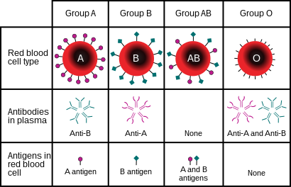
A blood type is a classification of blood, based on the presence and absence of antibodies and inherited antigenic substances on the surface of red blood cells (RBCs). These antigens may be proteins, carbohydrates, glycoproteins, or glycolipids, depending on the blood group system. Some of these antigens are also present on the surface of other types of cells of various tissues. Several of these red blood cell surface antigens can stem from one allele and collectively form a blood group system.

Rh disease is a type of hemolytic disease of the fetus and newborn (HDFN). HDFN due to anti-D antibodies is the proper and currently used name for this disease as the Rh blood group system actually has more than 50 antigens and not only the D-antigen. The term "Rh Disease" is commonly used to refer to HDFN due to anti-D antibodies, and prior to the discovery of anti-Rho(D) immune globulin, it was the most common type of HDFN. The disease ranges from mild to severe, and occurs in the second or subsequent pregnancies of Rh-D negative women when the biologic father is Rh-D positive.

<span class="mw-page-title-main">Duffy antigen system</span> Human blood group classification

Duffy antigen/chemokine receptor (DARC), also known as Fy glycoprotein (FY) or CD234, is a protein that in humans is encoded by the ACKR1 gene.

α-Galactosidase Enzyme

α-Galactosidase is a glycoside hydrolase enzyme that catalyses the following reaction:

The term human blood group systems is defined by the International Society of Blood Transfusion (ISBT) as systems in the human species where cell-surface antigens—in particular, those on blood cells—are "controlled at a single gene locus or by two or more very closely linked homologous genes with little or no observable recombination between them", and include the common ABO and Rh (Rhesus) antigen systems, as well as many others; 44 human systems are identified as of December 2022.

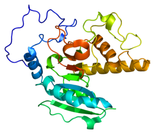
The Kell antigen system is a human blood group system, that is, a group of antigens on the human red blood cell surface which are important determinants of blood type and are targets for autoimmune or alloimmune diseases which destroy red blood cells. The Kell antigens are K, k, Kpa, Kpb, Jsa and Jsb. The Kell antigens are peptides found within the Kell protein, a 93-kilodalton transmembrane zinc-dependent endopeptidase which is responsible for cleaving endothelin-3.

<span class="mw-page-title-main">Rh blood group system</span> Human blood group system involving 49 blood antigens

The Rh blood group system is a human blood group system. It contains proteins on the surface of red blood cells. After the ABO blood group system, it is the most likely to be involved in transfusion reactions. The Rh blood group system consisted of 49 defined blood group antigens in 2005. As of 2023, there are over 50 antigens among which the five antigens D, C, c, E, and e are the most important. There is no d antigen. Rh(D) status of an individual is normally described with a positive (+) or negative (−) suffix after the ABO type. The terms Rh factor, Rh positive, and Rh negative refer to the Rh(D) antigen only. Antibodies to Rh antigens can be involved in hemolytic transfusion reactions and antibodies to the Rh(D) and Rh antigens confer significant risk of hemolytic disease of the newborn.

Animal erythrocytes have cell surface antigens that undergo polymorphism and give rise to blood types. Antigens from the human ABO blood group system are also found in apes and Old World monkeys, and the types trace back to the origin of humanoids. Other animal blood sometimes agglutinates with human blood group reagents, but the structure of the blood group antigens in animals is not always identical to those typically found in humans. The classification of most animal blood groups therefore uses different blood typing systems to those used for classification of human blood.

The MNS antigen system is a human blood group system based upon two genes on chromosome 4. There are currently 50 antigens in the system, but the five most important are called M, N, S, s, and U.

<span class="mw-page-title-main">P1PK blood group system</span> Human blood group system

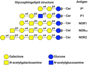
P1PK is a human blood group system based upon the A4GALT gene on chromosome 22. The P antigen was first described by Karl Landsteiner and Philip Levine in 1927. The P1PK blood group system consists of three glycosphingolipid antigens: Pk, P1 and NOR. In addition to glycosphingolipids, terminal Galα1→4Galβ structures are present on complex-type N-glycans. The GLOB antigen is now the member of the separate GLOB (globoside) blood group system.

The Lewis antigen system is a human blood group system. It is based upon two genes on chromosome 19: FUT3, or Lewis gene; and FUT2, or Secretor gene. Both genes are expressed in glandular epithelia. FUT2 has a dominant allele which codes for an enzyme and a recessive allele which does not produce a functional enzyme. Similarly, FUT3 has a functional dominant allele (Le) and a non-functional recessive allele (le).

<span class="mw-page-title-main">Ii antigen system</span> Human blood group system

The Ii antigen system is a human blood group system based upon a gene on chromosome 6 and consisting of the I antigen and the i antigen. The I antigen is normally present on the cell membrane of red blood cells in all adults, while the i antigen is present in fetuses and newborns.

<span class="mw-page-title-main">Diego antigen system</span> Human blood group system

The Diego antigen system is composed of 21 blood factors or antigens carried on the Band 3 glycoprotein, also known as Anion Exchanger 1 (AE1). The antigens are inherited through various alleles of the gene SLC4A1, located on human chromosome 17. The AE1 glycoprotein is expressed only in red blood cells and, in a shortened form, in some cells in the kidney. The Diegoa antigen is fairly common in Indigenous peoples of the Americas and East Asians, but very rare or absent in most other populations, supporting the theory that the two groups share common ancestry.

<span class="mw-page-title-main">Glycophorin A</span> Protein-coding gene in the species Homo sapiens

Glycophorin A (MNS blood group), also known as GYPA, is a protein which in humans is encoded by the GYPA gene. GYPA has also recently been designated CD235a (cluster of differentiation 235a).

<span class="mw-page-title-main">ABO (gene)</span> Protein-coding gene in the species Homo sapiens

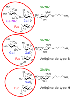
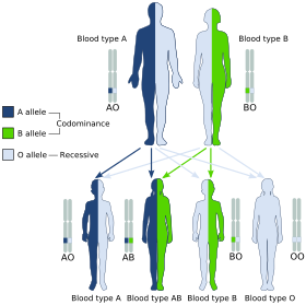
Histo-blood group ABO system transferase is an enzyme with glycosyltransferase activity, which is encoded by the ABO gene in humans. It is ubiquitously expressed in many tissues and cell types. ABO determines the ABO blood group of an individual by modifying the oligosaccharides on cell surface glycoproteins. Variations in the sequence of the protein between individuals determine the type of modification and the blood group. The ABO gene also contains one of 27 SNPs associated with increased risk of coronary artery disease.

Isoantibodies, formerly called alloantibodies, are antibodies produced by an individual against isoantigens produced by members of the same species. In the case of the species Homo sapiens, for example, there are a significant number of antigens that are different in every individual. When antigens from another individual are introduced into another's body, these isoantibodies immediately bind to and destroy them.

Cis AB is a type of rare mutation in the ABO gene. It happens when the transferase allele contains a mix of amino acids from either A or B alleles, producing a bifunctional enzyme that can produce both types of antigens, usually with one weaker than the other. This results in a serum test result much like the standard, separate (trans) AB phenotype, although the weaker antigen can occasionally fail to be detected. It complicates the basic inheritance pattern and blood-transfusion compatibility matching for ABO blood typing.

<span class="mw-page-title-main">Blood compatibility testing</span> Testing to identify incompatibilities between blood types

Blood compatibility testing is conducted in a medical laboratory to identify potential incompatibilities between blood group systems in blood transfusion. It is also used to diagnose and prevent some complications of pregnancy that can occur when the baby has a different blood group from the mother. Blood compatibility testing includes blood typing, which detects the antigens on red blood cells that determine a person's blood type; testing for unexpected antibodies against blood group antigens ; and, in the case of blood transfusions, mixing the recipient's plasma with the donor's red blood cells to detect incompatibilities (crossmatching). Routine blood typing involves determining the ABO and RhD type, and involves both identification of ABO antigens on red blood cells and identification of ABO antibodies in the plasma. Other blood group antigens may be tested for in specific clinical situations.

The Lan blood group system is a human blood group defined by the presence or absence of the Lan antigen on a person's red blood cells. More than 99.9% of people are positive for the Lan antigen. Individuals with the rare Lan-negative blood type, which is a recessive trait, can produce an anti-Lan antibody when exposed to Lan-positive blood. Anti-Lan antibodies may cause transfusion reactions on subsequent exposures to Lan-positive blood, and have also been implicated in mild cases of hemolytic disease of the newborn. However, the clinical significance of the antibody is variable. The antigen was first described in 1961, and Lan was officially designated a blood group in 2012.

The Sid blood group system is a human blood group defined by the presence or absence of the Sd(a) antigen on a person's red blood cells. About 96% of people are positive for the Sd(a) antigen, which is inherited as a dominant trait. Among Sd(a) positive individuals, the expression of the antigen ranges from extremely weak to extremely strong. Very strong expression of the antigen is referred to as a Sd(a++) phenotype. In addition to being expressed on red blood cells, Sd(a) is secreted in bodily fluids such as saliva and breast milk, and is found in the highest concentrations in urine. Urine testing is considered the most reliable method for determining a person's Sid blood type.

References

  1. Schreiber BA, Curley R, Gaur A, Rodriguez E, Rogers K, Sinha S (18 July 2017). "ABO blood group system". Encyclopædia Britannica. Encyclopædia Britannica, Inc. Retrieved 26 October 2017.
  2. "ISBT: Red Cell Immunogenetics and Blood Group Terminology". www.isbtweb.org. Archived from the original on 2 February 2022. Retrieved 25 April 2023.
  3. Storry JR, Castilho L, Chen Q, Daniels G, Denomme G, Flegel WA, et al. (August 2016). "International society of blood transfusion working party on red cell immunogenetics and terminology: report of the Seoul and London meetings". ISBT Science Series. 11 (2): 118–122. doi:10.1111/voxs.12280. PMC   5662010 . PMID   29093749.
  4. Muramatsu M, Gonzalez HD, Cacciola R, Aikawa A, Yaqoob MM, Puliatti C (March 2014). "ABO incompatible renal transplants: Good or bad?". World Journal of Transplantation. 4 (1): 18–29. doi: 10.5500/wjt.v4.i1.18 . PMC   3964193 . PMID   24669364.
  5. Maton A, Hopkins J, McLaughlin CW, Johnson S, Warner MQ, LaHart D, Wright JD (1993). Human Biology and Health . Englewood Cliffs, New Jersey, USA: Prentice Hall. ISBN   978-0-13-981176-0.
  6. Ségurel L, Thompson EE, Flutre T, Lovstad J, Venkat A, Margulis SW, et al. (November 2012). "The ABO blood group is a trans-species polymorphism in primates". Proceedings of the National Academy of Sciences of the United States of America. 109 (45): 18493–18498. arXiv: 1208.4613 . Bibcode:2012PNAS..10918493S. doi: 10.1073/pnas.1210603109 . PMC   3494955 . PMID   23091028.
  7. Landsteiner K (1900). "Zur Kenntnis der antifermentativen, lytischen und agglutinierenden Wirkungen des Blutserums und der Lymphe". Zentralblatt für Bakteriologie, Parasitenkunde und Infektionskrankheiten. 27: 357–362.
  8. Kantha SS (September 1995). "The blood revolution initiated by the famous footnote of Karl Landsteiner's 1900 paper" (PDF). The Ceylon Medical Journal. 40 (3): 123–125. PMID   8536328. Archived from the original (PDF) on 30 August 2018. Retrieved 1 June 2018.
  9. 1 2 Landsteiner K (1961) [1901]. "On agglutination of normal human blood". Transfusion. 1 (1): 5–8. doi:10.1111/j.1537-2995.1961.tb00005.x. PMID   13758692. S2CID   40158397.Originally published in German in Wiener Klinische Wochenschrift, 46, 1132–1134
  10. 1 2 3 Durand JK, Willis MS (2010). "Karl Landsteiner, MD: Transfusion Medicine". Laboratory Medicine. 41 (1): 53–55. doi: 10.1309/LM0MICLH4GG3QNDC . ISSN   0007-5027.
  11. Von Decastello AS, Sturli A (1902). "Concerning isoagglutinins in serum of healthy and sick humans". Munchener Medizinische Wochenschrift. 26: 1090–1095.
  12. 1 2 Farr AD (April 1979). "Blood group serology--the first four decades (1900--1939)". Medical History. 23 (2): 215–226. doi:10.1017/S0025727300051383. PMC   1082436 . PMID   381816.
  13. Farhud DD, Zarif Yeganeh M (2013). "A brief history of human blood groups". Iranian Journal of Public Health. 42 (1): 1–6. PMC   3595629 . PMID   23514954.)
  14. Von Dungern E, Hirschfeld L (1910). "Über Vererbung gruppenspezifischer Strukturen des Blutes". Zeitschrift für Immunitätsforschung und Experimentelle Therapie. G. Fischer.: 284–292. Die Vererbung der durch Isoagglutinine nachweisbaren spezifischen Strukturen A und B der menschlichen Blutkfirper erfolgt nach der Mendelschen Regel, wobei die Eigenschaft der Struktur dominant ist, das Fehlen der Struktur rezessiv. Die Tatsache, dafi die nachweisbaren Bestandteile der Blutkorper niemals rezessiv sind und daher bei den Kindern nie erscheinen, wenn nicht einer der Eltern sie enthait, ist forensisch zu verwerten.
  15. Janský J (1907). "Haematologick studie u. psychotiku". Sborn. Klinick (in Czech). 8: 85–139.
  16. Moss WL (1910). "Studies on isoagglutinins and isohemolysins". Bulletin of the Johns Hopkins Hospital. 21: 63–70.
  17. Doan CA (1927). "The Transfusion problem". Physiological Reviews. 7 (1): 1–84. doi:10.1152/physrev.1927.7.1.1. ISSN   0031-9333.
  18. 1 2 Schmidt P, Okroi M (2001). "Also sprach Landsteiner – Blood Group 'O' or Blood Group 'NULL'". Transfusion Medicine and Hemotherapy. 28 (4): 206–208. doi:10.1159/000050239. ISSN   1660-3796. S2CID   57677644.
  19. Garratty G, Dzik W, Issitt PD, Lublin DM, Reid ME, Zelinski T (April 2000). "Terminology for blood group antigens and genes-historical origins and guidelines in the new millennium". Transfusion. 40 (4): 477–489. doi:10.1046/j.1537-2995.2000.40040477.x. PMID   10773062. S2CID   23291031.
  20. Crow JF (January 1993). "Felix Bernstein and the first human marker locus". Genetics. 133 (1): 4–7. doi:10.1093/genetics/133.1.4. PMC   1205297 . PMID   8417988.
  21. Morgan WT, Watkins WM (January 1969). "Genetic and biochemical aspects of human blood-group A-, B-, H-, Le-a- and Le-b-specificity". British Medical Bulletin. 25 (1): 30–34. doi:10.1093/oxfordjournals.bmb.a070666. PMID   5782756.
  22. Watkins WM (1980). "Biochemistry and Genetics of the ABO, Lewis, and P Blood Group Systems". In Harris H, Hirschhorn K (eds.). Advances in Human Genetics 10. Vol. 10. New York: Plenum. pp. 1–136. doi:10.1007/978-1-4615-8288-5_1. ISBN   9781461582885. PMID   6156588.
  23. Watkins WM, Morgan WT (April 1959). "Possible genetical pathways for the biosynthesis of blood group mucopolysaccharides". Vox Sanguinis. 4 (2): 97–119. doi:10.1111/j.1423-0410.1959.tb04023.x. PMID   13669421. S2CID   45265348.
  24. Finne J, Krusius T, Rauvala H, Kekomaki R, Myllyla G (May 1978). "Alkali-stable blood group A- and B-active poly(glycosyl)-peptides from human erythrocyte membrane". FEBS Letters. 89 (1): 111–115. doi:10.1016/0014-5793(78)80534-1. PMID   26599. S2CID   825499.
  25. Krusius T, Finne J, Rauvala H (December 1978). "The poly(glycosyl) chains of glycoproteins. Characterisation of a novel type of glycoprotein saccharides from human erythrocyte membrane". European Journal of Biochemistry. 92 (1): 289–300. doi: 10.1111/j.1432-1033.1978.tb12747.x . PMID   729592.
  26. Järnefelt J, Rush J, Li YT, Laine RA (November 1978). "Erythroglycan, a high molecular weight glycopeptide with the repeating structure [galactosyl-(1 leads to 4)-2-deoxy-2-acetamido-glucosyl(1 leads to 3)] comprising more than one-third of the protein-bound carbohydrate of human erythrocyte stroma". The Journal of Biological Chemistry. 253 (22): 8006–8009. doi: 10.1016/S0021-9258(17)34351-X . PMID   711733.
  27. Laine RA, Rush JS (1988). "Chemistry of Human Erythrocyte Polylactosamine Glycopeptides (Erythroglycans) as Related to ABH Blood Group Antigenic Determinants". In Wu A, Kabat E (eds.). The Molecular Immunology of Complex Carbohydrates. Advances in Experimental Medicine and Biology. Vol. 228. Plenum Publishing Corporation. pp. 331–347. doi:10.1007/978-1-4613-1663-3_12. ISBN   9781461316633. PMID   2459929.
  28. Finne J (February 1980). "Identification of the blood-group ABH-active glycoprotein components of human erythrocyte membrane". European Journal of Biochemistry. 104 (1): 181–189. doi:10.1111/j.1432-1033.1980.tb04414.x. PMID   6768549.
  29. Yamamoto F, Clausen H, White T, Marken J, Hakomori S (May 1990). "Molecular genetic basis of the histo-blood group ABO system". Nature. 345 (6272): 229–233. Bibcode:1990Natur.345..229Y. doi:10.1038/345229a0. PMID   2333095. S2CID   4237562.
  30. Hara A, Imamura A, Ando H, Ishida H, Kiso M (December 2013). "A new chemical approach to human ABO histo-blood group type 2 antigens". Molecules. 19 (1): 414–437. doi: 10.3390/molecules19010414 . PMC   6270767 . PMID   24384923.
  31. Klug WS, Cummings MR (1997). Concepts of Genetics (5th ed.). Upper Saddle River, NJ: Prentice Hall. p. 83. ISBN   978-0135310625.
  32. Ferguson-Smith MA, Aitken DA, Turleau C, de Grouchy J (September 1976). "Localisation of the human ABO: Np-1: AK-1 linkage group by regional assignment of AK-1 to 9q34". Human Genetics. 34 (1): 35–43. doi:10.1007/BF00284432. PMID   184030. S2CID   12384399.
  33. Yazer MH, Olsson ML, Palcic MM (July 2006). "The cis-AB blood group phenotype: fundamental lessons in glycobiology". Transfusion Medicine Reviews. 20 (3): 207–217. doi:10.1016/j.tmrv.2006.03.002. PMID   16787828.
  34. "ABO inheritance patterns". Inheritance patterns of blood groups. Australian Red Cross Blood Service. Retrieved 30 October 2013.
  35. "ABO blood group system". Abobloodtypes.webnode.com. Retrieved 2 February 2015.
  36. Cited in "Misattributed paternity rates and non-paternity rates". Archived from the original on 26 August 2023. Retrieved 10 February 2022.
  37. "Understanding genetics". Stanford. Archived from the original on 10 August 2020.
  38. 1 2 Blood Group A Suptypes, The Owen Foundation. Retrieved 1 July 2008.
  39. Seltsam A, Hallensleben M, Kollmann A, Blasczyk R (October 2003). "The nature of diversity and diversification at the ABO locus". Blood. 102 (8): 3035–3042. doi: 10.1182/blood-2003-03-0955 . PMID   12829588.
  40. Ogasawara K, Bannai M, Saitou N, Yabe R, Nakata K, Takenaka M, et al. (June 1996). "Extensive polymorphism of ABO blood group gene: three major lineages of the alleles for the common ABO phenotypes". Human Genetics. 97 (6): 777–783. doi:10.1007/BF02346189. PMID   8641696. S2CID   12076999.
  41. Shastry S, Bhat S (October 2010). "Imbalance in A₂ and A₂B phenotype frequency of ABO group in South India". Blood Transfusion = Trasfusione del Sangue. 8 (4): 267–270. doi:10.2450/2010.0147-09. PMC   2957492 . PMID   20967168.
  42. Chun S, Choi S, Yu H, Cho D (March 2019). "Cis-AB, the Blood Group of Many Faces, Is a Conundrum to the Novice Eye". Annals of Laboratory Medicine. 39 (2): 115–120. doi:10.3343/alm.2019.39.2.115. PMC   6240514 . PMID   30430772.
  43. Potts WT (1979). "History and Blood Groups in the British Isles". In Sawyer PH (ed.). English Medieval Settlement. St. Martin's Press. ISBN   978-0-7131-6257-8.
  44. Cserti CM, Dzik WH (October 2007). "The ABO blood group system and Plasmodium falciparum malaria". Blood. 110 (7): 2250–2258. doi: 10.1182/blood-2007-03-077602 . PMID   17502454.
  45. 1 2 Calafell F, Roubinet F, Ramírez-Soriano A, Saitou N, Bertranpetit J, Blancher A (September 2008). "Evolutionary dynamics of the human ABO gene". Human Genetics. 124 (2): 123–135. doi:10.1007/s00439-008-0530-8. PMID   18629539. S2CID   12450238.
  46. van Oss CJ (August 2004). ""Natural" versus regular antibodies". The Protein Journal. 23 (6): 357, author reply 359-357, author reply 360. doi:10.1023/B:JOPC.0000039625.56296.6e. PMID   15517982. S2CID   189929325. Archived from the original on 14 July 2011. Retrieved 28 November 2009.
  47. Seymour RM, Allan MJ, Pomiankowski A, Gustafsson K (May 2004). "Evolution of the human ABO polymorphism by two complementary selective pressures". Proceedings. Biological Sciences. 271 (1543): 1065–1072. doi:10.1098/rspb.2004.2674. PMC   1691687 . PMID   15293861.
  48. Reid ME, Mohandas N (April 2004). "Red blood cell blood group antigens: structure and function". Seminars in Hematology. 41 (2): 93–117. doi:10.1053/j.seminhematol.2004.01.001. PMID   15071789.
  49. Mohandas N, Narla A (March 2005). "Blood group antigens in health and disease". Current Opinion in Hematology. 12 (2): 135–140. doi:10.1097/01.moh.0000153000.09585.79. PMID   15725904. S2CID   19866834.
  50. Sarode R, Goldstein J, Sussman II, Nagel RL, Tsai HM (June 2000). "Role of A and B blood group antigens in the expression of adhesive activity of von Willebrand factor". British Journal of Haematology. 109 (4): 857–864. doi:10.1046/j.1365-2141.2000.02113.x. PMID   10929042. S2CID   25343413.
  51. O'Donnell J, Laffan MA (August 2001). "The relationship between ABO histo-blood group, factor VIII and von Willebrand factor". Transfusion Medicine. 11 (4): 343–351. doi: 10.1046/j.1365-3148.2001.00315.x . PMID   11532189. S2CID   23603864.
  52. O'Donnell J, Boulton FE, Manning RA, Laffan MA (February 2002). "Amount of H antigen expressed on circulating von Willebrand factor is modified by ABO blood group genotype and is a major determinant of plasma von Willebrand factor antigen levels". Arteriosclerosis, Thrombosis, and Vascular Biology. 22 (2): 335–341. doi: 10.1161/hq0202.103997 . PMID   11834538. S2CID   325644.
  53. Gill JC, Endres-Brooks J, Bauer PJ, Marks WJ, Montgomery RR (June 1987). "The effect of ABO blood group on the diagnosis of von Willebrand disease". Blood. 69 (6): 1691–1695. doi: 10.1182/blood.V69.6.1691.1691 . PMID   3495304.
  54. Shima M, Fujimura Y, Nishiyama T, Tsujiuchi T, Narita N, Matsui T, et al. (1995). "ABO blood group genotype and plasma von Willebrand factor in normal individuals". Vox Sanguinis. 68 (4): 236–240. doi:10.1111/j.1423-0410.1995.tb02579.x. PMID   7660643. S2CID   11582510.
  55. Bowen DJ, Collins PW, Lester W, Cumming AM, Keeney S, Grundy P, et al. (March 2005). "The prevalence of the cysteine1584 variant of von Willebrand factor is increased in type 1 von Willebrand disease: co-segregation with increased susceptibility to ADAMTS13 proteolysis but not clinical phenotype". British Journal of Haematology. 128 (6): 830–836. doi: 10.1111/j.1365-2141.2005.05375.x . PMID   15755288. S2CID   45434815.
  56. Bongers TN, de Maat MP, van Goor ML, Bhagwanbali V, van Vliet HH, Gómez García EB, et al. (November 2006). "High von Willebrand factor levels increase the risk of first ischemic stroke: influence of ADAMTS13, inflammation, and genetic variability". Stroke. 37 (11): 2672–2677. doi: 10.1161/01.STR.0000244767.39962.f7 . PMID   16990571.
  57. Hollenhorst, Marie A.; Tiemeyer, Katherine H.; Mahoney, Keira E.; Aoki, Kazuhiro; Ishihara, Mayumi; Lowery, Sarah C.; Rangel-Angarita, Valentina; Bertozzi, Carolyn R.; Malaker, Stacy A. (April 2023). "Comprehensive analysis of platelet glycoprotein Ibα ectodomain glycosylation". Journal of Thrombosis and Haemostasis. 21 (4): 995–1009. doi:10.1016/j.jtha.2023.01.009. PMC   10065957 . PMID   36740532.
  58. Goldman M (February 2007). "Translational mini-review series on Toll-like receptors: Toll-like receptor ligands as novel pharmaceuticals for allergic disorders". Clinical and Experimental Immunology. 147 (2): 208–216. doi:10.1111/j.1365-2249.2006.03296.x. PMC   1810467 . PMID   17223960.
  59. Kay HE (November 1982). "Bone marrow transplantation: 1982". British Medical Journal. 285 (6351): 1296–1298. doi:10.1136/bmj.285.6351.1296. PMC   1500229 . PMID   6812684.
  60. Hakomori S (December 1999). "Antigen structure and genetic basis of histo-blood groups A, B and O: their changes associated with human cancer". Biochimica et Biophysica Acta (BBA) - General Subjects. 1473 (1): 247–266. doi:10.1016/s0304-4165(99)00183-x. PMID   10580143.
  61. 1 2 Dabelsteen E, Gao S (January 2005). "ABO blood-group antigens in oral cancer". Journal of Dental Research. 84 (1): 21–28. doi:10.1177/154405910508400103. PMID   15615870. S2CID   16975373.
  62. Dabelsteen E (February 2002). "ABO blood group antigens in oral mucosa. What is new?". Journal of Oral Pathology & Medicine. 31 (2): 65–70. doi:10.1046/j.0904-2512.2001.00004.x. PMID   11896825.
  63. Amundadottir LT (1 January 2016). "Pancreatic Cancer Genetics". International Journal of Biological Sciences. 12 (3): 314–325. doi:10.7150/ijbs.15001. PMC   4753160 . PMID   26929738.
  64. McGovern DP, Jones MR, Taylor KD, Marciante K, Yan X, Dubinsky M, et al. (September 2010). "Fucosyltransferase 2 (FUT2) non-secretor status is associated with Crohn's disease". Human Molecular Genetics. 19 (17): 3468–3476. doi:10.1093/hmg/ddq248. PMC   2916706 . PMID   20570966.
  65. de Lange KM, Moutsianas L, Lee JC, Lamb CA, Luo Y, Kennedy NA, et al. (February 2017). "Genome-wide association study implicates immune activation of multiple integrin genes in inflammatory bowel disease". Nature Genetics. 49 (2): 256–261. doi:10.1038/ng.3760. PMC   5289481 . PMID   28067908.
  66. Rühlemann MC, Hermes BM, Bang C, Doms S, Moitinho-Silva L, Thingholm LB, et al. (February 2021). "Genome-wide association study in 8,956 German individuals identifies influence of ABO histo-blood groups on gut microbiome". Nature Genetics. 53 (2): 147–155. doi:10.1038/s41588-020-00747-1. PMID   33462482. S2CID   231641761.
  67. Mega JL, Stitziel NO, Smith JG, Chasman DI, Caulfield M, Devlin JJ, et al. (June 2015). "Genetic risk, coronary heart disease events, and the clinical benefit of statin therapy: an analysis of primary and secondary prevention trials". Lancet. 385 (9984): 2264–2271. doi:10.1016/S0140-6736(14)61730-X. PMC   4608367 . PMID   25748612.
  68. Liu QP, Sulzenbacher G, Yuan H, Bennett EP, Pietz G, Saunders K, et al. (April 2007). "Bacterial glycosidases for the production of universal red blood cells". Nature Biotechnology. 25 (4): 454–464. doi:10.1038/nbt1298. PMID   17401360. S2CID   29804004.
  69. Kruskall MS, AuBuchon JP, Anthony KY, Herschel L, Pickard C, Biehl R, et al. (November 2000). "Transfusion to blood group A and O patients of group B RBCs that have been enzymatically converted to group O". Transfusion. 40 (11): 1290–1298. doi:10.1046/j.1537-2995.2000.40111290.x. PMID   11099655. S2CID   22740438.
  70. Rahfeld P, Withers SG (January 2020). "Toward universal donor blood: Enzymatic conversion of A and B to O type". J Biol Chem. 295 (2): 325–334. doi: 10.1074/jbc.REV119.008164 . PMC   6956546 . PMID   31792054.
  71. "Scientists create 'plastic' blood". BBC. 11 May 2007. Retrieved 7 April 2019.
  72. Southern California Blood Services Region. "Answers to Commonly Asked Questions About Blood and Blood Banking". Blood: The Basics (PDF). American Red Cross. p. 4. Archived (PDF) from the original on 29 November 2007. Retrieved 16 November 2007.
  73. Ryu SI, Sohn YW (2007). "A Review of sociocultural, behavioral, biochemical analyzes on ABO blood-groups typology". Korean Journal of Social and Personality Psychology. 21 (3): 27–55.
  74. Klein HG (7 March 2005). "Why Do People Have Different Blood Types?". Scientific American. Retrieved 16 November 2007.

Further reading