Angiocentric glioma

Last updated
Photomicrograph displaying the AG tumor cells by histological evaluation, H&E stain. The striking characteristic of AG is that cells tend to cluster around brain blood vessels. Neuropathology case V 03.jpg
Photomicrograph displaying the AG tumor cells by histological evaluation, H&E stain. The striking characteristic of AG is that cells tend to cluster around brain blood vessels.

Angiocentric glioma (AG) refers to a rare neuroepithelial tumor when the superficial brain malignant cells enclose the brain vessels, commonly found in children and young adults. Initially identified in 2005 by Wang and his team from the University of Texas, AG was classified as Grade I by 2007 WHO Classification of Tumors of the Central Nervous System due to its benign clinical behavior, low proliferation index, and curative properties. [2] AG primarily affects children and young adults at an average initial diagnosis age of 16 years old. Over 85% AG patients experience intractable seizures since childhood, especially partial epilepsy. [3]

Contents

Due to its short history of 15 years, the rarity of occurrence, and a lack of sufficient clinical trials, AG remains elusive on understanding symptoms, treatments, and long-term follow-up. Till now, scientists and researchers have not found the exact etiology, definitive pathological tests for identification, and the effect of radiation or chemotherapy on this rare indolent glioma. Yet, a series of suspected causes are under discussion, including the possible MYB-QKI protein fusion theory on AG etiology. Currently, the standard diagnostic tools are MRI (Magnetic Resonance Imaging) and Computed Tomography scan (CT scan). In terms of therapy, patients often undergo subtotal or total resection to remove the problematic lesion and have a relatively high likelihood of curing the disease. However, they still require more extended follow-up periods after surgery for monitoring tumor recurrence and assuring seizure-free.

History

Prior to AG's initiation, the most frequent and best-known types of rare glioneuronal tumors in the brain were dysembryoplastic neuroepithelial tumors and gangliogliomas. [4] In 2002, Wang and his group from the University of Texas recognised a novel neurological pattern in three cerebral hemispheric tumor cases and first published this rare subtype of AG tumor to the American Association of Neuropathologists. [5] In 2005, Lellouch-Tubiana and his team analyzed 204 epilepsy surgery specimens histologically from patients aged 2 to 14 and discovered three very similar cases shown by Wang et al. in terms of pathological appearance and MRI results. [4] AG manifests a distinct architectural neuron pattern and morphologic features, which were subsequently characterized as a new clinicopathologic entity named "angiocentric neuroepithelial tumors" within the spectrum of children glioneuronal tumors. [4] Simultaneously in the same month in 2005, Wang et al. also published their further discoveries of five AG cases and indicated the specified histologic, immunohistochemical, and ultrastructural properties of this seizure-producing low growing tumor. [5] In 2007, the Fourth Edition of the World Health Organization Classification of Tumors of the Central Nervous System recognized AG as a new clinicopathologic entity and designated it as an epilepsy-associated Grade I tumor with monomorphous bipolar cells and a unique perivascular growth arrangement in the category of "other neuroepithelial tumors". [2] In 2016, WHO made a few amendments in the tumor classification parameter and classified AG, choroid glioma of the third ventricle, and astroblastoma as Grade I "Other Gliomas" after revision. [6]

Signs and symptoms

Patients usually experience a history of intractable seizures at 3 to 14 years old. [7] The seizure severity often depends on the tumor locations rather than its size, as superficial lesions located in the frontal and temporal lobes often trigger a higher epileptogenic possibility than the deeper tumors. [8] Common symptoms also include headache, vision impairment, dizziness, otalgia, speech arrest, ataxia, and paresis, predominantly in children and young adults. [9] The symptoms of low-grade, slow-growing gliomas are more epileptogenic, whereas those high-grade gliomas manifest symptoms related to increased intracranial pressure. [10]

Causes

Many studies on AG have prioritized neuroradiological features, clinical highlights, pathophysiology, and surgical treatment of this rare disease while lacking discussion on its causes. AG's cytogenesis has remained elusive since 2005, and researchers only proposed several possible mechanisms for this low-grade child brain tumor.

As early as 2005, the first published research on AG postulated that ependymoma and variants of astroblastoma might contribute to the AG tumor formation. [5] In the same year, another group of researchers suggested AG possibly derives from the neoplastic transformation of radial glial cells during neuronal migration as they present a similar proliferation pattern. [4] However, these two publications have not provided further experiments to prove their rationales.

MYB-QKI Protein Fusion

In 2016, Bandopadhayay and his research team proposed a potential genetic abnormality attributor for AG formation. According to their proposal, gene fusions that activate the mitogen-associated protein kinase (MAPK) pathway can induce AG malignant cells. [11] After completing combined genomic analysis of whole-genome sequencing and RNA sequencing data from 172 paediatric low-grade gliomas (PLGG) subtypes, they recognized protein fusion of the MYB and QKI gene in most AG profiles, thus hypothesizing it to be the tumor-causing driver. MYB is a proto-oncogene, whereas QKI is a tumor-suppressor gene. The abnormal fusion of the two may impose a malignant effect on neurologic cells as the rearrangement of QKI and MYB can prompt an increased expression of in-frame MYB-QKI fusion protein than the normal paediatric cortical brain. The research group proved that this protein alternation could be the critical contributor to AG oncogenicity since mice showed an enhanced cell proliferation rate after intracranial injection in experiments. The same article also pointed out that MYB-QKI protein facilitates the growth of vessels around the glioma.

Low recurrence after surgery

AG often behaves as a low-grade indolent neoplasm and is curative after surgical resection. Researchers proposed that since AG does not acquire mutations of isocitrate dehydrogenase-1, making it have a lower recurrence potential after surgery compared to WHO grade II and III diffuse gliomas and secondary glioblastomas. [3]

Diagnosis

MRI scan of a four-year-old boy with AG. The light grey part indicated a hyperintense and elastic lesion in his left posterior frontal lobe. 4-year-old-boy-with-angiocentric-glioma-Axial-T2weighted-image-shows-a-hyperintense.png
MRI scan of a four-year-old boy with AG. The light grey part indicated a hyperintense and elastic lesion in his left posterior frontal lobe.
The Computed Tomography scan (CT scan) is medical imaging machinery for body diagnostic using X-rays. 13-11-12-rechtsmedizin-berlin-charite-by-RalfR-10.jpg
The Computed Tomography scan (CT scan) is medical imaging machinery for body diagnostic using X-rays.
Magnetic Resonance Imaging (MRI) is a medical technique for capturing body images using radio waves. MRI-Philips.JPG
Magnetic Resonance Imaging (MRI) is a medical technique for capturing body images using radio waves.

The neurological features of AG tumors are visible via CT scanning or MRI. A clear indication of AG may appear as well-delineated, solid, T2-hyperintense, non-enhancing cortical lesions located in the temporal or frontal lobes in MRI. [1] Another diagnostic trait is a stalk-like extension to adjacent brain ventricles. [13] These traits are similar to low-grade gliomas from a radiological perspective.

The results from CT scanning and MRI are different in terms of clarity and effectiveness of diagnosis. AG displays an expansive non-enhancing cortical tumor in CT scanning, whereas MRI shows a relatively clearer appearance of AG and the tumors appear to be infiltrative, well-defined, and hypointense with T1 lesion. [4] T2/FLAIR lesions indicate AG as a tumor tissue with some extension toward the ventricles along vessels. [4] The possibility of cystic-appearing areas exists as well. In some cases, MRI results show an increase in ribbon-like signal on T1W1 lesions. [4] The clear radiographic outcomes of MRI makes it the more widely used option in the diagnosis of AG. [4]

Nevertheless, precise diagnosis of AG from other phenotypically similar gliomas (such as astroblastoma or ganglioglioma) is a challenge merely based on MRI or CT scanning. [14] The main difference between AG and ganglioglioma could be only AG shows enhancement over time. Compared to AG, astroblastoma often has a discrete border in epithelioid cells and shows vascular sclerosis symptoms. [15]

For further confirmation, the clinicians require biopsy and immunohistochemical staining of the resected tumor after surgery. The infiltrative AG cells display positive results for several immunostaining s, especially the glial fibrillary acidic protein (GFAP) and epithelial membrane antigen (EMA). [1] Clinicians also observe a specific dot-like pattern from the stained EMA photomicrograph. [3] Other specific AG immunohistochemical tests include Ki-67 proliferative marker, neurospecific nucleoprotein (NeuN), protein 53, synaptophysin (Syn), oligodendrocyte transcription factor-2 (Olig-2) and creatine kinase (CK). [3] In the 2016 WHO classification of CNS tumors, AG is characterised as GFAP-positive, NeuN-positive and low Ki-67 proliferative rate with a perivascular growth pattern. [13]

Photomicrograph displaying the AG tumor cells by histological evaluation, H&E stain.Characteristics of AG: low Ki-67 proliferative rate, GFAP-positive, NeuN-positive, S-100-positive, Protein53-negative, Syn-negative, Olig-2-negative, CK-negative. Neuropathology case V 01.jpg
Photomicrograph displaying the AG tumor cells by histological evaluation, H&E stain.Characteristics of AG: low Ki-67 proliferative rate, GFAP-positive, NeuN-positive, S-100-positive, Protein53-negative, Syn-negative, Olig-2-negative, CK-negative.

Treatment

The definitive treatment for this low-grade entity is surgical resection. Given its rarity and short history, clinicians have not found other better treatment options for AG. [14] Nearly all AG patients either choose subtotal or total resection to remove the tumor tissue in the brain surgically. A total resection manages to regress epileptogenic growth and cure this brain neoplasm. [14] Subtotal resection shows a comparably higher recurrence rate and tumor progression with limited control of seizures. [14] The overall outcome of tumoral resection is highly ideal with a considerably favorable prognosis. [16] Additionally, the effectiveness of chemotherapy or radiation is yet to be known as these aggressive treatments are considered inappropriate for low-grade AG. [17] It is also rare to aid the treatment with extra radiotherapy. [14]

Epidemiology

By June 2020, the reported AG cases have reached 108 since the initial report from Wang et al. Within the reported cases, it happens mostly in children and young adults. [3] The average age of initial diagnosis is 16 year-old, and the prevalence in males to females is in a ratio of 1.5 to 1. [3] The initial diagnosis range varies from the age of 1.5 to 83, with a median of 13. [3]

102 out of 108 reported cases had AG tumors in a supratentorial location under the cerebral cortex (94.4%), and 88 out of 108 were found in a single lobe (81.5%). [3] 46 cases were in the left lobe of the brain and 43 in the right lobe. [3] The most common location where AG starts to grow is the temporal lobe with 39% of reported cases, followed by parietal (30%) and frontal lobes (15%). [9] The less common growth site is the thalamus, with only 1% of reported cases, and only six cases of brainstem lesions have been reported. [8]

94 out of 108 (87%) patients chose to conduct a certain degree of surgical removal: 61 patients took gross total resection (GTR, 64.9%), and 26 adopted subtotal resection (STR, 27.7%). [3] The resection outcome was highly ideal, with 93.1% of the patient completely free of seizure during the follow-up period. [3] The remaining six patients who chose to conduct STR experienced some degrees of reoccurred symptoms. [3]  This indicates that AG has a favourable prognosis with a comparatively low mortality rate and recurrence rate.

Related Research Articles

Brain tumor Neoplasm in the brain

A brain tumor occurs when abnormal cells form within the brain. There are two main types of tumors: malignant tumors and benign (non-cancerous) tumors. These can be further classified as primary tumors, which start within the brain, and secondary tumors, which most commonly have spread from tumors located outside the brain, known as brain metastasis tumors. All types of brain tumors may produce symptoms that vary depending on the size of the tumor and the part of the brain that is involved. Where symptoms exist, they may include headaches, seizures, problems with vision, vomiting and mental changes. Other symptoms may include difficulty walking, speaking, with sensations, or unconsciousness.

Glioma Tumour of the glial cells of the brain or spine

A glioma is a type of tumor that starts in the glial cells of the brain or the spine. Gliomas comprise about 30 percent of all brain tumors and central nervous system tumours, and 80 percent of all malignant brain tumours.

Oligodendroglioma Medical condition

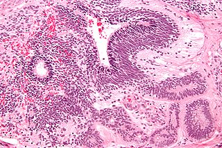
Oligodendrogliomas are a type of glioma that are believed to originate from the oligodendrocytes of the brain or from a glial precursor cell. They occur primarily in adults but are also found in children.

Meningioma Type of tumor

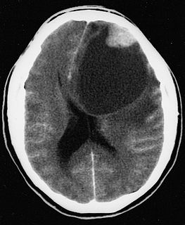
Meningioma, also known as meningeal tumor, is typically a slow-growing tumor that forms from the meninges, the membranous layers surrounding the brain and spinal cord. Symptoms depend on the location and occur as a result of the tumor pressing on nearby tissue. Many cases never produce symptoms. Occasionally seizures, dementia, trouble talking, vision problems, one sided weakness, or loss of bladder control may occur.

Glioblastoma Aggressive type of brain cancer

Glioblastoma, previously known as glioblastoma multiforme (GBM), is one of the most aggressive type of cancers that begin within the brain. Initially, signs and symptoms of glioblastoma are nonspecific. They may include headaches, personality changes, nausea, and symptoms similar to those of a stroke. Symptoms often worsen rapidly and may progress to unconsciousness.

Astrocytoma Medical condition

Astrocytomas are a type of brain tumor. They originate in a particular kind of glial cells, star-shaped brain cells in the cerebrum called astrocytes. This type of tumor does not usually spread outside the brain and spinal cord and it does not usually affect other organs. Astrocytomas are the most common glioma and can occur in most parts of the brain and occasionally in the spinal cord.

Oligoastrocytoma Medical condition

Oligoastrocytomas are a subset of brain tumors that present with an appearance of mixed glial cell origin, astrocytoma and oligodendroglioma. However, the term "Oligoastrocytoma" is now considered obsolete by the National Comprehensive Cancer Network stating "the term should no longer be used as such morphologically ambiguous tumors can be reliably resolved into astrocytomas and oligodendrogliomas with molecular testing."

Pilocytic astrocytoma Medical condition

Pilocytic astrocytoma is a brain tumor that occurs most commonly in children and young adults. They usually arise in the cerebellum, near the brainstem, in the hypothalamic region, or the optic chiasm, but they may occur in any area where astrocytes are present, including the cerebral hemispheres and the spinal cord. These tumors are usually slow growing and benign, corresponding to WHO malignancy grade 1.

Temporal lobe epilepsy Chronic focal seizure disorder

Temporal lobe epilepsy (TLE) is a chronic disorder of the nervous system which is characterized by recurrent, unprovoked focal seizures that originate in the temporal lobe of the brain and last about one or two minutes. TLE is the most common form of epilepsy with focal seizures. A focal seizure in the temporal lobe may spread to other areas in the brain when it may become a focal to bilateral seizure.

Electrocorticography

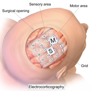
Electrocorticography (ECoG), or intracranial electroencephalography (iEEG), is a type of electrophysiological monitoring that uses electrodes placed directly on the exposed surface of the brain to record electrical activity from the cerebral cortex. In contrast, conventional electroencephalography (EEG) electrodes monitor this activity from outside the skull. ECoG may be performed either in the operating room during surgery or outside of surgery. Because a craniotomy is required to implant the electrode grid, ECoG is an invasive procedure.

Epilepsy surgery involves a neurosurgical procedure where an area of the brain involved in seizures is either resected, ablated, disconnected or stimulated. The goal is to eliminate seizures or significantly reduce seizure burden. Approximately 60% of all people with epilepsy have focal epilepsy syndromes. In 15% to 20% of these patients, the condition is not adequately controlled with anticonvulsive drugs. Such patients are potential candidates for surgical epilepsy treatment.

Dysembryoplastic neuroepithelial tumour Medical condition

Dysembryoplastic neuroepithelial tumour is a type of brain tumor. Most commonly found in the temporal lobe, DNTs have been classified as benign tumours. These are glioneuronal tumours comprising both glial and neuron cells and often have ties to focal cortical dysplasia.

Immature teratoma Medical condition

An immature teratoma is a teratoma that contains anaplastic immature elements, and is often synonymous with malignant teratoma. A teratoma is a tumor of germ cell origin, containing tissues from more than one germ cell line, It can be ovarian or testicular in its origin. and are almost always benign. An immature teratoma is thus a very rare tumor, representing 1% of all teratomas, 1% of all ovarian cancers, and 35.6% of malignant ovarian germ cell tumors. It displays a specific age of incidence, occurring most frequently in the first two decades of life and almost never after menopause. Unlike a mature cystic teratoma, an immature teratoma contains immature or embryonic structures. It can coexist with mature cystic teratomas and can constitute of a combination of both adult and embryonic tissue. The most common symptoms noted are abdominal distension and masses. Prognosis and treatment options vary and largely depend on grade, stage and karyotype of the tumor itself.

Pleomorphic xanthoastrocytoma Medical condition

Pleomorphic xanthoastrocytoma (PXA) is a brain tumor that occurs most frequently in children and teenagers. At Boston Children's Hospital, the average age at diagnosis is 12 years.

Neuro-oncology is the study of brain and spinal cord neoplasms, many of which are very dangerous and life-threatening. Among the malignant brain cancers, gliomas of the brainstem and pons, glioblastoma multiforme, and high-grade astrocytoma are among the worst. In these cases, untreated survival usually amounts to only a few months, and survival with current radiation and chemotherapy treatments may extend that time from around a year to a year and a half, possibly two or more, depending on the patient's condition, immune function, treatments used, and the specific type of malignant brain neoplasm. Surgery may in some cases be curative, but, as a general rule, malignant brain cancers tend to regenerate and emerge from remission easily, especially highly malignant cases. In such cases, the goal is to excise as much of the mass and as much of the tumor margin as possible without endangering vital functions or other important cognitive abilities. The Journal of Neuro-Oncology is the longest continuously published journal in the field and serves as a leading reference to those practicing in the area of neuro-oncology.

Tuber cinereum hamartoma Human disease

Tuber cinereum hamartoma is a benign tumor in which a disorganized collection of neurons and glia accumulate at the tuber cinereum of the hypothalamus on the floor of the third ventricle. It is a congenital malformation, included on the spectrum of gray matter heterotopias. Formation occurs during embryogenesis, typically between days 33 and 41 of gestation. Size of the tumor varies from one to three centimeters in diameter, with the mean being closer to the low end of this range. It is estimated to occur at a frequency of one in one million individuals.

Cortical stimulation mapping (CSM) is a type of electrocorticography that involves a physically invasive procedure and aims to localize the function of specific brain regions through direct electrical stimulation of the cerebral cortex. It remains one of the earliest methods of analyzing the brain and has allowed researchers to study the relationship between cortical structure and systemic function. Cortical stimulation mapping is used for a number of clinical and therapeutic applications, and remains the preferred method for the pre-surgical mapping of the motor cortex and language areas to prevent unnecessary functional damage. There are also some clinical applications for cortical stimulation mapping, such as the treatment of epilepsy.

Low-grade fibromyxoid sarcoma Medical condition

Low-grade fibromyxoid sarcoma (LGFMS) is a rare type of low-grade sarcoma first described by H. L. Evans in 1987. LGFMS are soft tissue tumors of the mesenchyme-derived connective tissues; on microscopic examination, they are found to be composed of spindle-shaped cells that resemble fibroblasts. These fibroblastic, spindle-shaped cells are neoplastic cells that in most cases of LGFMS express fusion genes, i.e. genes composed of parts of two different genes that form as a result of mutations. The World Health Organization (2020) classified LGFMS as a specific type of tumor in the category of malignant fibroblastic and myofibroblastic tumors.

Astroblastoma Medical condition

Astroblastoma is a rare glial tumor derived from the astroblast, a type of cell that closely resembles spongioblastoma and astrocytes. Astroblastoma cells are most likely found in the supratentorial region of the brain that houses the cerebrum, an area responsible for all voluntary movements in the body. It also occurs significantly in the frontal lobe, parietal lobe, and temporal lobe, areas where movement, language creation, memory perception, and environmental surroundings are expressed. These tumors can be present in major brain areas not associated with the main cerebral hemispheres, including the cerebellum, optic nerve, cauda equina, hypothalamus, and brain stem.

Myxofibrosarcoma (MFS), although a rare type of tumor, is one of the most common soft tissue sarcomas, i.e. cancerous tumors, that develop in the soft tissues of elderly individuals. Initially considered to be a type of histiocytoma termed fibrous histiocytoma or myxoid variant of malignant fibrous histiocytoma, Angervall et al. termed this tumor myxofibrosarcoma in 1977. In 2020, the World Health Organization reclassified MFS as a separate and distinct tumor in the category of malignant fibroblastic and myofibroblastic tumors.

References

  1. 1 2 3 Shakur SF, McGirt MJ, Johnson MW, Burger PC, Ahn E, Carson BS, Jallo GI (March 2009). "Angiocentric glioma: a case series". Journal of Neurosurgery. Pediatrics. 3 (3): 197–202. doi:10.3171/2008.11.PEDS0858. PMC   2675755 . PMID   19338465.
  2. 1 2 Brat DJ, Scheithauer BW, Fuller GN, Tihan T (July 2007). "Newly codified glial neoplasms of the 2007 WHO Classification of Tumours of the Central Nervous System: angiocentric glioma, pilomyxoid astrocytoma and pituicytoma". Brain Pathology. 17 (3): 319–324. doi:10.1111/j.1750-3639.2007.00082.x. PMC   8095654 . PMID   17598825.
  3. 1 2 3 4 5 6 7 8 9 10 11 12 13 Han G, Zhang J, Ma Y, Gui Q, Yin S (August 2020). "Clinical characteristics, treatment and prognosis of angiocentric glioma". Oncology Letters. 20 (2): 1641–1648. doi:10.3892/ol.2020.11723. PMC   7377082 . PMID   32724405.
  4. 1 2 3 4 5 6 7 8 Lellouch-Tubiana A, Boddaert N, Bourgeois M, Fohlen M, Jouvet A, Delalande O, et al. (October 2005). "Angiocentric neuroepithelial tumor (ANET): a new epilepsy-related clinicopathological entity with distinctive MRI". Brain Pathology. 15 (4): 281–286. doi:10.1111/j.1750-3639.2005.tb00112.x. PMC   8095937 . PMID   16389940.
  5. 1 2 3 Wang M, Tihan T, Rojiani AM, Bodhireddy SR, Prayson RA, Iacuone JJ, et al. (October 2005). "Monomorphous angiocentric glioma: a distinctive epileptogenic neoplasm with features of infiltrating astrocytoma and ependymoma". Journal of Neuropathology and Experimental Neurology. 64 (10): 875–881. doi:10.1097/01.jnen.0000182981.02355.10. PMID   16215459. S2CID   15439580.
  6. Louis DN, Perry A, Reifenberger G, von Deimling A, Figarella-Branger D, Cavenee WK, et al. (June 2016). "The 2016 World Health Organization Classification of Tumors of the Central Nervous System: a summary". Acta Neuropathologica. 131 (6): 803–820. doi:10.1007/s00401-016-1545-1. PMID   27157931. S2CID   3345100.
  7. Preusser M, Hoischen A, Novak K, Czech T, Prayer D, Hainfellner JA, et al. (November 2007). "Angiocentric glioma: report of clinico-pathologic and genetic findings in 8 cases". The American Journal of Surgical Pathology. 31 (11): 1709–1718. doi:10.1097/PAS.0b013e31804a7ebb. PMID   18059228. S2CID   41723081.
  8. 1 2 da Silva JF, de Souza Machado GH, Pedro MK, Vosgerau R, Hunhevicz SC, Ramina R (Dec 2019). "Angiocentric glioma: Literature review and first case in Brazil". Interdisciplinary Neurosurgery. 18: 100508. doi:10.1016/j.inat.2019.100508. ISSN   2214-7519. S2CID   196565334.
  9. 1 2 Alexandru D, Haghighi B, Muhonen MG (2013-03-01). "The treatment of angiocentric glioma: case report and literature review". The Permanente Journal. 17 (1): e100–e102. doi:10.7812/tpp/12-060. PMC   3627792 . PMID   23596378.
  10. Ampie L, Choy W, DiDomenico JD, Lamano JB, Williams CK, Kesavabhotla K, et al. (June 2016). "Clinical attributes and surgical outcomes of angiocentric gliomas". Journal of Clinical Neuroscience. 28: 117–122. doi:10.1016/j.jocn.2015.11.015. PMID   26778052. S2CID   1420908.
  11. Bandopadhayay P, Ramkissoon LA, Jain P, Bergthold G, Wala J, Zeid R, et al. (March 2016). "MYB-QKI rearrangements in angiocentric glioma drive tumorigenicity through a tripartite mechanism". Nature Genetics. 48 (3): 273–282. doi:10.1038/ng.3500. PMC   4767685 . PMID   26829751.
  12. Kumar M, Ramakrishnaiah R, Samant R (2013). "Angiocentric glioma, a recently added WHO grade-I tumor". Radiology Case Reports. 8 (4): 782. doi:10.2484/rcr.v8i4.782. PMC   4899553 . PMID   27330646.
  13. 1 2 Louis DN, Ohgaki H, Wiestler OD, Cavenee WK (2016). WHO classification of tumours of the central nervous system (Revised 4th ed.). Lyon: World Health Organization, International Agency for Research on Cancer. ISBN   978-92-832-4492-9. OCLC   951745876.
  14. 1 2 3 4 5 Gatto L, Franceschi E, Nunno VD, Tomasello C, Bartolini S, Brandes AA (2020-07-03). "Glioneuronal tumors: clinicopathological findings and treatment options". Future Neurology. 15 (3): FNL47. doi:10.2217/fnl-2020-0003. ISSN   1479-6708. S2CID   225450256.
  15. Zhou L, Burns DK, Cai C, eds. (2020). A Case-Based Guide to Neuromuscular Pathology. Springer International Publishing. doi:10.1007/978-3-030-25682-1. ISBN   978-3-030-25682-1. S2CID   204886503.
  16. Osborn AG, Salzman KL, Thurnher MM, Rees JH, Castillo M (May 2012). "The new World Health Organization Classification of Central Nervous System Tumors: what can the neuroradiologist really say?". AJNR. American Journal of Neuroradiology. 33 (5): 795–802. doi:10.3174/ajnr.A2583. PMC   7968804 . PMID   21835942.
  17. Mayor S (2005-05-05). "Aggressive treatment of low grade prostate cancer is unnecessary". BMJ. 330 (7499): 1042.2. doi:10.1136/bmj.330.7499.1042-a. ISSN   0959-8138. PMC   557253 .